. 2025 Apr 8;122(14):e2409729122.
doi: 10.1073/pnas.2409729122. Epub 2025 Mar 31.

A mechanically resilient soft hydrogel improves drug delivery for treating post-traumatic osteoarthritis in physically active joints

Affiliations

A mechanically resilient soft hydrogel improves drug delivery for treating post-traumatic osteoarthritis in physically active joints

Nitin Joshi et al. Proc Natl Acad Sci U S A. .

Abstract

Intra-articular delivery of disease-modifying osteoarthritis drugs (DMOADs) is likely to be most effective in the early stages of post-traumatic osteoarthritis (PTOA), when symptoms are minimal, and patients remain physically active. To ensure effective therapy, DMOAD delivery systems therefore must withstand repeated mechanical loading without altering the kinetics of drug release. While soft materials are typically preferred for DMOAD delivery, mechanical loading can compromise their structural integrity and disrupt controlled drug release. In this study, we present a mechanically resilient soft hydrogel that rapidly self-heals under conditions simulating human running while maintaining sustained release of the cathepsin-K inhibitor L-006235, used as a proof-of-concept DMOAD. This hydrogel demonstrated superior performance compared to a previously reported hydrogel designed for intra-articular drug delivery, which, in our study, neither recovered its structure nor maintained drug release under mechanical loading. When injected into mouse knee joints, the hydrogel provided consistent release kinetics of the encapsulated drug in both treadmill-running and nonrunning mice. In a mouse model of severe PTOA exacerbated by treadmill-running, the L-006235 hydrogel significantly reduced cartilage degeneration, whereas the free drug did not. Overall, our data underscore the hydrogel's potential for treating PTOA in physically active patients.

Keywords: disease modification; hydrogel; mechanical loading; osteoarthritis.


Conflict of interest statement

Competing interests statement:J.M.K. has been a paid consultant and or equity holder for multiple biotechnology companies including Alivio Therapeutics, Cobro Ventures, eClinical Solutions, Altrix Bio, Akita Bio, Eterna Tx, Sanofi, Celltex, Tissium, Corner Therapeutics, Katharos Labs, Triton Systems, Edge Immune, W. L. Gore, Camden Partners, Gyro Gear, Mirakel Labs, Pancryos, Quthero, and Vyome. The interests of J.M.K. were reviewed and are subject to a management plan overseen by his institutions in accordance with its conflict of interest policies. N.J., K.S., S.B., N.E.S., and J.M.K. have pending and issued patents on the hydrogel platform described in this manuscript.

Figures

Fig. 1.
Fig. 1.
DMOADs can be encapsulated into TG-18 hydrogel for the prevention of PTOA progression in active joints. (A) Schematic depicting intra-articular delivery of DMOAD-loaded TG-18 hydrogel as a strategy to prevent disease progression in young, active patients diagnosed with early PTOA. (B) The cathepsin-K inhibitor L-006235 was used as a proof of concept, hydrophobic DMOAD in this study. TG-18 self-assembles into lamellar structures and encapsulates L-006235 via noncovalent, hydrophobic interactions. High-resolution scanning electron microscopy (HR-SEM) image of L-006235 gel demonstrating higher-order fibrous assemblies that form the bulk hydrogel. (C) Schematic showing sustained release of the DMOAD from TG-18 hydrogel due to diffusion and gradual hydrolysis of TG-18. The viscoelastic properties of the intra-articular hydrogel recover rapidly after mechanical loading, with no premature release or loss of L-006235 during mechanical loading, and no impact on the release kinetics afterward. Parts of the figure were drawn by using pictures from Servier Medical Art. Servier Medical Art by Servier is licensed under a Creative Commons Attribution 3.0 Unported License (https://creativecommons.org/licenses/by/3.0/).
Fig. 2.
Fig. 2.
L-006235 gel recovers rapidly following mechanical loading conditions simulating human running. (A) Experimental outline to measure the effect of repeated mechanical loading conditions simulating interval running by humans on L-006235 gel, and release kinetics of L-006235. Using a rotational rheometer, the L-006235 gel was subjected to 30 cycles of alternating strain and frequency in the presence of PBS. Each cycle involved 1 min of high strain and high frequency relevant to a running human knee followed by 1 min of low strain and low frequency relevant to a resting human knee. (B) Shear storage modulus (G’) and shear loss modulus (G”) were recorded. (C and D) After 30 cycles, L-006235 gel was collected, evaluated for in vitro drug release in PBS (37 °C) and analyzed for morphology by HR-SEM. (E) Experimental outline to determine the effect of a single cycle of alternating strain and frequency on L-006235 gel at 37 °C in the presence of PBS. The cycle involved 2 min of low strain and low frequency followed by high strain and high frequency for 5, 10, or 15 min, and finally low strain and low frequency for 2 min. (F) Shear storage modulus (G’) and shear loss modulus (G”) were measured. (G) At the end of each cycle, L-006235 gel was collected and evaluated for in vitro drug release in PBS (37 °C). (H) Gel morphology was assessed using HR-SEM after a loading cycle with a 15 min step of high strain and high frequency. (I) Fraction of L-006235 or rhodamine-B (Rhod-B) released from TG-18 hydrogel in the presence of PBS during a single cycle of mechanical loading with a 15 min step of high strain and high frequency, and fraction of Rhod-B released in the absence of mechanical loading. (J) Shear storage modulus (G’) and shear loss modulus (G”) of L-006235-loaded gellan gel (L-006235 gellan gel) measured during 30 consecutive cycles of alternating strain and frequency at 37 °C. (K) After 30 cycles, L-006235 gellan gel was collected and evaluated for in vitro drug release in PBS at 37 °C (**P < 0.01 on day 28). Panels C, G, and K show individual data points (dots) overlaid on a line graph, (n = 3, each experiment performed at least twice). P value in K was determined using Student’s t test. Panel I shows means ± SD of technical repeats, represented as individual data points (dots), (n = 3, each experiment performed at least twice). Panels B, F, and J show data are from a single experiment (experiment repeated three times).
Fig. 3.
Fig. 3.
L-006235 gel is biocompatible with cells from human joints and safe for single and repeated intra-articular administration in mice. (AD) Primary human chondrocytes and fibroblast-like synoviocytes from healthy donors or OA patients were incubated in a 96-well plate in medium, PBS, or in medium with L-006235 gel or free L-006235 at a 2 nM final concentration of L-006235. Cells incubated in medium with blank TG-18 hydrogel (Blank gel) or DMSO/water at volumes equivalent to L-006235 gel were included as controls. After 24, 48, or 72 h of incubation, metabolic activity was determined by XTT assay. (E) Experimental outline to evaluate the safety of single or repeated intra-articular injections of L-006235 gel (4 µL, 60 µg L-006235) in mice. In the single dose study, healthy mice were injected on day 0 with 4 µL PBS, L-006235 gel, blank gel, or free L-006235. Two weeks later, injected knees were harvested and processed for histology. In the repeated dose study, knees were injected with 4 µL PBS or L-006235 gel on day 0, week 2, and week 4, and harvested for histology at week 6. (F) Representative images of histological sections stained with H&E or safranin O from each experimental group (Scale bar: 200 µm). Panels AD show means ± SD of technical repeats (n = 8 wells per condition, experiment performed at least twice).
Fig. 4.
Fig. 4.
Treadmill running does not impact release kinetics of encapsulated agents from TG-18 hydrogel injected into mouse knee joints. (A) Experimental outline to assess the impact of treadmill running on release of an encapsulated hydrophobic fluorescent dye (DiR) from TG-18 hydrogel. On day 0, mice were injected into both knees with TG-18 hydrogel coloaded with DiR and L-006235 (4 µL, 60 µg L-006235, and 0.4 µg DiR). Subsequently, one group ran on a treadmill at 400 m/30 min/d, 5 d/wk, while the other group did not run. Mice were imaged every other day using an IVIS. (B) IVIS images of one representative animal from each experimental group. (C and D) Fluorescence signal (normalized to day 0) measured over the injected knees and area under the curves (AUCs). (E) Experimental outline of the pharmacokinetic study. L-006235 gel or free L-006235 (4 µL, 60 µg L-006235) was injected into the right knee of healthy mice. Mice subsequently ran on a treadmill at 400 m/30 min/d, 5 d/wk. L-006235 level was quantified in the injected knee and in serum at 1 h, day 1, 6, and 14 post injection. (F and G) L-006235 level in the injected knee over time and at 1 h after injection. In mice injected with free L-006235, no drug was detected in the tissue beyond day 1 (*P < 0.05). (H and I) L-006235 serum level over time and 1 h after intra-articular injection (*P < 0.05). In mice injected with free L-006235, only two animals showed detectable drug levels on day 1, and subsequently no drug was detected. In mice injected with L-006235 gel, only two animals showed detectable drug levels on day 14. Panel C shows individual data points (dots) overlaid on a line graph, (n = 4 to 6 mice/group, experiment performed twice). Panel D shows means ± SD of individual data points (dots), (n = 4 to 6 mice/group, experiment performed twice). Panels F and H show individual data points (dots) overlaid on a line graph, (n = 3 to 6 mice/group, experiment performed twice). Panels G and I show means ± SD of individual data points (dots), (n = 3 to 6 mice/group, experiment performed twice). P values were determined using Student’s t test.
Fig. 5.
Fig. 5.
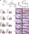
Intra-articular delivery of L-006235 in TG-18 hydrogel reduces OA severity in treadmill-running mice. (A) Experimental outline to determine the therapeutic efficacy of L-006235 gel in treadmill-running mice with surgically induced PTOA. DMM surgery or sham surgery was performed on day 0. Three weeks later, mice with DMM surgery were injected in the right knee with L-006235 gel, free L-006235, blank TG-18 hydrogel (Blank gel, 4 µL), or DMSO/water followed by repeat dosing at week 5 and 7. From week 3 to week 8, mice from all groups ran on a treadmill at 400 m/30 min/d, 5 d/wk. Mice were euthanized at week 9 and knees harvested for analysis by micro-computed tomography (µCT) and histology. (B) OARSI maximum scores for entire knee joints (*P < 0.05, **P < 0.01 and n.s. = not significant). (CF) OARSI maximum scores for each quadrant of the joint—medial and lateral tibial plateau (MTP and LTP), medial and lateral femoral condyle (MFC and LFC) (*P < 0.05, **P < 0.01 ***P < 0.001 and ****P < 0.0001 compared to DMSO/PBS). (GJ) Subchondral bone thickness for each quadrant of the joint measured from µCT images (*P < 0.05, **P < 0.01, ***P < 0.001 compared to DMSO/water). (K) Representative safranin O-stained sections from each experimental group (Scale bar: 200 µm). Arrows indicate cartilage degeneration of varying severity ranging from fibrillations in the cartilage to loss of surface lamina and vertical clefts/erosion to the calcified cartilage. Panel B shows means ± SD of individual data points (dots), (n = 10 mice/group). Panels CJ show means ± SD (n = 10 mice/group). P-values for BF were determined using the Kruskal–Wallis nonparametric test. P-values for GJ were determined using one-way ANOVA with Tukey’s post hoc analysis, after confirming normality using the Kolmogorov–Smirnov test.

Update of

References

    1. Thomas A. C., Hubbard-Turner T., Wikstrom E. A., Palmieri-Smith R. M., Epidemiology of posttraumatic osteoarthritis. J. Athl. Train 52, 491–496 (2017), 10.4085/1062-6050-51.5.08. - DOI - PMC - PubMed
    1. Bowman S., Awad M. E., Hamrick M. W., Hunter M., Fulzele S., Recent advances in hyaluronic acid based therapy for osteoarthritis. Clin. Transl. Med. 7, 6–17 (2018), 10.1186/s40169-017-0180-3. - DOI - PMC - PubMed
    1. Little C. B., Hunter D. J., Post-traumatic osteoarthritis: From mouse models to clinical trials. Nat. Rev. Rheumatol. 9, 485–497 (2013), 10.1038/nrrheum.2013.72. - DOI - PubMed
    1. Murtha A. S., Johnson A. E., Buckwalter J. A., Rivera J. C., Total knee arthroplasty for posttraumatic osteoarthritis in military personnel under age 50. J. Orthop. Res. 35, 677–681 (2017), 10.1002/jor.23290. - DOI - PubMed
    1. Oo W. M., Yu S. P. C., Daniel M. S., Hunter D. J., Disease-modifying drugs in osteoarthritis: Current understanding and future therapeutics. Expert Opin. Emerg. Drugs 23, 331–347 (2018), 10.1080/14728214.2018.1547706. - DOI - PubMed

LinkOut - more resources