. 2025 Mar 31;9(13):CASE24777.
doi: 10.3171/CASE24777. Print 2025 Mar 31.

Can posterior fossa decompression alone effectively treat Chiari malformation type I patients with even severe syringes? Illustrative cases

Affiliations

Can posterior fossa decompression alone effectively treat Chiari malformation type I patients with even severe syringes? Illustrative cases

Amro H Mohammad et al. J Neurosurg Case Lessons. .

Abstract

Background: The benefit of posterior fossa decompression (PFD) alone (without duraplasty) for Chiari malformation type I (CM-I) remains controversial. Many neurosurgeons still open the dura when a syrinx is present, particularly for what appears to be a severe syrinx. Based on the existing literature, the authors hypothesized that even a severe syrinx would respond well to a PFD alone.

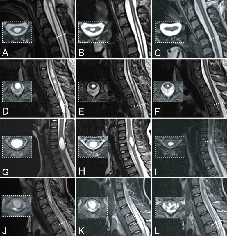
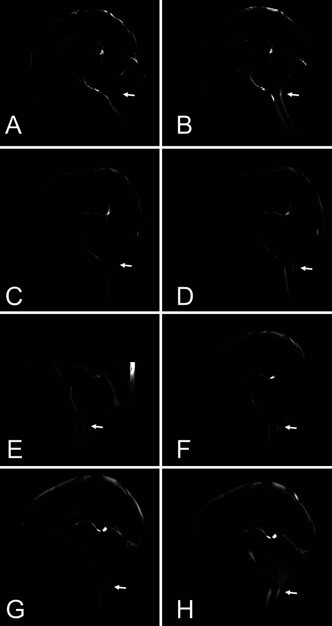
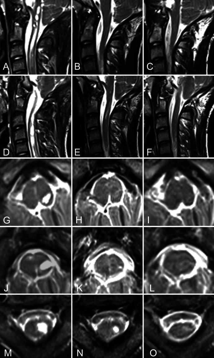
Observations: In a proof-of-principle study, the authors prospectively included 4 patients with CM-I, aged 3-15 years, with severe symptomatic syringes (spanning multiple cervical/thoracic levels and/or focally expanding the spinal cord) to undergo PFD with pre- and postdissection intraoperative MRI (iMRI). In all cases, same-day postdissection iMRI cine images showed enhanced CSF flow compared with predissection iMRI findings. Three of the 4 patients showed early syrinx and symptom improvement (≤ 6 weeks); all showed improvements at long-term follow-up (≥ 2 years). The mean hospital length of stay (LOS) was 1.25 days, with no complications or repeat surgery needed, and the mean follow-up was 37.5 months.

Lessons: CM-I can be treated via PFD alone without duraplasty, even with a severe syrinx. Improved CSF flow is achieved intraoperatively, and the syrinx can significantly decrease over a 6-week to 1-year period postoperatively. Additional benefits include a low risk of complications and a short LOS. https://thejns.org/doi/10.3171/CASE24777.

Keywords: Chiari malformation type I; duraplasty; syringomyelia.


Figures

FIG. 1.
FIG. 1.
Sagittal and axial (insets) T2-weighted spine MR images. Case 1. A: Preoperative images demonstrating a syrinx spanning C2–7 with syrinx dimensions measuring 6.0 × 5.0 mm (anteroposterior × coronal) at C2 and 8.2 × 4.8 mm at C5. B: Postoperative month 1. Images demonstrating a significant decrease in the size of the syrinx, measuring 2.5 × 6.0 mm at C5. C: Postoperative month 20. Images demonstrating a total resolution of the previously seen cervical syrinx. Case 2. D:Preoperative images demonstrating a syrinx extending from C4 to T6 with dimensions of 9.0 × 8.0 mm (anteroposterior × coronal) in width at C7 and 7.0 × 7.0 mm at T4. E:Postoperative month 12. Images demonstrating significant improvement with the size of the cervical cord syrinx measuring 5.2 × 6.7 mm and 4.2 × 4.8 mm at C7 and T4, respectively. F: Postoperative month 30. The syrinx spanned C5–T6 and measured 3.4 × 2.7 mm in width at the C7 level and almost negligible syrinx dimensions at T4. Case 3. G: Preoperative images demonstrating an ovoid-shaped syrinx between C5 and C7 measuring 21.7 × 10.0 × 13.3 mm (craniocaudal [CC] × anteroposterior × coronal) at C6. H:Postoperative month 10. Images showing an improvement in the size of the cervical syrinx measuring 17.3 × 6.0 × 8.8 mm. I:Postoperative month 24. Images demonstrating continued improvement in the size of the syrinx measuring 14.0 × 5.0 × 7.0 mm. Case 4. J: Preoperative images demonstrating a holocord syrinx with 2 components combining the central ependymal canal from C6 to T1 (measuring 5.9 × 5.0 mm in width at C6–7) and a loculated left hemicord syrinx that extended from the medulla oblongata to T2 (measuring 7.0 × 6.0 mm at T1–2). K: Postoperative month 9. Images showing slight improvement in the cervical syrinx despite worsening at the thoracic segment, measuring 9.7 × 8.8 mm at T1–2 and 11.0 × 9.0 mm at T5–6. L: Postoperative month 24. Images demonstrating reduction in the syrinx segments, with 3.6 × 3.0 mm at T2 (compared with 7.0 × 6.0–mm dimensions at postoperative month 12) and 7.6 × 5.9 mm at T5–6. The scale is the same for each triad but different across the triads. Each unit is 10.0 mm (1.0 cm) regardless of the scale.
FIG. 2.
FIG. 2.
Predissection and immediate postdissection MRI cine CSF flow (indicated by arrows) studies. Case 1. A: Predissection image showing blunting of CSF flow posterior to the CMJ. B: Immediate postoperative image showing improved CSF flow at the CMJ in both the anterior and posterior aspects. Case 2. C: Preoperative image showing significant narrowing of the anterior and posterior columns. D: Immediate postoperative image showing enhanced CSF flow at the caudal aspect of the cerebellum and posterior to the CMJ with some persistent but decreased hindrance to anterior flow. Case 3. E: Because of technical difficulties, the cine CSF flow study could not be performed. The postdissection CSF flow study was compared with the patient’s preoperative MRI cine study performed 2 months earlier. This image showed reduced CSF flow at the anterior and lateral CMJ and no flow posterior to the cerebellar tonsils. F: Postdissection cine imaging, in comparison, showed significantly increased flow at the anterior and posterior aspects of the CMJ. There was also increased flow anterior to the brainstem and within the cerebral aqueduct and fourth ventricle. Case 4. G:Predissection image showing blunting of CSF flow posterior to the CMJ. H: Postdissection image showing new prominent flow at the level of the anterior and posterior columns.
FIG. 3.
FIG. 3.
Case 4. Sagittal and axial T2-weighted spine MR images of the patient’s syringobulbia. A and D: Preoperative sagittal images of syringobulbia showing a significant cranial extension. B and E: Significant improvement in the CC extension of the syringobulbia at the CMJ 12 months postoperatively. C and F: Images obtained 24 months postoperatively, showing total regression of the CC extension of the syringobulbia. G–I: Level of the midbrain. Preoperative left syringobulbia cavity measuring 2.5 × 5.4 mm in the anteroposterior and coronal dimensions (G). Nine months postoperatively, imaging showed redemonstration of the syringobulbia at the respective level (H). Twenty-four months postoperatively, imaging at the same level showed total resolution of the syringobulbia (I). J–L: Level of the pons. Preoperative left syringobulbia cavity (J). Nine months postoperatively, imaging demonstrated interval worsening, measuring 4.1 × 9.2 mm (K). Twenty-four months postoperatively, imaging at the same level showed total resolution (L). M–O: Level of the medulla. Preoperative left syringobulbia cavity (M). Nine months postoperatively, imaging demonstrated a slight interval decrease (N). Twenty-four months postoperatively, imaging at the same level showed total resolution of the syringobulbia (O).
FIG. 4.
FIG. 4.
Cases 1–4. Postoperative MSD evolution and MSD distribution relative to other reports studying the efficacy of PFD versus PFDD. A: MSD changes on spine MR images obtained at different follow-ups (vertical dashed gray lines). Case 1 (blue) had MR images obtained preoperatively (MSD, 8.2 mm) and at 1 month (6.0 mm), 8 months (2.5 mm), and 20 months (0.0 mm) postoperatively. Case 2 (yellow) had MR images obtained preoperatively (9.0 mm) and at 1 month (8.8 mm), 6 months (7.6 mm), 12 months (6.7 mm), 24 months (4.8 mm), and 36 months (3.4 mm) postoperatively. Case 3 (green) had MR images obtained preoperatively (13.3 mm) and at 1 month (9.2 mm), 12 months (8.8 mm), 24 months (7.0 mm), and 36 months (6.4 mm) postoperatively. Case 4 (red) had MR images obtained preoperatively (10.0 mm) and at 1 month (11.0 mm), 6 months (11.0 mm), 12 months (7.0 mm), and 24 months (3.6 mm) postoperatively. The mean MSD regression rate in the first 12 ± 2 months was 3.9 mm (case 1, 5.7 mm; case 2, 2.3 mm; case 3, 4.5 mm; and case 4, 3.0 mm). B: MSD distribution (mean 10.1 mm; case 1, 8.2 mm; case 2, 9.0 mm; case 3, 13.3 mm; case 4, 10.0 mm) relative to Strahle et al. (mean 7.8 ± 3.9 mm [±SD]), Lee et al. (all patients: mean 5.5 ± 2.5 mm; PFD: 4.9 ± 2.4 mm, n = 29; PFDD: mean 5.9 ± 2.5 mm, n = 36), Jiang et al. (all patients: mean 5.6 mm [range 2–15 mm]; PFD: 5.2 mm [range 2–11 mm], n = 40; PFDD: mean 5.9 mm [range 2–15 mm], n = 42), and Limbrick et al. (all patients: mean 5.7 ± 2.1 mm; PFD: 5.1 ± 1.8 mm, n = 81; PFDD: mean 6.2 ± 2.2 mm, n = 81).

Similar articles

References

    1. Hankinson T Tubbs RS Wellons JC.. Duraplasty or not? An evidence-based review of the pediatric Chiari I malformation. Childs Nerv Syst. 2011;27(1):35-40. - PubMed
    1. Yahanda AT Limbrick DD Jr.. Posterior fossa decompression with or without duraplasty for Chiari I malformation. Neurosurg Clin N Am. 2023;34(1):105-111. - PubMed
    1. Durham SR Fjeld-Olenec K.. Comparison of posterior fossa decompression with and without duraplasty for the surgical treatment of Chiari malformation Type I in pediatric patients: a meta-analysis. J Neurosurg Pediatr. 2008;2(1):42-49. - PubMed
    1. Tavallaii A Keykhosravi E Ghorbanpour A Shahriari A Rezaee H.. Clinical and radiological outcomes of dura-splitting versus duraplasty techniques in pediatric Chiari I malformation: a systematic review and meta-analysis. Asian J Neurosurg. 2023;18(3):423-436. - PMC - PubMed
    1. Limonadi FM Selden NR.. Dura-splitting decompression of the craniocervical junction: reduced operative time, hospital stay, and cost with equivalent early outcome. J Neurosurg. 2004;101(2suppl):184-188. - PubMed

LinkOut - more resources