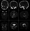
Giant pediatric brainstem cavernoma: illustrative case
- PMID: 40164000
- PMCID: PMC11959640
- DOI: 10.3171/CASE24814
Giant pediatric brainstem cavernoma: illustrative case
Abstract
Background: Cerebral cavernous malformations are low-flow vascular lesions and the most common of all vascular pathologies encountered by neurosurgeons. The location of these lesions is critically important in determining the symptomatology and whether intervention is indicated. Cavernous malformations localized to the brainstem are often the most complex of these to manage given the density of critical structures and tracts found within the brainstem.
Observations: Here, the authors present the case of a 3-year-old male who presented with hemiparesis and eventually developed hydrocephalus from a giant brainstem cavernous malformation. The patient initially did well on steroids but had a recurrent hemorrhage that led to worsening hemiparesis and hydrocephalus. The authors then elected to resect the cavernous malformation via a supracerebellar infratentorial approach utilizing intraoperative MRI to ensure complete resection. Postoperatively, the patient returned to near baseline neurological function.
Lessons: The authors describe an uncommon approach to the management of a pediatric patient with a giant brainstem cavernous malformation by carefully examining the potential approaches and utilizing available technologies to ensure an excellent outcome for the patient. https://thejns.org/doi/10.3171/CASE24814.
Keywords: brainstem cavernoma; brainstem cavernous malformation; pediatric cavernoma; supracerebellar infratentorial approach.
Figures


References
-
- Gross BA Du R.. Hemorrhage from cerebral cavernous malformations: a systematic pooled analysis. J Neurosurg. 2017;126(4):1079-1087. - PubMed
-
- Batra S Lin D Recinos PF Zhang J Rigamonti D.. Cavernous malformations: natural history, diagnosis and treatment. Nat Rev Neurol. 2009;5(12):659-670. - PubMed
-
- Al-Holou WN, O’Lynnger TM, Pandey AS.Natural history and imaging prevalence of cavernous malformations in children and young adults. J Neurosurg Pediatr. 2012;9(2):198-205. - PubMed
-
- Gross BA Batjer HH Awad IA Bendok BR Du R.. Brainstem cavernous malformations: 1390 surgical cases from the literature. World Neurosurg. 2013;80(1-2):89-93. - PubMed
-
- Paddock M Lanham S Gill K Sinha S Connolly DJA.. Pediatric cerebral cavernous malformations. Pediatr Neurol. 2021;116:74-83. - PubMed
LinkOut - more resources
Full Text Sources

