. 2025 Mar 31;13(3):e010699.
doi: 10.1136/jitc-2024-010699.

Genome-wide CRISPR/Cas9 screen reveals factors that influence the susceptibility of tumor cells to NK cell-mediated killing

Affiliations

Genome-wide CRISPR/Cas9 screen reveals factors that influence the susceptibility of tumor cells to NK cell-mediated killing

Sophie Guia et al. J Immunother Cancer. .

Abstract

Background: Natural killer (NK) cells exhibit potent cytotoxic activity against various cancer cell types. Over the past five decades, numerous methodologies have been employed to elucidate the intricate molecular mechanisms underlying NK cell-mediated tumor control. While significant progress has been made in elucidating the interactions between NK cells and tumor cells, the regulatory factors governing NK cell-mediated tumor cell destruction are not yet fully understood. This includes the diverse array of tumor ligands recognized by NK cells and the mechanisms that NK cells employ to eliminate tumor cells.

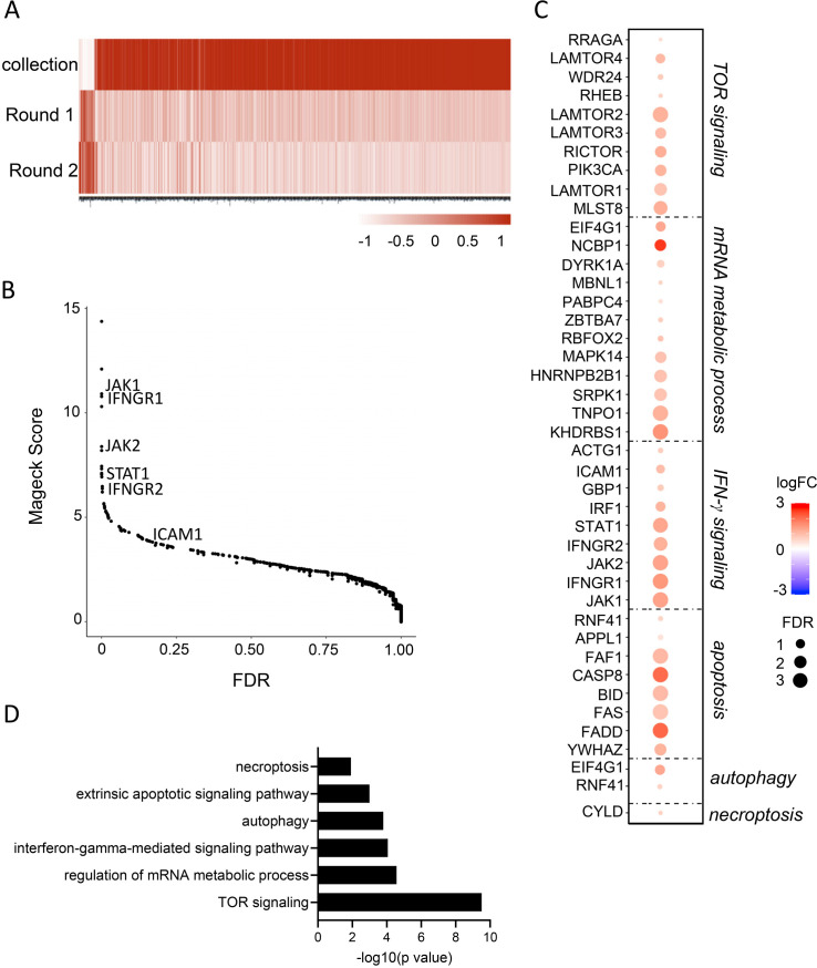
Methods: In this study, we employed a genome-wide CRISPR/Cas9 screening approach in conjunction with functional cytotoxicity assays to delineate the pathways modulating the susceptibility of colon adenocarcinoma HCT-116 cells to NK cell-mediated cytotoxicity.

Results: Analysis of guide RNA distribution in HCT-116 cells that survived co-incubation with NK cells identified ICAM-1 as a pivotal player in the NKp44-mediated immune synapse, with NKp44 serving as an activating receptor crucial for the elimination of HCT-116 tumor cells by NK cells. Furthermore, disruption of genes involved in the apoptosis or interferon (IFN)-γ signaling pathways conferred resistance to NK cell attack. We further dissected that NK cell-derived IFN-γ promotes mitochondrial apoptosis in vitro and exerts control over B16-F10 lung metastases in vivo.

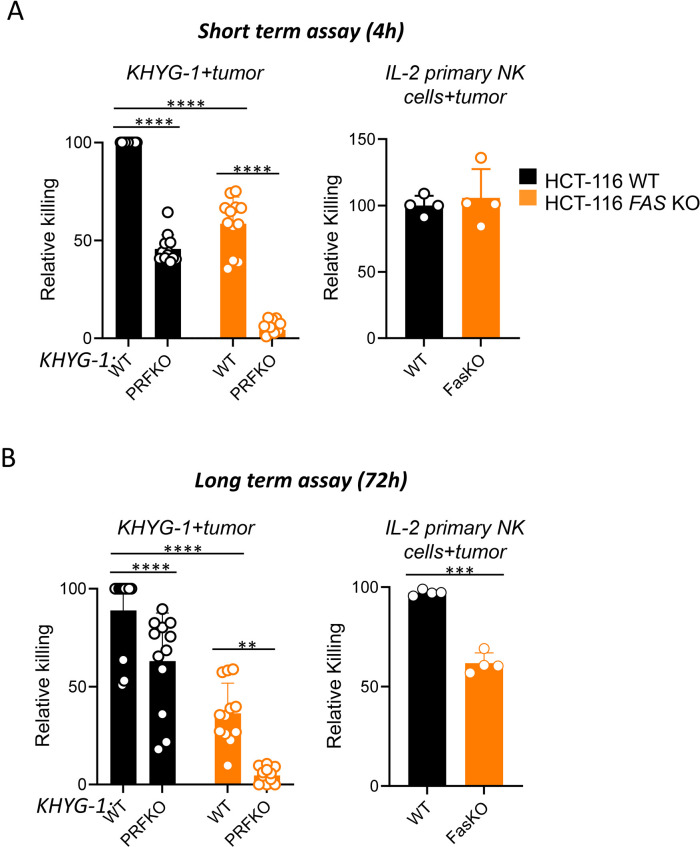
Conclusion: Monitoring ICAM-1 levels on the surface of tumor cells or modulating its expression should be considered in the context of NK cell-based therapy. Furthermore, promoting FasL expression on the NK cell surface is reaffirmed as an important strategy to enhance NK cell-mediated tumor killing, offering an additional avenue for therapeutic optimization. Additionally, considering the diffusion properties of IFN-γ, our findings highlight the potential of leveraging NK cell-derived IFN-γ to enhance direct tumor cell killing and facilitate bystander effects via cytokine diffusion, warranting further investigation.

Keywords: Natural killer - NK.


Conflict of interest statement

Competing interests: EV, AF, JG, LG and BR are employees of Innate Pharma. The other authors declare no competing interests.

Figures

Figure 1
Figure 1. Genome-wide CRISPR/Cas9 screening identifies genes involved in NKp44-mediated specific killing of tumor cells by KHYG-1 cells. (A) Genome-wide CRISPR/Cas9 screening design. HCT-116 Cas9+ cells were transduced with the knockout sgRNA Brunello library. Collection of mutant cells was subjected to lysis by WT KHYG-1 or NKp44-deficient KHYG-1 in the presence of α-NKp44 mAb or not (Round 1). Mutant cells that survived lysis by KHYG-1 were subjected to a second round (Round 2) of co-culture with WT KHYG-1 (condition 1), in the presence of α-NKp44 mAb (condition 2) or with NKp44-deficient KHYG-1 (condition 3). The abundance of sgRNA in the collection, in the surviving cells in Round 1 and in the surviving cells in Round 2 were determined by sequencing (see Material and methods). (B) Venn diagram of hits obtained with KHYG-1 co-culture (condition 1, see online supplemental table 1), KHYG-1+αNKp44mAb (condition 2, see online supplemental table 2) and NKp44-deficient KHYG-1 cells (condition 3, see online supplemental table 3). (B) Scatter plot showing the ranking of hits enriched in the NKp44-dependent killing of tumor cells by KHYG-1 cells by MAGeCK score and false discovery rate (see online supplemental table 4). FDR, false discovery rate; mAb, monoclonal antibody; sgRNA, single guide RNA; WT, wild-type.
Figure 2
Figure 2. Knockout of ICAM-1 in HCT-116 cells impairs NKp44-mediated lysis. (A) Enrichment of surface-encoding genes. (B–C) Indicated mutant cells were subjected to KHYG-1 killing in a standard 4-hour chromium release assay in the presence or not of α-NKp44 mAb. The frequencies of specific lysis normalized to WT were shown in (B) and the frequencies of inhibition by α-NKp44 mAb were shown in (C). P value<0.0001, unpaired t-test. mAb, monoclonal antibody; WT, wild-type.
Figure 3
Figure 3. Expression of ICAM-1 is required but not sufficient for NKp44-mediated natural killer cell lysis of tumors. HCT-116 cells (A), ICAM-1-deficient HCT-116 cells (A), HEK 293T cells (B, C) and K562 cells (D) were cultured with KHYG-1 in the presence or not of α-NKp44, α-NKp30 or α-ICAM-1 mAbs as indicated. The frequencies of specific lysis were determined in a standard 4-hour chromium release assay. Data are representative of two to three independent experiments. KO, knockout; mAb, monoclonal antibody; SNP, single nucleotide polymorphism; WT, wild-type.
Figure 4
Figure 4. Identification of tumor signaling pathways involved in sensitivity to NK cells. (A) Heatmap of gene enrichment in rounds 1 and 2 in the KHYG-1+HCT-116 co-culture (condition 1 in figure 1, see online supplemental table 1). (B) Scatter plot showing the ranking of hits enriched in the NK cell-dependent killing of tumor cells by KHYG-1 cells by MAGeCK score and false discovery rate. (C–D). Selected enrichment of hits in the indicated pathways. FDR, false discovery rate; IFN, interferon; mRNA, messenger RNA; NK, natural killer; TOR, target of rapamycin.
Figure 5
Figure 5. Relative requirement of NK cell effector functions for NK cell-mediated tumor lysis over time. WT and Fas-deficient HCT-116 Cas9+ cells were co-cultured with WT KHYG-1 or perforin-deficient KHYG-1 cells or with IL-2 activated primary NK cells from four donors, as indicated. The extent of specific lysis was determined in a standard 4-hour assay (short-term) (A) or as the CRISPR screen condition (long-term, 72 hours) (B). P values: ****<0.0001; **=0.024 in (A) and **=0.0037 in (B). Unpaired t-test. Data result from the pool of two to three independent experiments. IL, interleukin; KO, knockout; NK, natural killer; WT, wild-type.
Figure 6
Figure 6. IFN-γ from NKp46+ cells is required to control the metastasis of B16F10 in the lung. Wild-type mice treated with depleting α-NK1.1 mAb or with neutralizing IFN-γ mAb, Ncr1iCre/+R26DTA/+ mice, Ncr1iCre/+Ifngflfl/fl) and their respective controls (mice treated with isotype controls, Ncr1iCre/+R26+/+ or Ncr1iCre/+Ifng+/+) were injected intravenously with B16F10 cells. The number of foci in the lungs was determined 7 days later. Data represent one to four independent experiments; mean±SD; Student’s t-test with Welsh correction ***p<0.005. IFN, interferon; mAb, monoclonal antibody.
Figure 7
Figure 7. IFN-γ can directly kill tumor cells in part by triggering mitochondrial apoptosis. (A) WT or IFNGR1 KO HCT-116 cells were cultured in the presence of the indicated amount of recombinant IFN-γ for 72 hours. The number of live tumor cells was estimated using an IncuCyte Imaging System. Two-way analysis of variance with Dunnett’s comparison test. KHYG-1 cells (B) or IL-2-activated primary NK cells from four donors (C) were co-cultured with WT or IFNGR1 KO HCT-116 cells for 24 hours. The amount of cytochrome C released in target cells was determined by flow cytometry. Data are representative of two independent experiments. Mean±SD; Student’s t-test. E:T, effector-to-target ratio; FACS, flow cytometry; IFN, interferon; IL, interleukin; NK, natural killer; WT, wild-type.

References

    1. Vivier E, Artis D, Colonna M, et al. Innate Lymphoid Cells: 10 Years On. Cell. 2018;174:1054–66. doi: 10.1016/j.cell.2018.07.017. - DOI - PubMed
    1. Vivier E, Raulet DH, Moretta A, et al. Innate or adaptive immunity? The example of natural killer cells. Science. 2011;331:44–9. doi: 10.1126/science.1198687. - DOI - PMC - PubMed
    1. Gonzalvez F, Ashkenazi A. New insights into apoptosis signaling by Apo2L/TRAIL. Oncogene. 2010;29:4752–65. doi: 10.1038/onc.2010.221. - DOI - PubMed
    1. Kägi D, Ledermann B, Bürki K, et al. Cytotoxicity mediated by T cells and natural killer cells is greatly impaired in perforin-deficient mice. Nature New Biol. 1994;369:31–7. doi: 10.1038/369031a0. - DOI - PubMed
    1. Wallin RPA, Screpanti V, Michaëlsson J, et al. Regulation of perforin-independent NK cell-mediated cytotoxicity. Eur J Immunol. 2003;33:2727–35. doi: 10.1002/eji.200324070. - DOI - PubMed

LinkOut - more resources