Review
. 2025 Oct;132(10):1515-1523.
doi: 10.1007/s00702-025-02916-0. Epub 2025 Mar 31.

Alzheimer mimicry: LATE and PART

Affiliations
Review

Alzheimer mimicry: LATE and PART

Nenad Bogdanovic et al. J Neural Transm (Vienna). 2025 Oct.

Abstract

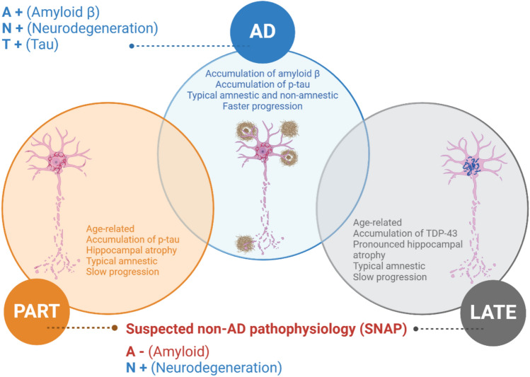
Alzheimer's disease (AD) is the main cause of dementia and accounts for 60% of dementia syndromes in people older than 75 years. The correct classification of AD and non-AD cases is mandatory to study disease mechanisms or new treatment possibilities. A typical clinical picture of AD consists of a progressive cognitive decline, with primary memory impairment. Structural, functional, and molecular brain imaging, along with CSF biomarkers of amyloid pathology, neurodegeneration, and the presence of a vulnerability-associated APOE genotype, support the diagnosis of AD. Use of biomarkers have led to the identification of individuals with mild cognitive impairment who are amyloid-negative addressing a conceptually separate clinical entity named suspected non-Alzheimer disease pathophysiology (SNAP). Clinical presentation and progression of SNAP can mimic AD which makes the final diagnosis and possible treatment uncertain in up to 30% of cases in clinical centers that are not using biomarkers. These non-AD pathologies are common with advancing age both in cognitively impaired and clinically normal elderly people and include Argyrophilic Grain Disease (ARG), Tangle Predominant Dementia and TDP-43 proteinopathy. The terms Primary age-related tauopathy (PART) and Limbic-dominant TDP-43 age-related encephalopathy (LATE) have been proposed as the most common and useful biological and emerging clinical construct to describe this phenomenon in > 80 years old individuals. Current evidence underlines the limitations of existing diagnostic tools, which remain inadequate for fully capturing the complexities of these conditions. Addressing these diagnostic ambiguities is crucial for assigning accurate diagnoses, reducing frequent misdiagnoses of AD, and implementing appropriate therapeutic strategies for elderly patients with mild cognitive impairment and dementia.

Keywords: Atypical Alzheimer; Dementia; Elderly; LATE; PART; SNAP.


Figures

Fig. 1
Fig. 1
Overview of basic differences between ADNC, PART-NC and LATE-NC. AD = Alzheimer’s disease, LATE-NC = Limbic-predominant age-related TDP-43 encephalopathy neuropathologic change; PART-NC = primary age-related tauopathy neuropathologic change
Fig. 2
Fig. 2
The human hippocampus has anatomically distinct anterior and posterior segments that are associated with different functional specializations, morphology, and connectivity patterns. The anterior and posterior hippocampal projection areas are shown in the left (A = anterior) and right (P = posterior) blue squares
Fig. 3
Fig. 3
An 86-year-old patient presents with an MMSE score of 25/30 and APOE ε3/3. Cerebrospinal fluid analysis reveals no amyloid or tau pathology. MRI shows severe atrophy in both hippocampi, graded as MTA stage 4*, while cortical atrophy is minimal and notably disproportionate to the hippocampal changes. This presentation strongly resembles the clinical and pathological features associated with “pure” LATE-NC. *MTA (medial temporal lobe atrophy) visually rated by a semiquantitative scale (Scheltens et al. 1992) ranging from 0 to 4, where 4 stand for end stage hippocampal atrophy
Fig. 4
Fig. 4
The nucleus basalis of Meynert (nbM), a key cholinergic nucleus, exhibits distinct patterns of involvement in LATE and PART. In LATE, TDP-43 pathology does not affect the nbM, whereas in PART, the nbM is notably impacted by phospho-tau pathology. On the other hand, TDP-43 pathology is commonly found in the amygdala in LATE. In cognitively unimpaired older individuals, minimal tau pathology can often be observed in the brain, which may represent a preclinical or pre-PART stage
Fig. 5
Fig. 5
CT image of the 80-year-old patient who has an amnestic cognitive impairment accompanied by behavioral symptoms. Cognitive impairment has developed slowly over several years. Neuropsychological examination has revealed the cognitive impairment in more detail than the screening MMSE test. During the last 3 years, the atrophic changes accelerated mostly in the anterior hippocampal regions (upper row), L > R, in comparison with cortex. The patient did not have amyloid or tau pathology in the CSF and had APOE ε2/3. The clinical picture and limbic atrophy correspond more to PART. For the final diagnosis, neuropathological analysis is mandatory
Fig. 6
Fig. 6
Baseline (left) and 7 years follow up CT of the brain (right) indicating accelerated atrophy of anterior hippocampus but not the cortex. [18F]FDG-PET showed accelerated hypometabolism (blue color) in the temporal lobe and medial frontal/cingulate cortex but not in the typical AD regions as precuneus (red ring)

References

    1. ALZforum (2023) https://www.alzforum.org/news/research-news/alzheimers-primary-age-relat...
    1. Amador-Ortiz C, Lin W-L, Ahmed Z et al (2007) TDP-43 immunoreactivity in hippocampal sclerosis and Alzheimer’s disease. Ann Neurol 61(5):435–445 - PMC - PubMed
    1. Aoki N, Murray ME, Ogaki K et al (2015) Hippocampal sclerosis in Lewy body disease is a TDP-43 proteinopathy similar to FTLD-TDP Type A. Acta Neuropathol 129:53–64 - PMC - PubMed
    1. Braak H, Braak E (1991) Neuropathological stageing of Alzheimer-related changes. Acta Neuropathol 82:239–259 - PubMed
    1. Braak H, Thal DR, Ghebremedhin E et al (2011) Stages of the pathologic process in Alzheimer disease: age categories from 1 to 100 years. J Neuropathol Exp Neurol 70:960–969 - PubMed

LinkOut - more resources