. 2025 Mar 31;27(1):47.
doi: 10.1186/s13058-025-02005-w.

A ROR1 targeted bispecific T cell engager shows high potency in the pre-clinical model of triple negative breast cancer

Affiliations

A ROR1 targeted bispecific T cell engager shows high potency in the pre-clinical model of triple negative breast cancer

Fan Wang et al. Breast Cancer Res. .

Abstract

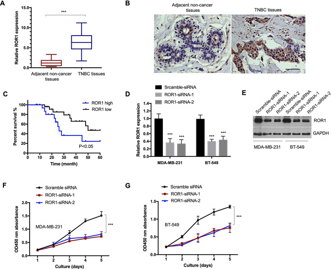
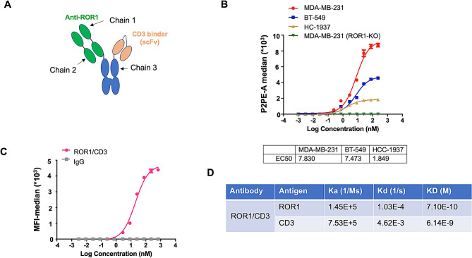
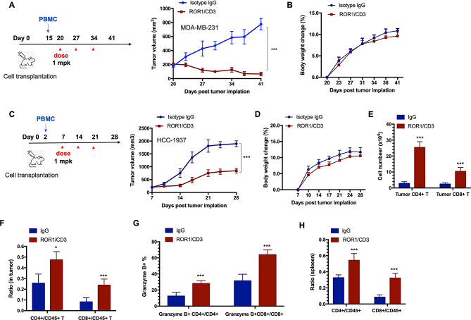
Background: Triple negative breast cancer (TNBC) is an aggressive breast cancer subtype characterized with poor prognosis and high metastatic potential. Although traditional chemotherapy, radiation, and surgical resection remain the standard treatment options for TNBC, bispecific antibody-based immunotherapy is emerging as new strategy in TNBC treatment. Here, we found that the receptor tyrosine kinase-like Orphan Receptor 1 (ROR1) was highly expressed in TNBC but minimally expressed in normal tissue. A bispecific ROR1-targeted CD3 T cell engager (TCE) was designed in IgG-based format with extended half-life.

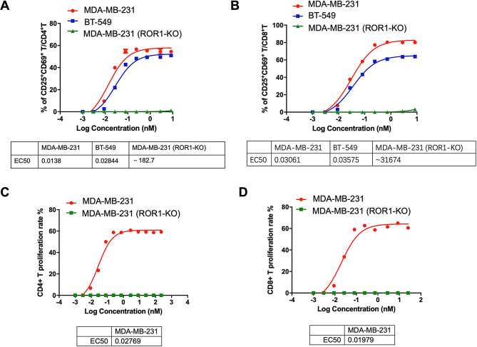
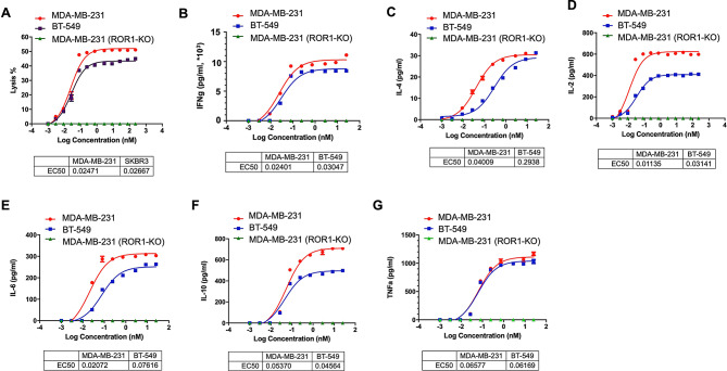
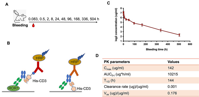
Method: The expression of ROR1 in TNBC was detected by RT-qPCR and immunohistology analysis. The killing of ROR1/CD3 antibody on TNBC cells was determined by the in vitro cytotoxicity assay and in vivo PBMC reconstituted mouse model. The activation of ROR1/CD3 on T cells was analyzed by the flow cytometry and ELISA assay. Pharmacokinetics study of ROR1/CD3 was performed in mouse.

Results: The ROR1/CD3 TCE triggered T cell activation and proliferation, which showed potent and specific killing to TNBC cells in ROR1-depedent manner. In vivo mouse model indicated that ROR1/CD3 TCE redirected the cytotoxic activity of T cells to lyse TNBC cells and induced significant tumor regression. Additionally, the ROR1/CD3 bispecific antibody exhibited an extended half-life in mouse, which may enable intermittent administration in clinic.

Conclusions: Collectively, these results demonstrated that ROR1/CD3 TCE has a promising efficacy profile in preclinical studies, which suggested it as a possible option for the treatment of ROR1-expressing TNBC.

Keywords: Immunotherapy; ROR1; T cell engager; TNBC.


Conflict of interest statement

Declarations. Ethics approval and consent to participate: Not applicable. Competing interests: The authors declare no competing interests.

Figures

Fig. 1
Fig. 1
ROR1 was highly enriched in TNBC and essential for TNBC cell growth. (A) ROR1 expression in TNBC and adjacent normal tissues was detected by RT-qPCR, which showed the significantly increased expression of ROR1. The PCR reaction was performed in duplicates. Data analysis was done by student’s t tset. (B) Representative IHC staining of ROR1 in TNBC and adjacent normal tissues. Scale bar, 100 μm. (C) High levels of ROR1 were correlated with the shorter 5-year survival rate of TNBC patients. Patients were classified as ROR1-high (n = 29) or -low (n = 21) according to the median expression value of ROR1 in these 50 TNBC tissues. Log rank test was performed for data analysis. (D, E) TNBC cells were transfected with siRNA-control or siRNA-ROR1, and the knockdown efficacy was confirmed by RT-qPCR after transfection for 48 h. The experiment was repeated three times and PCR reaction was performed in triplicates. One-way ANOVA test was performed for data analysis (D) and western blot analysis (E). (F, G) CCK-8 assay showed the significantly reduced TNBC cell proliferation upon ROR1 down-regulation. The experiment was performed with three technological replicates and validated by other two biological replicates. Two-way ANOVA was performed for data analysis
Fig. 2
Fig. 2
ROR1/CD3 TCE shows high binding affinity to ROR1 and CD3. (A) The schematic diagram of ROR1/CD3 antibody. The green part is the Fab targeting ROR1, and the right yellow part is the single-chain variable fragment (scFv) targeting CD3. ROR1/CD3 TCE molecule consists of three chains: anti-ROR1 antibody heavy chain (chain 1), anti-ROR1 antibody light chain (chain2) and an anti-CD3 antibody ScFv-Fc fused chain (Chain 3). (B, C) Cell-based binding of ROR1/CD3 TCE molecule to ROR1-expressing MDA-MB-231 cell and CD3-expressing human T cells. Cells were incubated with a serially diluted ROR1/CD3 antibody followed by staining with PE-conjugated anti-human IgG. The MFI was determined by flow cytometry. The experiment was performed with three technological replicates and validated by other two biological replicates. (D) The binding affinity of ROR1/CD3 to ROR1 and CD3 was evaluated by the surface plasma resonance. The experimemnt was performed with three technological replicates. KD, binding dissociation equilibrium constants; Ka, kinetic association rate; Kd, kinetic dissociation rate
Fig. 3
Fig. 3
ROR1/CD3 TCE induced T cell activation in a ROR1-dependent manner. (A, B) The activation of CD4+, CD8+ T cells were determined by flow cytometry via analyzing the activation marker CD25 and CD69. (C, D) The proliferation of CD4+ and CD8+ T cells was measured after co-cultured with a serially diluted ROR1/CD3 antibody for 96 h. The experiment was performed with three tecnological replicates, and validated by another PBMC donor
Fig. 4
Fig. 4
ROR1/CD3 TCE killed ROR1-expressing TNBC cells. (A) The cytotoxic lysis of ROR1/CD3 antibody to TNBC cells were detected by measuring the lactate dehydrogenase release. (B-G) Human PBMCs were incubated with ROR1/CD3 antibody molecule and the cytokine release in the supernatants was detected by ELISA. Both the cytotoxic and cytokine release experiments were performed with three tecnological replicates, and validated by another PBMC donor
Fig. 5
Fig. 5
ROR1/CD3 inhibited tumor growth in TNBC xenograft mouse model. (A) The schematic diagram for the in vivo efficacy study (left panel). Administration of 1 mpk ROR1/CD3 antibody induced significantly tumor growth of NOG mice bearing MDA-MB-231 compared with mice treated with control IgG (right panel). N = 8 mice for each group. Two-way ANOVA was performed for data analysis. (B) No body weight loss was observed within mice treated with ROR1/CD3 antibody. (C, D) ROR1/CD3 suppressed the tumor growth of HCC1937 model on NOG mice. N = 8 mice for each group. Two-way ANOVA was performed for data analysis (C). No significant body weight loss was observed with the administration of ROR1/CD3 molecule (D). ROR1/CD3 engaged tumor-infiltrating T cells. Mice were treated with 1 mpk ROR1/CD3 after 20 days’ of tumor implantation. At day 27, tumors were isolated and analyzed for T cell activation and infiltration by flow cytometry. The percentage of CD4+, CD8+ T cells in tumors (E), the ratio of CD4+ or CD8+ in CD45+ T cell populations (F), granzyme B+ positive CD4+/CD8+ in CD4+ or CD8+ T cell (G), the ratio of CD4+ or CD8+ T cells in CD45+ population of the spleen (H) was determined. S student’s t test was performed for data analysis
Fig. 6
Fig. 6
ROR1/CD3 TCE shows acceptable PK parameters in mice. (A) The schematic diagram of dosing and bleeding schedule of the PK study. (B) The format of ELISA used for detecting the blood concentration of ROR1/CD3. N = 3 mice for each time points. (C) The PK profiles of ROR1/CD3. BALB/c mouse was intravenously injected with ROR1/CD3 as mentioned in panel (A) and the serum concentration of ROR1/CD3 was detected at the indicated time points after dosing. (D) The PK parameters of ROR1/CD3 molecule in BALB/c mouse

Similar articles

References

    1. Elias AD. Triple-negative breast cancer: a short review. Am J Clin Oncol. 2010;33(6):637–45. - PubMed
    1. Ismail-Khan R, Bui MM. A review of triple-negative breast cancer. Cancer Control. 2010;17(3):173–6. - PubMed
    1. Jamdade VS, Sethi N, Mundhe NA, Kumar P, Lahkar M, Sinha N. Therapeutic targets of triple-negative breast cancer: a review. Br J Pharmacol. 2015;172(17):4228–37. - PMC - PubMed
    1. Zeichner SB, Terawaki H, Gogineni K. A review of systemic treatment in metastatic Triple-Negative breast cancer. Breast Cancer (Auckl). 2016;10:25–36. - PMC - PubMed
    1. da Silva JL, Cardoso Nunes NC, Izetti P, de Mesquita GG, de Melo AC. Triple negative breast cancer: A thorough review of biomarkers. Crit Rev Oncol Hematol. 2020;145:102855. - PubMed

MeSH terms

Substances

LinkOut - more resources