. 2025 Apr;358(4):e2400889.
doi: 10.1002/ardp.202400889.

New potent N-hydroxycinnamamide-based histone deacetylase inhibitors suppress proliferation and trigger apoptosis in THP-1 leukaemia cells

Affiliations

New potent N-hydroxycinnamamide-based histone deacetylase inhibitors suppress proliferation and trigger apoptosis in THP-1 leukaemia cells

Magdalena Onuscakova et al. Arch Pharm (Weinheim). 2025 Apr.

Abstract

A new group of potent histone deacetylase inhibitors (HDACis) capable of inhibiting cell growth and affecting cell-cycle progression in Tohoku Hospital Pediatrics-1 (THP-1) monocytic leukaemia cells was synthesized. The inhibitors belong to a series of hydroxamic acid derivatives. We designed and synthesized a series of 22 N-hydroxycinnamamide derivatives, out of which 20 are new compounds. These compounds contain various substituted anilides as the surface recognition moiety (SRM), a p-hydroxycinnamate linker, and hydroxamic acids as the zinc-binding group (ZBG). The whole series of synthesized hydroxamic acids inhibited THP-1 cell proliferation. Compounds 7d and 7p, which belong to the category of derivatives with the most potent antiproliferative properties, exert a similar effect on cell-cycle progression as vorinostat and induce apoptosis in THP-1 cells. Furthermore, compounds 7d and 7p were demonstrated to inhibit HDAC class I and II in THP-1 cells with comparable potency to vorinostat and increase acetylation of histones H2a, H2b, H3, and H4. Molecular modelling was used to predict the probable binding mode of the studied HDACis in class I and II histone deacetylases in terms of Zn2+ ion chelation by the hydroxamate group.

Keywords: HDACi; anticancer agents; haematological malignancies; hydroxamic acid; inhibitors of histone deacetylases.


Conflict of interest statement

The authors declare no conflicts of interest.

Figures

Figure 1
Figure 1
Structures of approved histone deacetylase (HDAC) inhibitors and pracinostat as drug previously developed under Orphan Designation.
Figure 2
Figure 2
A typical three‐feature pharmacophore of histone deacetylase inhibitors (HDACis) contains a zinc‐binding group (ZBG), a linker, and a surface recognition moiety (SRM).
Scheme 1
Scheme 1
The first attempts for the synthesis of the target histone deacetylase (HDAC) inhibitors.
Scheme 2
Scheme 2
Convergent variants of the synthesis of inhibitors 7a–v b .
Scheme 3
Scheme 3
Two routes for the synthesis of vorinostat (SAHA) c .
Figure 3
Figure 3
A library of compounds studied.
Figure 4
Figure 4
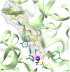
Binding mode of the most potent hydroxamic acid derivative 7d and three‐dimensional (3D) structure of the HDAC8 binding site predicted by molecular docking using the Glide XP protocol (release 2022‐3, Schrödinger LLC, USA, 2022[ 40 , 41 ]). The protein backbone is shown as a green ribbon, the mesh type molecular surface of 7d is shown in grey colour, the atom colouring: ligand C – yellow, protein C – green, N – blue, O – red, H – light grey, Zn – purple.
Figure 5
Figure 5
Hydroxamic acid derivatives 7d and 7p induce inhibition of histone deacetylase (HDAC) class I and II enzyme activity in THP‐1 cells. (a) The inhibitory effects of vorinostat 1, 7d, and 7p on the enzyme activity of HDAC class I and II were determined using the HDAC‐Glo™ I/II Assay. The results are expressed as the mean ± SD of three independent experiments. The levels of histone H2A, H2B, H3, or H4 acetylation after (b) 24 h or (c) 48 h of treatment with vorinostat 1, 7d, and 7p were determined by immunoblot analysis using appropriate antibodies. Representative immunoblots of one of three experiments are shown. CTRL is a drug‐free control.
Figure 6
Figure 6
Compounds 7d and 7p induce changes in the cell‐cycle progression of THP‐1 cells. THP‐1 cells were treated with indicated concentrations of compounds vorinostat 1, 7d, or 7p for 48 h. (a) The results of the distribution of THP‐1 cells in the cell‐cycle phases are expressed as mean ± SD from three independent experiments. *p < 0.05, **p < 0.01, ***p < 0.001, significantly different from drug‐free control (CTRL). (b) Cell‐cycle regulator levels were determined by immunoblot analysis using appropriate antibodies. Representative immunoblots of one of three experiments are shown. CTRL is a drug‐free control.
Figure 7
Figure 7
Compounds 7d and 7p induce apoptosis in THP‐1 cells. (a) THP‐1 cells were incubated with indicated concentrations and vorinostat 1, 7d, or 7p. Cell viability was determined by LDH assay after 24 and 48 h of incubation. (b, c) THP‐1 cells were treated with compound and vorinostat 1, 7d, or 7p, incubated for 48 h, and analysed by flow cytometry after staining with annexin V‐FITC conjugate and propidium iodide (PI). Here are shown the representative flow cytometry plots of the results of the control sample and the results of samples treated with 2.5 µM of the compounds tested. (d) THP‐1 cells were treated with compounds vorinostat 1, 7d, or7p, incubated for 48 h, and caspase 3 activation was evaluated by flow cytometry. (e) After 48 h of incubation of THP‐1 cells with and vorinostat 1, 7d, or 7p cleaved poly(ADP‐ribose)polymerase (PARP) levels were detected by immunoblot analysis. The results are shown as the mean ± SD of three independent experiments. *p < 0.05, ***p < 0.001, significantly different from drug‐free control (CTRL). LDF, lactate dehydrogenase.
Figure 8
Figure 8
Three types of chelation of the Zn2+ ion by hydroxamate group of HDACi 7d. Type A – bidenate neutral, type B – monodenate neutral and type C – bidenate anionic hydroxamate group. Imidazole Nδ of His180 coordinates the zinc ion from above.

References

    1. Nepali K., Liou J. P., J. Biomed. Sci. 2021, 28(1), 27. - PMC - PubMed
    1. Putiri E. L., Robertson K. D., Clin. Epigenetics 2011, 2(2), 299. - PMC - PubMed
    1. Lu Y., Chan Y. T., Tan H. Y., Li S., Wang N., Feng Y., Mol. Cancer 2020, 19(1), 79. - PMC - PubMed
    1. Sharma S., Kelly T. K., Jones P. A., Carcinogenesis 2010, 31(1), 27. - PMC - PubMed
    1. Llinàs‐Arias P., Esteller M., Open Biol. 2017, 7(9), 170152. - PMC - PubMed

MeSH terms

LinkOut - more resources