. 2025 Mar 17:16:1540754.
doi: 10.3389/fimmu.2025.1540754. eCollection 2025.

Sodium citrate pretreatment enhances CAR-T cell persistence and anti-tumor efficacy through inhibition of calcium signaling

Affiliations

Sodium citrate pretreatment enhances CAR-T cell persistence and anti-tumor efficacy through inhibition of calcium signaling

Xuechen Yin et al. Front Immunol. .

Abstract

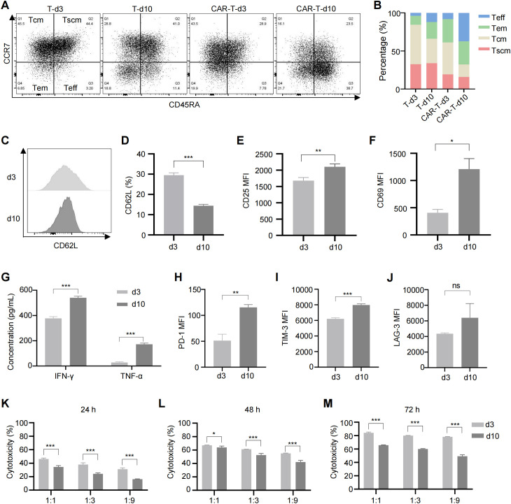
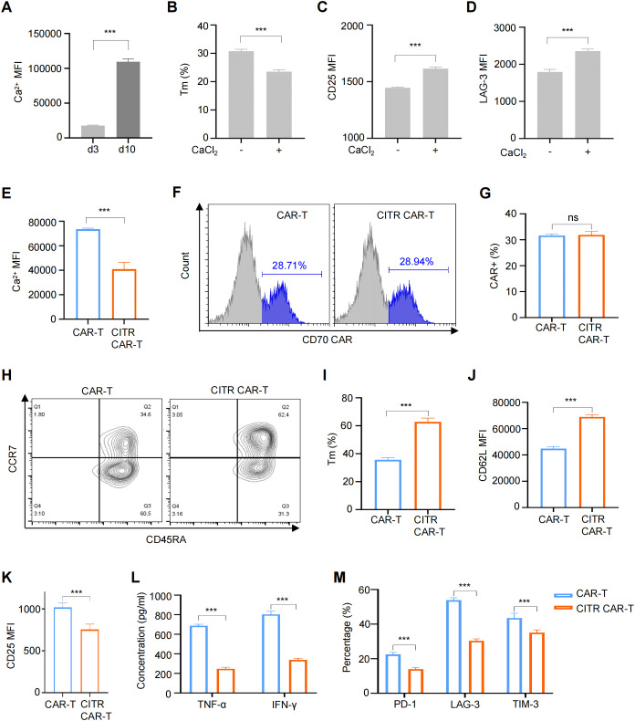
Introduction: Chimeric antigen receptor T cell (CAR-T) therapy has shown success in treating hematological malignancies, but its effectiveness against solid tumors is hindered by T cell exhaustion. During in vitro expansion, tonic signaling induced by CAR expression contributes to CAR-T cell exhaustion, which can be mitigated by inhibiting calcium signaling. Given that sodium citrate can chelate calcium ions and inhibit calcium signaling, in this study, we investigated whether sodium citrate could reduce exhaustion and enhance CAR-T cell function.

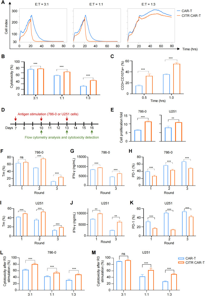
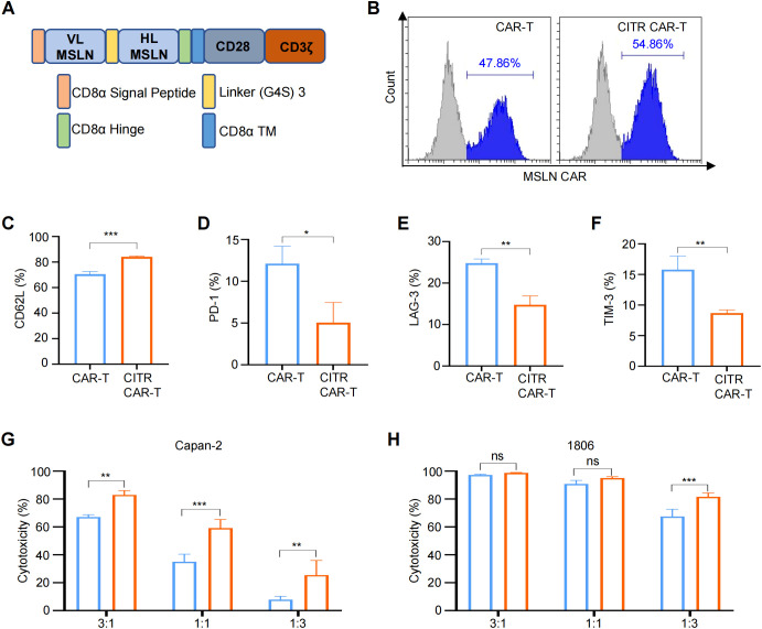
Methods: We constructed anti-CD70 CAR-T cells and cultured them in the presence of sodium citrate. The characteristics and functionality of sodium citrate-pretreated CAR-T cells were assessed through in vitro and in vivo experiments. To further validate our observation, we also treated anti-mesothelin (MSLN) CAR-T cells with sodium citrate and detected the phenotypes and anti-tumor function of CAR-T cells.

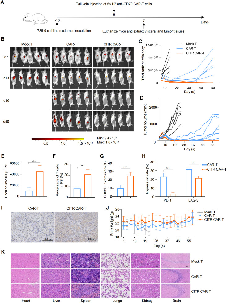
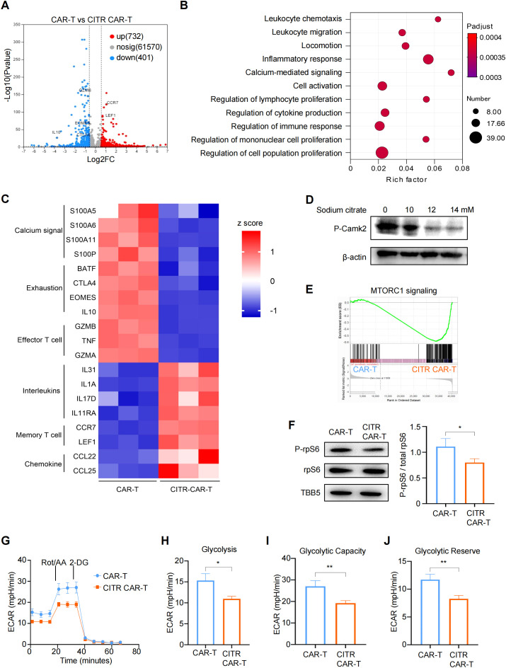
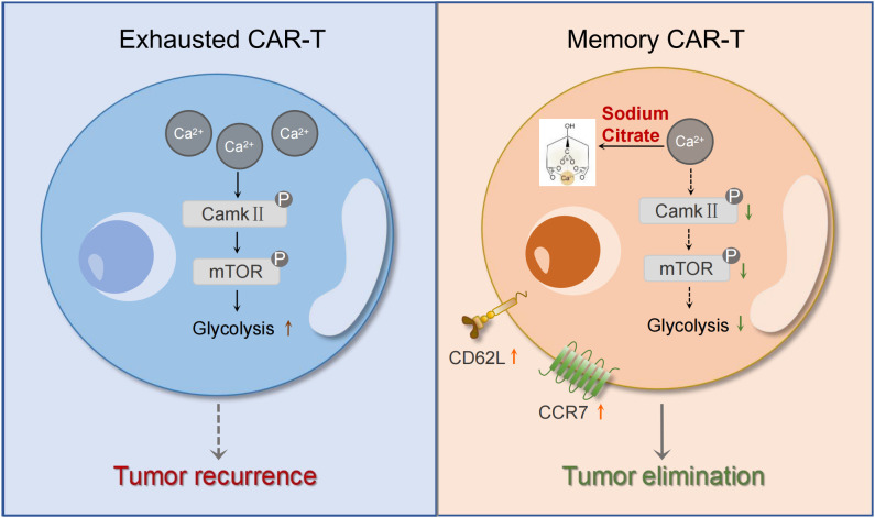
Results: We found that sodium citrate-pretreated anti-CD70 CAR-T cells exhibited reduced exhaustion, increased memory T cell proportions, and enhanced anti-tumor efficacy both in vitro and in vivo. Notably, sodium citrate treatment improved the in vivo persistence of CAR-T cells and prevented tumor recurrence. These beneficial effects were also observed in anti-MSLN CAR-T cells. Transcriptomic and metabolite analyses revealed that sodium citrate inhibited calcium signaling, mTORC1 activity, and glycolysis pathways, thus modulating T cell exhaustion and differentiation.

Discussion: Our findings suggest that sodium citrate supplementation during CAR-T cell expansion could be a promising strategy to improve CAR-T therapy for solid tumors by preventing exhaustion and promoting memory T cell formation.

Keywords: CAR-T; T cell exhaustion; calcium ions; cancer immunotherapy; sodium citrate; solid tumors.


Conflict of interest statement

ZG is one of the inventors of a patent on the anti-CD70 CAR sequence employed in this work and the application number is 202410495812.1. The remaining authors declare that the research was conducted in the absence of any commercial or financial relationships that could be construed as a potential conflict of interest.

Figures

Figure 1
Figure 1
Long-term culture induced CAR-T cell exhaustion, excessive activation and reduced cytotoxicity. (A) Representative flow cytometric profile showing the expression of CCR7 and CD45RA on CAR-T cells. (B) Proportions of Tscm (CCR7+CD45RA+), Tcm (CCR7+CD45RA-), Tem (CCR7-CD45RA-), and Teff (CCR7-CD45RA+) subsets in anti-CD70 CAR-T cells as measured by flow cytometry. (C) Representative flow cytometric profile showing the expression level of CD62L on CAR-T cells. (D) Histogram plot showing the percentage of CD62L+ CAR-T cells. (E, F) Mean fluorescence intensity (MFI) of CD25 (E) and CD69 (F) expression on CAR-T cells. (G) Levels of TNF-α and IFN-γ released by CAR-T cells at different time points, measured by ELISA. (H–J) MFI of exhaustion markers PD-1 (H), TIM-3 (I), and LAG-3 (J) in anti-CD70 CAR-T cells. (K–M) Cytotoxicity of anti-CD70 CAR-T cells against 786-0 cells after co-culture at the indicated effector-to-target (E:T) ratios for 24 hours (K), 48 hours (L), and 72 hours (M). Data were expressed as mean ± SD from at least 3 independent donors. Statistical significance was determined by t-test (D–F, H–J) and two-way ANOVA (G, K–M). ns, not significant, *p < 0.05, **p < 0.01, ***p < 0.001.
Figure 2
Figure 2
Sodium citrate treatment reduced exhaustion and preserved the memory phenotype of CAR-T cells. (A) Intracellular Ca2+ levels in CAR-T cells cultured for 3 or 10 days. (B to D) CAR-T cells treated with 6 mM CaCl2 for 72 hours, with flow cytometry analysis of memory phenotype (B), excessive activation (C), and exhaustion (D) markers. (E) Intracellular Ca2+ levels in CAR-T cells and CAR-T cells cultured with the supplementation of 12 mM sodium citrate (CITR CAR-T) measured by flow cytometry. (F, G) Representative flow cytometric profile (F) and histogram (G) of CAR expression on CAR-T cells. (H, I) Representative flow cytometric profile (H) and histogram (I) showing the proportion of Tm cells in CAR-T cells (Tscm + Tcm). (J, K) Histogram plots showing the MFI of CD62L (J) and CD25 (K) in CAR-T cells. (L) TNF-α and IFN-γ release levels by CAR-T cells measured by ELISA. (M) Positive rates of exhaustion markers in CAR-T and CITR CAR-T cells. Results were expressed as mean ± SD from at least 3 independent donors. Statistical significance was determined by t-test (A–E, G, I–K) and two-way ANOVA (L, M). ns, not significant, ***p < 0.001.
Figure 3
Figure 3
Sodium citrate pretreatment enhanced the anti-tumor cytotoxicity of CAR-T cells. (A) Real-time cell analysis was performed to monitor the cytotoxicity of CAR-T cells. Cell index value reflects the viability of tumor cells. 786-0 cells were allowed to attach for 24 hours, and mock T or CAR-T cells were then added and co-cultured with 786-0 cells until 80 hours. (B) Cytotoxicity of CAR-T or mock T cells co-cultured with 786-0 cells at E:T ratios of 3:1, 1:1, and 1:3 for 24 hours, measured by One-Lite Luciferase Assay. (C) CD107a expression in CAR-T cells after 0.5 and 1 hour of co-culture with 786-0 cells. (D) Schematic of multiple rounds of antigen stimulation. (E–K) Proliferation (E), memory phenotype (CCR7+) (F, I), IFN-γ secretion (G, J), and exhaustion markers (H, K) of CAR-T cells after each round of stimulation. (L, M) Cytotoxicity of CAR-T cells after three rounds of stimulation, co-cultured with 786-0 (L) or U251 (M) cells at indicated E:T ratios, assessed by One-Lite Luciferase Assay. Results were expressed as mean ± SD from at least 3 independent donors and statistical significance was determined by two-way ANOVA (B, C, F–M) and t-test (E). ns, not significant, **p < 0.01, ***p < 0.001.
Figure 4
Figure 4
Sodium citrate-pretreated CAR-T cells exhibited enhanced anti-solid tumor activity and reduce cancer relapse in mice. (A) Experimental setup: 786-0 tumor cells were subcutaneously injected into NKG mice on day 0. PBS, mock T cells, untreated anti-CD70 CAR-T cells, or sodium citrate-pretreated anti-CD70 CAR-T cells were injected via tail vein on day 16. (B, C) BLI images (B) and tumor growth quantification (C) at indicated time points (n = 5). (D) Tumor volumes over time for each group (n = 6). (E, F) Flow cytometry analysis of CAR-T cell number (E) and percentage (F) in peripheral blood at day 6 (n = 6). (G) CD62L expression in CAR-T cells from peripheral blood (n = 6). (H) PD-1 and LAG-3 expression in CAR-T cells from peripheral blood (n = 6). (I) Representative IHC staining for CD3 in tumor tissues from NKG mice treated with untreated or sodium citrate-pretreated CAR-T cells. Scale bar = 100 μm. (J) Body weight changes of mice throughout the experiment (n = 6). (K) Representative H&E staining of heart, liver, spleen, lungs, kidneys and brain from mock T, untreated CAR-T, and sodium citrate-pretreated CAR-T groups. Scale bar = 100 μm. Results were expressed as mean ± SD. Statistical significance was determined by t-test (E–G) and two-way ANOVA (H). ***p < 0.001.
Figure 5
Figure 5
Sodium citrate alleviated exhaustion and enhanced anti-tumor activity of anti-MSLN CAR-T cells. (A) Schematic representation of the anti-MSLN CAR construct. (B) Representative flow cytometric analysis of anti-MSLN CAR expression in CAR-T cells with or without sodium citrate treatment. (C–F) Positive rates of CD62L (C), PD-1 (D), LAG-3 (E), and TIM-3 (F) in CAR-T and CITR CAR-T cells. (G, H) Cytotoxicity of CAR-T and CITR CAR-T cells co-cultured with Capan-2 (G) and 1806 (H) cells at indicated E:T ratios for 6 hours, assessed by one-lite luciferase assay system. Results were expressed as mean ± SD from at least 3 independent donors. Statistical significance was determined by t-test (C–F) and two-way ANOVA (G, H). ns, not significant, *p < 0.05, **p < 0.01, ***p < 0.001.
Figure 6
Figure 6
Sodium citrate blocked mTOR signaling and glycolysis pathways through calcium inhibition in CAR-T cells. (A) Volcano plot showing differentially expressed genes (DEGs) in sodium citrate-pretreated CAR-T cells compared with untreated CAR-T cells, with significance defined by fold change >2 or <0.5 and -Log10Pvalue > 2. (B) Gene ontology (GO) term enrichment analysis of DEGs between CAR-T and CITR CAR-T cells. (C) Heatmap of DEGs involved in calcium signaling and T cell function. (D) Western blot analysis of Camk2 phosphorylation in CAR-T cells stimulated with PMA and ionomycin, treated with PBS or sodium citrate (10 mM, 12 mM, 14 mM) for 3 days. (E) Gene set enrichment analysis (GSEA) plot showing mTORC1 signaling enrichment in CITR CAR-T cells. (F) (left) Western blot analysis of rpS6 and P-rpS6 levels in untreated and sodium citrate-pretreated CAR-T cells. (right) The relative level of P-rpS6 to total rpS6 was quantified using ImageJ. (G–J) Extracellular acidification rate (ECAR) measurement in CAR-T cells with or without sodium citrate pretreatment, with calculated values for glycolysis (H), glycolytic capacity (I), and glycolytic reserve (J). Results were expressed as mean ± SD from at least 3 independent donors. Statistical significance was determined by t-test. *p < 0.05, **p < 0.01.
Figure 7
Figure 7
Mechanism diagram of the effects of sodium citrate on CAR-T cells. Sodium citrate chelates Ca2+, reducing intracellular Ca2+ levels. This inhibition of calcium signaling decreases CamkII phosphorylation, which in turn blocks mTORC1 activity and glycolysis. These effects prevent T cell exhaustion and promote the generation of memory T cells, enhancing CAR-T cell function.

Similar articles

References

    1. Rahbarizadeh F, Ahmadvand D, Moghimi SM. CAR T-cell bioengineering: Single variable domain of heavy chain antibody targeted CARs. Adv Drug Delivery Rev. (2019) 141:41–6. doi: 10.1016/j.addr.2019.04.006 - DOI - PubMed
    1. Li R, Ma C, Cai H, Chen W. The CAR T-cell mechanoimmunology at a glance. Adv Sci (Weinh). (2020) 7:2002628. doi: 10.1002/advs.202002628 - DOI - PMC - PubMed
    1. Depil S, Duchateau P, Grupp SA, Mufti G, Poirot L. [amp]]lsquo;Off-the-shelf’ allogeneic CAR T cells: development and challenges. Nat Rev Drug Discovery. (2020) 19:185–99. doi: 10.1038/s41573-019-0051-2 - DOI - PubMed
    1. Brentjens RJ, Davila ML, Riviere I, Park J, Wang X, Cowell LG, et al. . CD19-targeted T cells rapidly induce molecular remissions in adults with chemotherapy-refractory acute lymphoblastic leukemia. Sci Transl Med. (2013) 5:177ra38. doi: 10.1126/scitranslmed.3005930 - DOI - PMC - PubMed
    1. Grupp SA, Kalos M, Barrett D, Aplenc R, Porter DL, Rheingold SR, et al. . Chimeric antigen receptor-modified T cells for acute lymphoid leukemia. N Engl J Med. (2013) 368:1509–18. doi: 10.1056/NEJMoa1215134 - DOI - PMC - PubMed