[Preprint]. 2025 Mar 17:2025.03.14.642805.
doi: 10.1101/2025.03.14.642805.

High-throughput single cell -omics using semi-permeable capsules

Affiliations

High-throughput single cell -omics using semi-permeable capsules

Denis Baronas et al. bioRxiv. .

Abstract

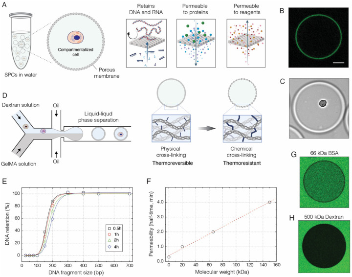
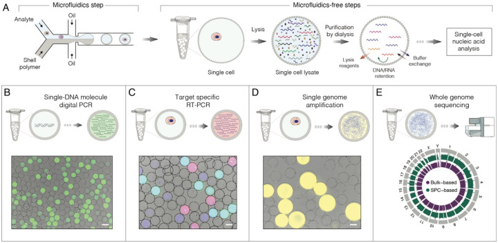
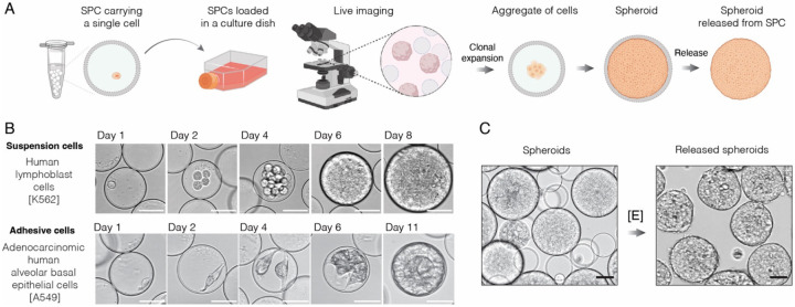
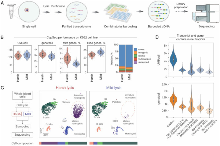
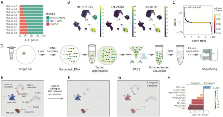
Biological systems are inherently complex and heterogeneous. Deciphering this complexity increasingly relies on high-throughput analytical methods and tools that efficiently probe the cellular phenotype and genotype. While recent advancements have enabled various single-cell -omics assays, their broader applications are inherently limited by the challenge of efficiently conducting multi-step biochemical assays while retaining various biological analytes. Extending on our previous work (1) here we present a versatile technology based on semi-permeable capsules (SPCs), tailored for a variety of high-throughput nucleic acid assays, including digital PCR, genome sequencing, single-cell RNA-sequencing (scRNA-Seq) and FACS-based isolation of individual transcriptomes based on nucleic acid marker of interest. Being biocompatible, the SPCs support single-cell cultivation and clonal expansion over long periods of time - a fundamental limitation of droplet microfluidics systems. Using SPCs we perform scRNA-Seq on white blood cells from patients with hematopoietic disorders and demonstrate that capsule-based sequencing approach (CapSeq) offers superior transcript capture, even for the most challenging cell types. By applying CapSeq on acute myeloid leukemia (AML) samples, we uncover notable changes in transcriptomes of mature granulocytes and monocytes associated with blast and progenitor cell phenotypes. Accurate representation of the entirety of the cellular heterogeneity of clinical samples, driving new insights into the malfunctioning of the innate immune system, and ability to clonally expand individual cells over long periods of time, positions SPC technology as customizable, highly sensitive and broadly applicable tool for easy-to-use, scalable single-cell -omics applications.


Conflict of interest statement

Conflict of interests Some of the results presented in this work have been filed in patent applications PCT/EP2022/084074; PCT/EP2022/084071; PCT/EP2022/084066; PCT/EP2022/084063. LM is a shareholder of Atrandi Biosciences.

Figures

Figure 1.
Figure 1.
The generation and biochemical properties of semi-permeable capsules (SPCs). A) Conceptually, the SPCs represent liquid droplets enveloped by a thin semi-permeable shell, which is reminiscent of a dialysis membrane. This membrane is permeable to assay reagents, oligonucleotides, and enzymes, yet it efficiently retains longer nucleic acid molecules inside the capsule. B) The confocal image of a SPC prepared with fluorescently labelled gelatin, which forms a thin outer layer surrounding the liquid core. Scale bar 50 μm. C) The bright field image of a SPC suspended in phosphate buffered saline (1x PBS) and carrying a single cell. Scale bar 50 μm. D) The generation of SPCs relies on droplet microfluidics and aqueous two-phase system (ATPS), which in this work comprises high molecular weight dextran (500 kDa) and methacryloyl-modified gelatin (GelMA). Upon liquid-liquid phase separation, the GelMA forms a shell, which is then solidified by cooling (physical-crosslinking) followed by photopolymerization (chemical-crosslinking). E) Selective retention of nucleic acid fragments by SPCs suspended in 1x PBS, over the course of 4 hours at 22 °C. F) The permeability of SPCs to proteins and biopolymers at 22 °C. G) Fluorescence image of a randomly selected capsule after 3 min incubation with 0.1 μM FITC-BSA (MW 66 kDa) at 22 °C. H) Fluorescence image of a randomly selected capsule after 60 min incubation with 0.2 μM FITC-Dextran (MW 500 kDa) at 22 °C.
Figure 2.
Figure 2.
Selected examples of nucleic acid applications using SPCs. A) The general strategy for conducting nucleic acid assays on single cells, or single DNA molecules, encapsulated in SPCs. At first, analytes of interest are isolated in SPCs using high-throughput droplet microfluidics followed by subsequent steps that are microfluidics-free and are conducted in a regular laboratory tube by processing all SPCs in bulk. B) Single DNA molecule amplification and analysis by digital PCR (staining dye: SYBR Green I). C) Multiplex single-cell RT-PCR targeting transcripts encoding CD45, yes-associated protein 1 and β-actin. The SPCs carrying cells expressing both CD45 and ACTB appear as cyan, YAP and ACTB as pink, and ACTB alone as blue. D) Single genome amplification of human lymphoblast cells (K-562) by phi29 DNA polymerase and staining with SYTO dye (yellow). The SPCs containing single amplified genomes appear larger due to swelling. E) The circular map of a whole genome sequencing of SPC-encapsulated lymphoblast cells. Scale bars, 50 μm
Figure 3.
Figure 3.
The use of SPCs for live cell cultivation and expansion. A) The schematics of conducting 3D cell cultivation and clonal expansion with SPCs. The single cells isolated in SPCs are placed in a suitable growth medium to allow clonal cell expansion and spheroid formation. B) The cell culture in SPCs using suspension cells (top row) and adhesive cells (bottom row) over the course of 8 and 11 days, respectively. C) The spheroids enclosed in SPCs are released upon addition of collagenase enzyme [E]. Scale bars, 50 μm.
Figure 4.
Figure 4.
CapSeq performance. A) Schematics of capsule-based single-cell RNA-Seq (CapSeq) approach. B) CapSeq performance on K-562 cell line at a sequencing depth 20.000 reads per cell. C) Left: Flowchart detailing harsh and mild lysis workflow. Right: White blood cells from a healthy individual, prepared for scRNA-Seq under harsh and mild lysis conditions, projected on a UMAP. Note, the reduced recovery of neutrophils and basophils (Bsph) under mild lysis conditions. D) Comparative analysis of transcript and gene recovery in healthy human neutrophils with CapSeq, 10X Genomics (V3.1) and BD Rhapsody platforms (downsampled to 15’000 reads per cell).
Figure 5.
Figure 5.
Transcriptional profiling of hematopoietic disorders using CapSeq. A) Single cell transcriptomes from 25 samples embedded on UMAP and colored according to annotated cell phenotypes. B) Force directed graph layout of the leukemic compartment. C) Gene set enrichment analysis of Hallmark pathways (MSigDB) was performed using ranked gene lists based on differential expression and statistical significance, comparing each AML cell cluster against all others (cluster vs. rest) or myeloid cell types between healthy donors and AML patients in pseudobulk analysis. Normalized enrichment score (NES) is represented by a color gradient, with blue indicating depletion (negative values) and red indicating enrichment (positive values). Statistically significant pathways (FDR < 0.05) are marked by a white dot. D) Upset plot showing shared differentially expressed genes among innate immune cell types when comparing AML patients and healthy donors. The bottom panel represents the intersecting cell types, while the top panel displays the size of the intersection (number of shared differentially expressed genes). Colored dots in the top panel indicate the most frequently shared genes across all intersections. E) A volcano plot illustrating all significantly differentially expressed genes in basophils from AML patients compared to healthy donors, with upregulated events shown in red and downregulated events in blue.
Figure 6.
Figure 6.. Single cell transcriptome enrichment by FACS for expression of a nucleic marker of interest.
A) Gene biotype distribution among the top 100 differentially expressed genes in AML cells compared to all other cells. B) Single cell transcriptomes embedded on UMAP and colored for MIR181A1HG, LNCAROD and LINC02147 expression. C) Differentially expressed genes in AML cells, compared to all other cells, ranked by a combined score of differential expression and statistical significance. Each dot represents a gene and is colored by the adjusted P-value. Positive score values indicate upregulated genes, while negative values correspond to downregulated genes in AML blast cells. D) Experimental design for sorting and sequencing single cell transcriptomes based on the nucleic acid marker of interest. E) Single cell transcriptomes of 4 AML patients (# 2, 4, 6 and 7) embedded on UMAP and colored according to the cell type. F) Sorted single cell transcriptomes for expression of MIR181A1HG embedded on UMAP G) Positive and negative fractions of FACS-sorted transcriptomes based on MIR181A1HG expression H) Differential abundance analysis of cell types between the positive and negative fractions of FACS-sorted cells based on MIR181A1HG gene expression. Log2 fold change is shown on the x-axis and indicated by a color gradient, with blue representing depletion and red representing enrichment of a given cell type in the positive fraction of FACS-sorted cells.

Similar articles

References

    1. Leonaviciene G., Mazutis L., RNA cytometry of single-cells using semi-permeable microcapsules. Nucleic Acids Res 51, e2 (2023). - PMC - PubMed
    1. Laks E. et al., Clonal Decomposition and DNA Replication States Defined by Scaled Single-Cell Genome Sequencing. Cell 179, 1207–1221.e1222 (2019). - PMC - PubMed
    1. Chen C. et al., Single-cell whole-genome analyses by Linear Amplification via Transposon Insertion (LIANTI). Science 356, 189–194 (2017). - PMC - PubMed
    1. Vitak S. A. et al., Sequencing thousands of single-cell genomes with combinatorial indexing. Nature Methods 14, 302–308 (2017). - PMC - PubMed
    1. Klein A. M. et al., Droplet barcoding for single-cell transcriptomics applied to embryonic stem cells. Cell 161, 1187–1201 (2015). - PMC - PubMed

Publication types