[Preprint]. 2025 Mar 19:2025.03.19.644215.
doi: 10.1101/2025.03.19.644215.

ATP hydrolysis-driven structural transitions within the S. cerevisiae Rad51 and Dmc1 nucleoprotein filaments

Affiliations

ATP hydrolysis-driven structural transitions within the S. cerevisiae Rad51 and Dmc1 nucleoprotein filaments

Yeonoh Shin et al. bioRxiv. .

Update in

Abstract

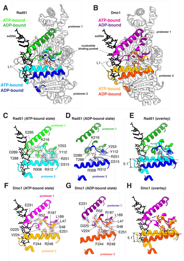
Homologous recombination (HR) is essential for the maintenance of genome stability and for generating genetic diversity during meiosis. The eukaryotic protein Rad51 is member of the Rad51/RecA family of DNA recombinases and is responsible for guiding the DNA pairing reactions that take place in HR during mitosis. Dmc1 is a meiosis-specific paralog of Rad51 and is responsible for the DNA pairing reactions that take place in HR during meiosis. Rad51 and Dmc1 are both ATP-dependent DNA-binding proteins and both form extended helical filaments on ssDNA which are key intermediates in HR. The stability of these nucleoprotein filaments is highly regulated and is also tightly coupled to nucleotide binding and hydrolysis. ATP binding promotes filament assembly whereas the hydrolysis of ATP to ADP reduces filament stability to promote filament disassembly. Here, we present CryoEM structures of the Saccharomyces cerevisiae recombinases Rad51 and Dmc1 in the ADP-bound states and provide a detailed structural comparison to the ATP-bound filaments. Our findings yield insights into the structural transitions that take place during the hydrolysis of ATP to ADP and suggest a new model for how these structural changes may be linked to nucleoprotein filament disassembly.


Figures

Figure 1.
Figure 1.. Overview of Rad51 and Dmc1 filament structures in the ADP-bound states.
(A) Our previously published CryoEM structure of S. cerevisiae Rad51 nucleoprotein filament in the ATP-bound state (PDB ID: 9D46)[44]. A subsection of the nucleoprotein comprised of six Rad51 monomers is shown, the different protein monomers are highlighted in alternating cyan and light green, and the bound ssDNA is shown in black. (B) CryoEM structure of S. cerevisiae Rad51 bound in the ADP-bound state (PDB ID: 9NJK). A subsection of the nucleoprotein comprised of six Rad51 monomers is shown, and the different protein monomers are highlighted in alternating blue and green; the ssDNA is disordered within the structure. (C) Our previously published CryoEM structure of S. cerevisiae Dmc1 nucleoprotein filament in the ATP-bound state (PDB ID: 9D4N)[44]. A subsection of the nucleoprotein comprised of six Dmc1 monomers is shown, the different protein monomers are highlighted in alternating magenta and orange, and the bound ssDNA is shown in black. (D) CryoEM structure of S. cerevisiae Dmc1 bound in the ADP-bound state (PDB ID: 9NJR). A subsection of the nucleoprotein comprised of six Dmc1 monomers is shown, and the different protein monomers are highlighted in alternating purple and dark orange; the ssDNA is disordered within the structure.
Figure 2.
Figure 2.. Comparison of the nucleotide-binding pockets in the ATP and ADP-bound states.
(A) Nucleotide-binding pocket of S. cerevisiae Rad51 in the ATP-bound state. The Rad51 promoter in which the Walker A and Walker B motifs interact with the bound nucleotide is shown in light green and the second protomer is shown in cyan. The locations of important side chain residues and the two metal ions are indicated. (B) Nucleotide-binding pocket of S. cerevisiae Rad51 in the ADP-bound state. The Rad51 promoter in which the Walker A and Walker B motifs interact with the bound nucleotide is shown in green and the second protomer is shown in blue. The locations of important side chain residues and the two metal ions are indicated. (C) Overlay of Rad51 in the ATP- and ADP-bound states. (D) Nucleotide-binding pocket of S. cerevisiae Dmc1 in the ATP-bound state. The Dmc1 promoter in which the Walker A and Walker B motifs interact with the bound nucleotide is shown in magenta and the second protomer is shown in orange. The locations of important side chain residues and the two metal ions are indicated; here, the metal ions are indicated as Me2+ because we cannot distinguish between Mg2+ and Ca2+, both of which were present in samples with Dmc1 (E) Nucleotide-binding pocket of S. cerevisiae Dmc1 in the ADP-bound state. The Dmc1 promoter in which the Walker A and Walker B motifs interact with the bound nucleotide is shown in purple and the second protomer is shown in dark orange. The locations of important side chain residues and the two metal ions are indicated. (F) Overlay of Dmc1 in the ATP- and ADP-bound states.
Figure 3.
Figure 3.. The FxxA polymerization interface remains largely unaltered in the ATP and ADP-bound states.
(A) Overlay of two adjacent Rad51 protomers in the ATP- and ADPbound states, as indicated, highlighting the location of the FxxA polymerization motif in one protomer and its binding cleft on the adjacent protomer. (B) Overlay of two adjacent Dmc1 protomers in the ATP- and ADP-bound states, as indicated, highlighting the location of the FxxA polymerization motif in one protomer and its binding cleft on the adjacent protomer. (C) Close-up view of the Rad51 FxxA polymerization motif interaction in the ATP-bound state. (D) Close-up view of the Rad51 FxxA polymerization motif interaction in the ADP-bound state. (E) Overlay of the Rad51 FxxA polymerization motif interaction in the ATP- and ADP-bound states. (F) Close-up view of the Dmc1 FxxA polymerization motif interaction in the ATP-bound state. (G) Close-up view of the Dmc1 FxxA polymerization motif interaction in the ADP-bound state. (H) Overlay of the Rad51 Dmc1 polymerization motif interaction in the ATP- and ADP-bound states.
Figure 4.
Figure 4.. Loss of trans L1-L1 contacts between adjacent protomers in the ADP-bound state.
(A) Overlay of two adjacent Rad51 protomers in the ATP- and ADP-bound states, as indicated, highlighting the location of the L1 DNA-binding loop connect to helix 13. (B) Overlay of two adjacent Dmc1 protomers in the ATP- and ADP-bound states, as indicated, highlighting the location of the L1 DNA-binding loop connect to helix 13. (C) Close-up view of the Rad51 trans interactions between L1 and helix 13 interaction in the ATP-bound state. (D) Close-up view of the Rad51 trans interactions between L1 and helix 13 interaction in the ADP-bound state. (E) Overlay of the Rad51 DNA-binding loop L1 and helix 13 in the ATP- and ADP-bound states. (F) Close-up view of the Dmc1 trans interactions between L1 and helix 13 interaction in the ATP-bound state. (G) Close-up view of the Dmc1 trans interactions between L1 and helix 13 interaction in the ADPbound state. (H) Overlay of the Dmc1 DNA-binding loop L1 and helix 13 in the ATP- and ADP-bound states.
Figure 5.
Figure 5.. Loss of L2 ssDNA- and inter-protomer contacts in the ADP-bound state.
(A) Overlay of two adjacent Rad51 protomers in the ATP- and ADP-bound states, as indicated, highlighting the location of the L2 DNA-binding loop, α helix 14, and β strands 6 and 7. (B) Overlay of two adjacent Dmc1 protomers in the ATP- and ADP-bound states, as indicated, highlighting the location of the L2 DNA-binding loop, α helix 14, and β strands 6 and 7. (C) Close-up view of the Rad51 L2 contacts with the bound ssDNA substrate and inter-protomer interactions in the ATP-bound state. (D) Close-up view showing the loss of Rad51 L2 contacts with the bound ssDNA substrate and inter-protomer interactions in the ADP-bound state. (E) Overlay of the Rad51 region encompassing the L2 DNA-binding loop, α helix 14, and β strands 6 and 7 in the ATP- and ADP-bound states. (F) Close-up view of the Dmc1 L2 contacts with the bound ssDNA substrate and inter-protomer interactions in the ATP-bound state. (G) Close-up view showing the loss of Dmc1 L2 contacts with the bound ssDNA substrate and inter-protomer interactions in the ADP-bound state. (H) Overlay of the Dmc1 region encompassing the L2 DNA-binding loop, α helix 14, and β strands 6 and 7 in the ATP- and ADP-bound states.
Figure 6.
Figure 6.. Model for Rad51 and Dmc1 nucleoprotein filament disassembly.
(A) Rad51 and Dmc1 in the ATP-bound states give exhibit extended nucleoprotein filaments and the bound ssDNA is organized into base triplets with an axial rise of ~7.2 Å between the last base of one triplet and the first base of the next triplet. (B) Conversion to the ADP-bound state results in nucleoprotein filament length, which would require even greater extension of the phosphate backbone in order the base triples to maintain correct register with the protein protomers. (C) The nucleoprotein filaments begin in the ATP-bound state and conversion of end-bound protomers leads to loss of contacts with the ssDNA and disruption of the protein-protein interfaces leading to protein dissociation from the ssDNA. Successive rounds of ATP hydrolysis can lead to complete disassembly of the nucleoprotein filaments.

References

    1. Coop G. and Przeworski M., An evolutionary view of human recombination. Nat Rev Genet, 2007. 8(1): p. 23–34. - PubMed
    1. Ortiz-Barrientos D., Engelstadter J., and Rieseberg L.H., Recombination Rate Evolution and the Origin of Species. Trends Ecol Evol, 2016. 31(3): p. 226–36. - PubMed
    1. Paques F. and Haber J.E., Multiple pathways of recombination induced by double-strand breaks in Saccharomyces cerevisiae. Microbiol Mol Biol Rev, 1999. 63(2): p. 349–404. - PMC - PubMed
    1. Symington L.S., Rothstein R., and Lisby M., Mechanisms and regulation of mitotic recombination in Saccharomyces cerevisiae. Genetics, 2014. 198(3): p. 795–835. - PMC - PubMed
    1. Cox M.M., et al. , The importance of repairing stalled replication forks. Nature, 2000. 404(6773): p. 37–41. - PubMed

Publication types