[Preprint]. 2025 Mar 19:2025.03.19.644175.
doi: 10.1101/2025.03.19.644175.

Tracing LYVE1+ peritoneal fluid macrophages unveils two paths to resident macrophage repopulation with differing reliance on monocytes

Affiliations

Tracing LYVE1+ peritoneal fluid macrophages unveils two paths to resident macrophage repopulation with differing reliance on monocytes

Alexandre Gallerand et al. bioRxiv. .

Abstract

Mouse resident peritoneal macrophages, called large cavity macrophages (LCM), arise from embryonic progenitors that proliferate as mature, CD73+Gata6+ tissue-specialized macrophages. After injury from irradiation or inflammation, monocytes are thought to replenish CD73+Gata6+ LCMs through a CD73-LYVE1+ LCM intermediate. Here, we show that CD73-LYVE1+ LCMs indeed yield Gata6+CD73+ LCMs through integrin-mediated interactions with mesothelial surfaces. CD73-LYVE1+ LCM repopulation of the peritoneum was reliant upon and quantitatively proportional to recruited monocytes. Unexpectedly, fate mapping indicated that only ~10% of Gata6-dependent LCMs that repopulated the peritoneum after injury depended on the LYVE1+ LCM stage. Further supporting nonoverlapping lifecycles of CD73-LYVE1+ and CD73+Gata6+ LCMs, in mice bearing a paucity of monocytes, Gata6+CD73+ LCMs rebounded after ablative irradiation substantially more efficiently than their presumed LYVE1+ or CD73- LCM upstream precursors. Thus, after inflammatory insult, two temporally parallel pathways, each generating distinct differentiation intermediates with varying dependencies on monocytes, contribute to the replenish hment of Gata6+ resident peritoneal macrophages.


Conflict of interest statement

Competing interests: none

Figures
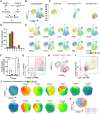
Figure 1.
Figure 1.. Phenotyping and comparison of ICAM2+ macrophages from the omentum and peritoneal fluid.
(A) Experimental scheme used to acquire data for subsequent unsupervised analysis. CD45.2+ cells retrieved from the peritoneal wash of Gata6fl/fl (WT) or Lyz2ΔGata6 mice were added to the minced CD45.1 omental tissue, and cells were digested at 37°C for 30 min. (B, C) UMAPs showing unsupervised clustering of omental and peritoneal wash macrophages analyzed via flow cytometry. (D) Proportions of each cluster’s contribution arising from the omentum. (E) UMAP plots representing the expression of macrophage markers used to define cluster identity. (F) Proportions of WT and Lyz2ΔGata6 cells found among clusters 2 and 3. (G) Flow cytometry plot showing expression of LYVE1 and ICAM2 in macrophages from clusters 2, 3, and 4. (H) UMAP showing the distribution of ICAM2+ LYVE1+ macrophages (red) compared with all other macrophages (gray) contained in the dataset. (I) Flow cytometry plot showing expression of CD62P and CD73 in macrophages from clusters 2, 3, and 4. (J) UMAPs displaying expression of the indicated surface markers by mesenteric macrophages. (K) Overlay of manually gated mesenteric macrophage populations. Panels A-I contain concatenated data from CD45.1 mice (n=3), CD45.2 WT mice (n=3), CD45.2 Lyz2ΔGata6 mice (n=3) and represent three similar experiments. Panels J-K are representative of two independent experiments. An ANOVA was used for statistical analysis in panel F. See also Extended Data Figure 1. *P<0.05.
Figure 2.
Figure 2.. Peritoneal surface-associated ICAM2+ peritoneal macrophages contribute to the peritoneal fluid compartment and mature into CD62P+ CD73+ LCMs.
(A, B) Representative flow cytometry plots and quantification of CD73 LYVE1+ cells among CD11b+ ICAM2+ LCMs in (A) the peritoneal fluid and mesothelial surfaces or (B) omentum and mesentery. (C) Representative flow cytometry plots showing the expression of TdTomato in omental (top row) and peritoneal (bottom row) macrophages from Lyve1creER x R26TdTomato mice at day 1 or day 7 after tamoxifen gavage. (D) Proportions of TdTomato+ cells among CD11b+ ICAM2+ macrophages in peritoneal fluid and omentum 1 (n=4) and 7 (n=3) days post tamoxifen gavage. Data were pooled from two independent experiments. (E) Proportions of CD62P+CD73+ macrophages among TdTomato+ CD11b+ICAM2+ macrophages in peritoneal fluid 1 (n=4) and 7 (n=3) days following tamoxifen oral gavage. Data were pooled from two independent experiments. (F) Experimental scheme used to photoconvert and chase omental cells for 5 days (top) and representative stereoscope pictures showing the detection of KikumeGreen and KikumeRed in the omentum before and after photoconversion (bottom). (G) Representative flow cytometry plots showing the detection of KikumeGREEN and KikumeRED in peritoneal macrophages (top row) and B cells (bottom row) over time after photoconversion of the omentum. (H) Representative flow cytometry plot (left) and proportions (right) of CD62P+ CD73+ cells among KikumeRED+ peritoneal macrophages 2 (n=6) and 5 (n=6) days after photoconversion. Data were pooled from two independent experiments. Mann-Whitney tests were used for statistical analysis. See also Extended Data Figure 2. *P<0.05, ***P<0.001, ****P<0.0001.
Figure 3.
Figure 3.. Integrin signaling disruption promotes peritoneal fluid accumulation of ICAM2+CD73LYVE1+ LCMs.
(A) UMAP projection of peritoneal CD11b+ CD115+ cells concatenated from WT (Lyz2+/+ Talin1fl/fl) and Lyz2ΔTalin (Lyz2cre/+ Talin1fl/fl) mice. (B) UMAP representation of surface marker expression in the concatenated dataset from (A). (C, D) Representative flow cytometry plots and quantification CD73 LYVE1+ (C), CD73 LYVE1 and CD62P+ CD73+ (D) macrophages in peritoneal fluid from WT (male n=14, female n=7) and Lyz2ΔTalin (male n=24, female n=3) mice. Data were pooled from three independent experiments. (E, F) Quantification of CD73 LYVE1+ LCMs, previously described as converting macrophages (convMacs), in the omentum (E) and mesothelial surfaces (F) from WT (male n=9, female n=8) and Lyz2ΔTalin (male n=8, female n=7) mice. (G) Experimental scheme used for competitive transplant of WT (ActinmTFP) and Lyz2ΔTalin bone marrow into lethally irradiated CD45.1 recipients. (H, I) Proportions of WT and Lyz2ΔTalin donor-derived cells in monocytes and SCMs (H), and CD11b+ICAM2+ LCM subsets (I). Data from recipient mice (n=8) were analyzed and combined across two independent experiments. ANOVA (panels C, D, E, F, and H) and Mann-Whitney (panel I) tests were used for statistical analysis. See also Extended Data Figure 3. *P<0.05, **P<0.01,***P<0.001.
Figure 4.
Figure 4.. LYVE1+ LCMs are minor precursors of CD73+CD62P+ LCMs even after bone marrow transplant.
(A) Experimental scheme used to generate BMTs for fate mapping cells that express LYVE1 during differentiation after transplant. (B, C) (B) Quantification of donor SCMs and ICAM2+ LCM subsets and (C) YFP expression from CD45.1 mice transplanted with YFP bone marrow cells from Lyve1cre x R26YFP mice with conditional deletion of Gata6 (Gata6fl/fl, n=3) or control littermates (Gata6fl/+, n=7). (D) Quantification of donor YFP+ and YFP cells among SCMs and ICAM2+ LCM subsets from CD45.1 mice transplanted with YFP bone marrow cells from Lyve1cre x R26YFP mice. Each symbol represents data from an independent mouse. An ANOVA was used for statistical analysis. (E) Quantification of CD62P+CD73+ cells in the peritoneal cavity of mice administrated with low dose (10ug) zymosan with (OMX) or without (Sham) omentectomy. *P<0.05, **P<0.01, ***P<0.001, ****P<0.0001, n.s. not significant.
Figure 5.
Figure 5.. Reconstitution of different peritoneal macrophage subsets across time after BMT.
(A-B) Quantification of peritoneal (A) monocytes, and (B) SCMs in WT control (n=7) and Zeb2 (n=4) mice at steady state. Data are representative of 3 independent experiments. (C) Quantification of total CD73 LCM (left), CD73 LYVE1+ LCM (middle), and CD62P+ CD73+ (right) LCMs in WT control (n=15) and Zeb2 (n=9) mice at steady state. Data were pooled from two independent experiments. Mann-Whitney tests were used for statistical analysis. (D) Quantification of CD73LYVE1 (left), CD73LYVE1+ (middle) and CD62P+ CD73+ (right) LCMs at 14 days (WT n=2, Zeb2 n=4), 21 days (WT n=3, Zeb2 n=4), 29 days (WT n=3, Zeb2 n=4), 51 days (WT n=4, Zeb2 n=3), and 60 days (WT n=9, Zeb2 n=6) post BMT. WT group (blue dots) are CD45.1 recipient mice transplanted with 50:50 Lyz2cre x R26TdTomato and CCR2gfp/gfp bone marrow after lethal irradiation. Zeb2 group (green dots) are CD45.1 recipient mice transplanted with Zeb2 bone marrow after lethal irradiation. (E-G) Quantification of (E) Bronchoalveolar lavage alveolar macrophages (BAL AM) and monocytes, (F) liver Kupffer cells and monocytes, and (G) heart macrophages 4 weeks after transplantation of lethally irradiated CD45.1 mice with WT or Zeb2TM bone marrow. (H-I) The cell number quantification (H) and percentage of total ICAM2+ LCMs (I) of the CD73 LCMs and CD62P+ CD73+ LCMs in the peritoneal cavity of lethally irradiated CD45.1 recipient mice 21 days post transplantation with bone marrow of different genotypes. Multiple two-tailed t-tests followed by FDR correction; **P < 0.01, ***P < 0.001, ****P < 0.0001, ns. not significant. (J) Representative plots and bar plot showing the proportions of TdTomato+ cells among CD73−LYVE1+, CD73+CD62P+ LCMs or blood monocytes in mice transplanted with Ms4a3CreR26TdTamato donor BM. (K) Proportions of TdTomato+ versus GFP+ cells out of the total CD73LYVE1+ or CD62P+CD73P+ LCMs in CD45.1 recipient mice transplanted with 50:50 Lyz2cre x R26TdTomato and CCR2gfp/gfp bone marrow after lethal irradiation. *P<0.05, **P<0.01, ***P<0.001, ****P<0.0001.

References

    1. Zhang N. et al., Expression of factor V by resident macrophages boosts host defense in the peritoneal cavity. J Exp Med 216, 1291–1300 (2019). - PMC - PubMed
    1. Zindel J. et al., Primordial GATA6 macrophages function as extravascular platelets in sterile injury. Science 371, (2021). - PubMed
    1. Vega-Perez A. et al., Resident macrophage-dependent immune cell scaffolds drive anti-bacterial defense in the peritoneal cavity. Immunity 54, 2578–2594 e2575 (2021). - PubMed
    1. Davies L. C. et al., A quantifiable proliferative burst of tissue macrophages restores homeostatic macrophage populations after acute inflammation. Eur J Immunol 41, 2155–2164 (2011). - PubMed
    1. Jenkins S. J. et al., Local macrophage proliferation, rather than recruitment from the blood, is a signature of TH2 inflammation. Science 332, 1284–1288 (2011). - PMC - PubMed

Publication types