[Preprint]. 2025 Mar 21:2025.03.17.25324099.
doi: 10.1101/2025.03.17.25324099.

Leveraging tissue-resident memory T cells for non-invasive immune monitoring via microneedle skin patches

Affiliations

Leveraging tissue-resident memory T cells for non-invasive immune monitoring via microneedle skin patches

Sasan Jalili et al. medRxiv. .

Abstract

Detecting antigen-specific lymphocytes is crucial for immune monitoring in the setting of vaccination, infectious disease, cancer, and autoimmunity. However, their low frequency and dispersed distribution across lymphoid organs, peripheral tissues, and blood pose challenges for reliable detection. To address this issue, we developed a strategy exploiting the functions of tissue-resident memory T cells (TRMs) to concentrate target circulating immune cells in the skin and then sample these cells non-invasively using a microneedle (MN) skin patch. TRMs were first induced at a selected skin site through initial sensitization with a selected antigen. Subsequently, these TRMs were restimulated by intradermal inoculation of a small quantity of the same antigen to trigger the "alarm" and immune recruitment functions of these cells, leading to accumulation of antigen-specific T cells from the circulation over several days. In mouse models of vaccination, we show that application of MN patches coated with an optimized hydrogel layer for cell and fluid sampling to this skin site allowed effective isolation of thousands of live antigen-specific lymphocytes as well as innate immune cells. In a human subject with allergic contact dermatitis, stimulation of TRMs with allergen followed by MN patch application allowed the recovery of diverse lymphocyte populations that were absent from untreated skin sites. These results suggest that TRM restimulation coupled with microneedle patch sampling can be used to obtain a window into both local and systemic antigen-specific immune cell populations in a noninvasive manner that could be readily applied to a wide range of disease or vaccination settings.


Conflict of interest statement

Competing interests S.J., P.T.H. and D.J.I. have submitted a patent application filed by MIT related to the data presented in this work. M.R. is principal or co-investigator of studies sponsored by Pfizer, Biogen, AbbVie, Incyte, LEO Pharma, Abeona Therapeutics, Dermavant, and Target RWE; and M.R. provides consulting for Pfizer, Biogen, Incyte, Takeda, Inzen, ROME Therapeutics, Almirall, Medicxi, Related Sciences, and VisualDx. The other authors declare no interests.

Figures

Figure 1.
Figure 1.. Identifying properties of hydrogel-coated microneedles for optimized cell sampling.
a, Photographs and scanning electron micrographs of the hydrogel-coated MN patch. b, Schematic view of cell and interstitial fluid sampling process with MN array applied to the skin. c, Molecular weight, G/M ratio, and viscosity of alginates tested as MN coatings. d, Still frames from timelapse microscopy of activated T cells incubated in SLG20 and SLM20 hydrogels in vitro showing tracked individual cell paths as colored lines. e, Quantification of T cell motility length and average track speed inside different hydrogels. f, Comparison of interstitial fluid sampling capacity between microneedles coated with different alginates. g, Optical and scanning electron micrographs of the patches after 18 hr of in vivo sampling on the skin of OVA-immunized mice and sampled following the scheme of Figure 2b (TRM recall), showing the retention of the alginate layer (labeled with Alexa647 for visualization) and collected cells on the patch. Data shown are means±SEM. ns, nonsignificant; *P<0.05, **P<0.01, ***P<0.001, ****P<0.0001 analyzed by ANOVA, followed by Tukey’s HSD.
Figure 2.
Figure 2.. Enhancing microneedle cell sampling via tissue-resident T cell restimulation.
a, Schematic view of TRM establishment, recall, and recruitment of antigen-specific tissue-resident memory T cells for MN patch sampling at a selected site on the skin. b, Study design for establishing TRM populations in the skin and subsequent TRM recall in mice immunized with OVA protein (10 ug per dose) and Lipo-CpG (1.24 nmol per dose). Blood and MN samples were collected 7 days after TRM recall. c-e, Enumeration of recovered total live leukocytes, CD3+ T cells, TRM cells, and antigen-specific CD8+ T cells recovered by MN patches under different sampling regimens. f, Representative flow cytometry plots showing SIINFEKL peptide-MHC tetramer staining to identify antigen-specific CD8+ T cells collected using MN patches 7 days after TRM recall. g, Enumeration of recovered antigen-specific CD8+ T cells in the MN patches at TRM establishment and recall steps in comparison with No TRM (n=10 animals per group). h, study design for longitudinal cytokine sampling post TRM recall step. i, Expression of inflammatory cytokines and chemokines induced in the skin measured using multiplexed ELISA analysis of interstitial fluid samples recovered by MN patches following TRM recall. Data shown are means±SEM. ns, nonsignificant; *P<0.05, **P<0.01, ***P<0.001, ****P<0.0001 analyzed by ANOVA, followed by Tukey’s HSD.
Figure 3.
Figure 3.. TRM recall leads to recruitment of antigen-specific T cells to the skin from circulation for recovery by MN patches.
a, Schematic representation of temporal labeling of the skin of the C57BL/6 KikGR mice, photoconverted right before the TRM recall dose (day 28) by violet light exposure on the skin site. b, c, Representative flow cytometry plots showing KikGR Red and Green gene expression in skin before and immediately after photoconversion. d, Representative flow cytometry plots showing KikGR Red and Green expression by skin infiltrating myeloid cells, CD3, CD4, CD8, and antigen-specific T cells on Day 34. e, Quantitation of frequencies of KikGR red+green+ and green-only+ cells by subtype recovered from MN patch sampling following the timeline in 3a. f-j, Enumeration of recovered live leukocytes (f), myeloid cells (g), CD4+ T cells (h), CD8+ T cells (i), and antigen-specific CD8+ T cells (j) 7 days after TRM recall dose. Data shown are means±SEM. ns, nonsignificant, *P<0.05, **P<0.01, ***P<0.001, ****P<0.0001 analyzed by ANOVA, followed by Tukey’s HSD.
Figure 4.
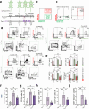
Figure 4.. TRM-enabled MN patch sampling of virus-specific T cells primed by mRNA vaccination.
a, ELISPOT analysis of antigen-specific IFN-γ-producing T cells from spleens of mice immunized with mRNA encoding SIV epitopes on day 21 post immunization (n = 5 animals/group). b, Study timeline comparing MN patch sampling under TRM recall vs. “TRM, no recall” conditions. c, Representative flow cytometry plots showing expression of tissue residence phenotypic markers for cells recovered from MN patches 7 days after TRM establishment. d, Enumeration of tissue-resident (CD103+CD69+, CD103+CD69−, CD103−CD69+) or non-tissue-resident recruited (CD103−CD69−) CD8+ T cells recovered from MN patches with or without TRM recall stimulation. e-i, Quantitation of total live leukocytes (e), total T cells (f), CD4+ T cells (g), CD8+ T cells (h), and antigen-specific CD8+ T cells recovered under recall or no-recall conditions. j, Quantitative comparison of antigen-specific CD8+ T cells recovered via blood draw (100 ul blood draw), skin biopsy (6 mm punch biopsy), or MN patches. Each data point represents an individual mouse. Data shown are means±SEM. *P<0.05, **P<0.01, analyzed by Student’s t-test or ANOVA, followed by Tukey’s HSD.
Figure 5.
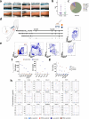
Figure 5.. Cell and cytokine sampling in human patient with allergic contact dermatitis.
a, Representative photographs of pre- and post-MN patch application on the forearm of human volunteers. b, c. Tolerability of MN sampling was assessed on a cohort of volunteers. Shown is the breakdown of volunteer gender and age (b) and skin areas tested (c). d, Human patient undergo SADBE-induced allergic contact dermatitis with sites previously exposed (14 days before reexposure) to SADBE. Suction blister and microneedle samples were collected 2 and 4 days after SADBE treatment. A non-SADBE exposed site was selected as nonlesional skin. e, Representative flow cytometry plots showing expression of immune cell markers in ISF collected from MN patches and suction blisters. f, Enumeration of recovered total live, CD45, CD3, CD4, CD8, CD4+ TRM and CD8+ T cells as recovered by MN patches or suction blister sampling on day 4 of the “TRM recall” condition. g, Comparison of cell yields from MN patches applied at 2 vs. 4 days post-TRM recall, alongside nonlesional skin and no-recall control groups. h, Olink proteomics data showing temporal changes in skin-associated proteins collected via MN patches.

References

    1. Sun L., Su Y., Jiao A., Wang X. & Zhang B. T cells in health and disease. Sig Transduct Target Ther 8, 235 (2023). - PMC - PubMed
    1. Koh C.-H., Lee S., Kwak M., Kim B.-S. & Chung Y. CD8 T-cell subsets: heterogeneity, functions, and therapeutic potential. Exp Mol Med 55, 2287–2299 (2023). - PMC - PubMed
    1. Mizukoshi E. et al. Peptide vaccine-treated, long-term surviving cancer patients harbor self-renewing tumor-specific CD8+ T cells. Nat Commun 13, 3123 (2022). - PMC - PubMed
    1. Philpott J. D. et al. Antigen-specific T cell responses in SARS-CoV-2 mRNA-vaccinated children. Cell Reports Medicine 4, 101298 (2023). - PMC - PubMed
    1. Bacher P. & Scheffold A. Flow cytometric analysis of rare antigen specific T cells. Cytometry Pt A 83A, 692–701 (2013). - PubMed

Publication types

LinkOut - more resources