. 2025 Feb 22;36(2):102491.
doi: 10.1016/j.omtn.2025.102491. eCollection 2025 Jun 10.

Reverse-phase chromatography removes double-stranded RNA, fragments, and residual template to decrease immunogenicity and increase cell potency of mRNA and saRNA

Affiliations

Reverse-phase chromatography removes double-stranded RNA, fragments, and residual template to decrease immunogenicity and increase cell potency of mRNA and saRNA

Andreja Krušič et al. Mol Ther Nucleic Acids. .

Abstract

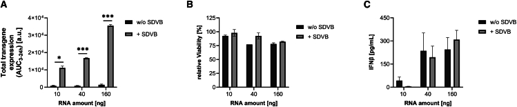
mRNA is produced by in vitro transcription reaction, which also leads to formation of immuno-stimulatory impurities, such as double-stranded RNA (dsRNA). dsRNA leads to activation of innate immune response linked to inhibition of protein synthesis. Its removal from mRNA preparations increases efficiency of protein translation. Previous studies identified ion-pair reverse-phase high-performance liquid chromatography as a highly efficient approach for dsRNA removal. Here, we present a comprehensive study of IP-RP LC purification on monolith chromatographic supports for mRNA polishing, demonstrating its ability to remove dsRNA, as well as hybridized RNA fragments and residual DNA template, which are not fully removed by mRNA capture methods. We develop step elution methodology, including at microgram scale with novel spin columns operated by centrifugation. We demonstrate SDVB efficiency across a range of molecular sizes and explore the necessity for temperature control for effective dsRNA removal from self-amplifying RNA. SDVB-purified mRNA and saRNA showed significantly increased transgene expression in cell-based assays and reduced the activation of cell autonomous innate immunity in A549 at early time points. Our findings highlight the importance of IP-RP purification for high-quality mRNA production, while simplifying the technological requirements for its adoption in clinical mRNA and saRNA manufacturing processes.

Keywords: HPLC; MT: Delivery Strategies; chromatography; dsRNA; mRNA; saRNA; vaccines.


Conflict of interest statement

A.K., N.M., M.L., P.M., A.Š., and R.S. are employees of Sartorius BIA Separations, d.o.o., which provided columns and chromatography systems used for this work.

Figures

None
Graphical abstract
Figure 1
Figure 1
Ion-pair reverse-phase polishing of mRNA on CIMmultus SDVB with linear gradient removes dsRNA and truncated RNA (A) A mass of 0.5 mg of Oligo dT pre-purified mRNA (eGFP) was loaded onto 1 mL CIMmultus SDVB column at room temperature and eluted in linear gradient from 7.5% to 18% ACN over 95 CV at 1 CV/min. (B) Fragment Analyzer electropherograms of selected fractions show lower molecular weight (MW) impurities eluting in fractions 1 and 2, with higher MW impurities observable in fractions 5–7 and significant in fraction 11. LM: Low molecular weight marker. (C) Elution fractions were analyzed on J2 dot blot, which confirmed the presence of dsRNA sub-populations in fractions 1, 2, 12, and strip.
Figure 2
Figure 2
Analytical characterization of RNA fractions collected from SDVB linear gradient purification A mass of 0.5 mg of ARCA-capped mRNA eGFP, pre-purified with CIM Oligo dT, was loaded onto CIM SDVB column at room temperature and eluted in a linear gradient (from 7.5% to 18% ACN over 95 CV) at 1 CV/min. The first 10 mL of elution peak was collected in 20 fractions (1-20), and the remaining 30 mL was collected as one fraction (21). (A) Fragment Analyzer results show RNA size distribution in fractionated elution peak. LM: molecular weight marker. (B) Capping efficiency analysis of selected elution fractions. (C) CIMac Oligo dT chromatographic analysis indicates % polyadenylation of elution fractions. Relative amounts of unbound, non-specifically bound, and bound RNA are presented in the bar chart, from integration of CIMac Oligo dT flow-through (FT), wash (W), and elution (E) peaks, respectively.
Figure 3
Figure 3
Ion-pair reverse-phase polishing of mRNA with CIMmultus SDVB operated with step elution removes dsRNA, truncated RNA, and residual pDNA template at least to 80-mL column scale Purification of (A.i) 0.6 mg and (B.i) 4.8 mg of Oligo dT-prepurified mRNA eGFP on 1 mL and 8 mL CIMmultus SDVB column, respectively, at room temperature and at 1 CV/min. Step elutions of 8.5% ACN, 11.7% ACN, and 18% ACN were applied. (A.ii and B.ii) Fragment Analyzer results of corresponding fractions. (A.iii and B.iii) J2 dot blot confirmed clearance of truncated sequences and dsRNA species in main mRNA fractions (“2”). (C.i) Scale-up preparative chromatogram of Oligo dT-prepurified mRNA (48 mg) on 80 mL CIMmultus SDVB column at room temperature and at 2 CV/min operated with Hipersep Flowdrive Pilot HPLC chromatography system. Step elutions were fractionated in five fractions (1–4 and strip). (C.ii) Fragment Analyzer and (C.iii) J2 dot blot analytics of collected fractions. (C.iv) AGE residual DNA template analysis and (C.v) CIMac pDNA quantification of residual DNA template in collected fractions.
Figure 4
Figure 4
mRNA polishing to remove dsRNA performed with CIM SDVB spin (0.1 mL) column in a benchtop centrifuge (A) A mass of 95 μg of Oligo dT-prepurified mRNA eGFP was diluted 10 times in loading buffer containing 7.5% ACN and applied onto a CIM SDVB spin (0.1 mL) column. Wash with 8.5% ACN eluted truncated species (fraction 1), elution with 11.7% ACN yielded full-length RNA (fraction 2) and strip with 18% ACN eluted dsRNA (strip). Recovery of mRNA in fraction 2 was 77%. (B) Fragment Analyzer of load, fractions 1, 2, and strip. (C) J2 dot blot of collected fractions.
Figure 5
Figure 5
Linear gradient SDVB purification of long saRNA construct on CIMmultus SDVB column (A) A mass of 1.5 mg of saRNA mCHIK(S27) (pre-purified with CIM Swiper multimodal column) was loaded onto a 4-mL SDVB column at 50°C and eluted in linear gradient from 7.5% to 18% ACN over 25 CV at 1 CV/min. (B) Fragment Analyzer electropherograms of sample load and elution fractions 1–9. (C) J2 dot blot analysis of collected fractions.
Figure 6
Figure 6
Purification of mRNA and saRNA with CIM SDVB does not compromise functionality in immuno-incompetent BHK-21 cells (A) A mass of 100 ng mRNA purified with Oligo dT (“w/o SDVB”) or Oligo dT + SDVB with linear gradient elution strategy (“linear-SDVB”) or Oligo dT + SDVB with step elution strategy (“step-SDVB”) was transfected into BHK-21 cells. After 24 h, GFP expression was measured by flow cytometry. The mean fluorescence intensity was multiplied with the percentage of GFP expressing cells to receive the total GFP expression levels. (B) Relative viability of cells was assessed by measuring viability after 48 h and normalized to the mock control. (C) BHK-21 cells were lipofected with the indicated doses of either Swiper (“w/o SDVB”) or Swiper and SDVB-purified saRNA (“+SDVB”). Luciferase expression was measured 6, 24, 48, and 72 h after transfection. Area under the curve (AUC) was calculated to determine the total luciferase expression. (D) Viability was measured 48 h after transfection and normalized to the mock control. Mean and standard error of the mean (SEM) of three independent experiments, each performed in technical triplicates, are shown.
Figure 7
Figure 7
SDVB purification of mRNA and saRNA improves transgene expression in A549 cells, where IFNβ response limits expression (A) A549 cells were lipofected with 100 ng of mRNA purified with Oligo dT (“w/o SDVB”) or Oligo dT plus SDVB with linear gradient elution strategy (“linear-SDVB”) or Oligo dT plus step elution strategy (“step-SDVB”). Twenty-four hours after transfection, GFP expression was detected by flow cytometry. (B) Viability of cells was detected 48 h after lipofection and normalized to untreated cells. (C) IFNβ release into the supernatant was quantified by ELISA 6 and 24 h after transfection. (D) 10, 40, or 160 ng of saRNA without (“w/o SDVB”) or with (“+ SDVB”) SDVB purification was transfected into A549 cells. Luciferase expression was measured at 6, 24, 48, and 72 h after transfection and area under the curve was calculated to assess total transgene expression. (E) After 48 h, ATP levels were quantified and normalized to the levels of untreated cells to determine relative viability. (F) 6 and 24 h after lipofection, IFNβ levels in the supernatant were quantified by ELISA. Mean and standard error of the mean of three independent experiments, each done in technical triplicates, are shown. Statistical differences were calculated using two-sided unpaired t tests with p values lower than 0.05 considered as significant and classified into ∗: p < 0.05; ∗∗: p < 0.01; ∗∗∗: p < 0.001; ∗∗∗∗: p < 0.0001.
Figure 8
Figure 8
CIM SDVB purification of saRNA allows transgene expression in PBMCs saRNA mCHIK(S27) without (“w/o SDVB”) or with (“+ SDVB”) SDVB purification was lipofected at 10, 40, and 160 ng into PBMCs. Luciferase expression was measured at 3, 6, 9, and 24 h after transfection. (A) Area under the curve was calculated to assess total transgene expression. (B) Viability was measured 24 h after lipofection and normalized to the untreated cells to assess relative viability. (C) Secreted IFNβ levels in the supernatant were quantified by ELISA 6 h after transfection. Error bars depict standard error of the mean from three independent experiments with triplicates each. Statistical differences were calculated using two-sided unpaired t tests with p values lower than 0.05 considered as significant and classified into ∗: p < 0.05; ∗∗: p < 0.01; ∗∗∗: p < 0.001; ∗∗∗∗: p < 0.0001.

Similar articles

References

    1. Sahin U., Karikó K., Türeci Ö. mRNA-based therapeutics — developing a new class of drugs. Nat. Rev. Drug Discov. 2014;13:759–780. doi: 10.1038/nrd4278. - DOI - PubMed
    1. Kozlova N.V., Pichon C., Rahmouni A.R. mRNA with Mammalian Codon Bias Accumulates in Yeast Mutants with Constitutive Stress Granules. Int. J. Mol. Sci. 2020;21:1234. doi: 10.3390/ijms21041234. - DOI - PMC - PubMed
    1. Curry E., Muir G., Qu J., Kis Z., Hulley M., Brown A. Engineering an Escherichia coli based in vivo mRNA manufacturing platform. Biotechnology and Bioengineering n/a. Biotechnol. Bioeng. 2024;121:1912–1926. doi: 10.1002/bit.28684. - DOI - PubMed
    1. Rosa S.S., Prazeres D.M.F., Azevedo A.M., Marques M.P.C. mRNA vaccines manufacturing: Challenges and bottlenecks. Vaccine. 2021;39:2190–2200. doi: 10.1016/j.vaccine.2021.03.038. - DOI - PMC - PubMed
    1. Lenk R., Kleindienst W., Szabó G.T., Baiersdörfer M., Boros G., Keller J.M., Mahiny A.J., Vlatkovic I. Understanding the impact of in vitro transcription byproducts and contaminants. Front. Mol. Biosci. 2024;11 doi: 10.3389/fmolb.2024.1426129. - DOI - PMC - PubMed

LinkOut - more resources