. 2025 Mar 20;53(6):gkaf229.
doi: 10.1093/nar/gkaf229.

N4-acetylcytidine coordinates with NP1 and CPSF5 to facilitate alternative RNA processing during the replication of minute virus of canines

Affiliations

N4-acetylcytidine coordinates with NP1 and CPSF5 to facilitate alternative RNA processing during the replication of minute virus of canines

Xueyan Zhang et al. Nucleic Acids Res. .

Abstract

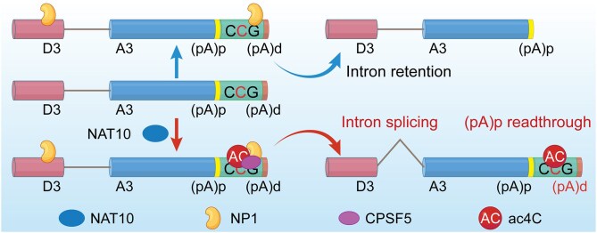
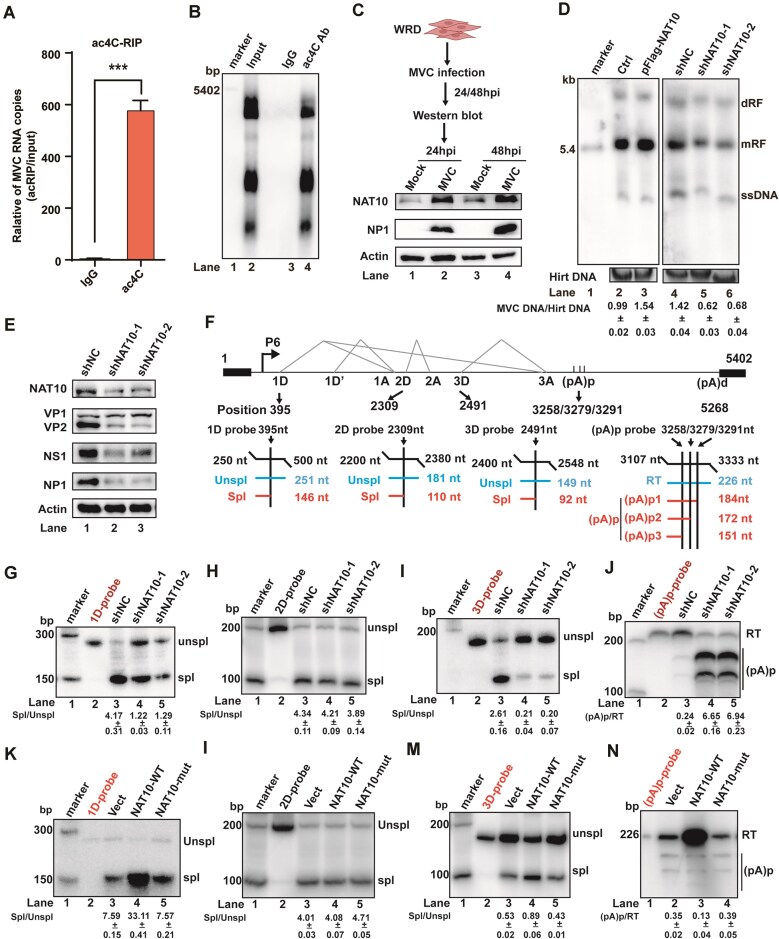
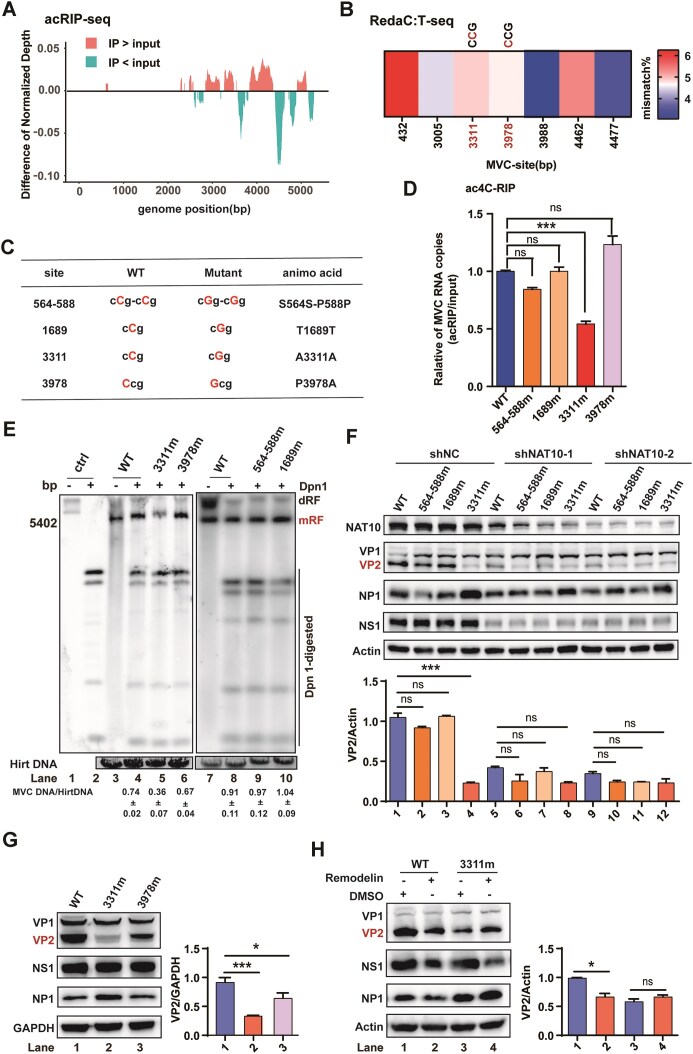
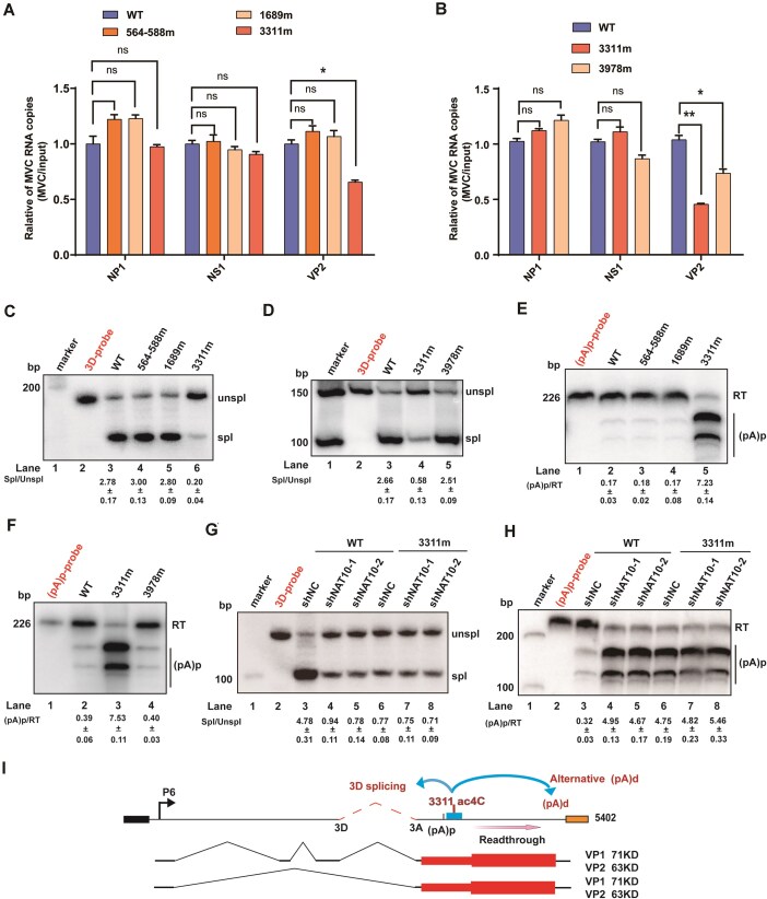
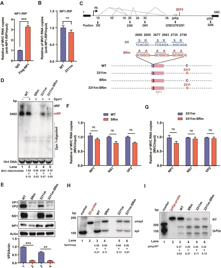
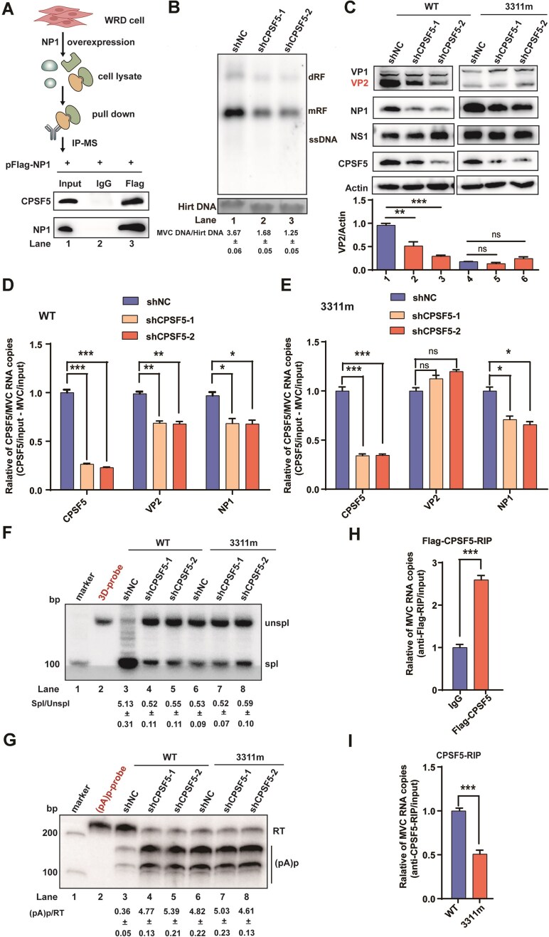
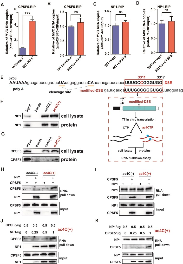
RNA modifications play crucial roles in RNA metabolism, structure, and functions. N4-acetylcytidine (ac4C) modifications have been shown to enhance stability and translation efficiency of messenger RNAs and viral RNAs. However, the relationship between ac4C and alternative RNA processing remains unexplored. Here, N-acetyltransferase 10 (NAT10) and its catalyzed ac4C modifications on minute virus of canines (MVC) were shown to regulate viral DNA replication and RNA processing, including both the alternative RNA splicing and polyadenylation. Through acRIP-seq and RedaC:T-seq, functional ac4C-modified residue 3311 was identified and characterized, which affected MVC RNA processing rather than altered the viral RNA stability. Ac4C modification at nt 3311 was revealed to participate in NP1-mediated viral RNA processing without influencing RNA affinity of NP1. Meanwhile, CPSF5 was identified to interact with NP1 and mediate viral RNA processing in an ac4C-dependent manner. Further in vitro assays showed that NP1 recruited CPSF5 to MVC RNAs, and the ac4C modification promoted specific binding of CPSF5 to the target region, which ensured precise alternative MVC RNA processing. This study not only reveals the functions of NAT10 and ac4C but also elucidates the mechanisms by which RNA modifications orchestrate MVC proteins and host factors for efficient viral replication and alternative RNA processing.


Conflict of interest statement

None declared.

Figures

Graphical Abstract
Graphical Abstract
Figure 1.
Figure 1.
NAT10 promotes MVC replication and affects the alternative viral RNA processing. (A) Identification of ac4C on MVC RNA via acRIP (RNA-immunoprecipitation)-qRT-PCR. RNAs from MVC-infected cells were incubated with IgG or anti-ac4C antibodies, followed by IP and qRT-PCR. Data are the means ± SEMs (n = 3). ***P≤ 0.001, unpaired student’s t-tests. (B) The acRIP and Northern blotting. RNAs from MVC-infected WRD cells were incubated with IgG or anti-ac4C specific antibodies as indicated. Immunoprecipitated RNAs were resolved on 1% agarose gels containing 2.2 M formaldehyde and transferred to Hybond-N+ membranes, followed by RNA signal detection with MVC-specific probes spanning from nt 1 to nt 5402. (C) Western blot analysis of NAT10 protein expression in MVC-infected cells at 24 h and 48 h post-infection. Actin served as an internal control. (D) Southern blot analysis of MVC DNA replication. Hirt DNA was extracted from infected cells with NAT10 overexpression or knockdown and subjected to Southern blot analysis at 48 h post-infection. The MVC full-length probe targeting the 1–5402 region was used for hybridization. EB staining of Hirt DNA served as an internal control. dRF, double replicative form; mRF, monomer replicative form; and ssDNA, single-stranded DNA. (E) Western blot analysis of MVC protein expression with NAT10 knockdown from MVC-infected cells at 48 h post-infection. (F) The top panel is a diagram of MVC genomic structure, including P6 promoter, four splicing donor sites (1D, 1D′, 2D, and 3D), three accept sites (1A, 2A, and 3A), the proximal polyadenylation cleavage site [(pA)p], and distal polyadenylation cleavage site [(pA)d]. The bottom panel shows the positions and sizes of probes used in RPA. The size of expected RPA products from unspliced RNA (unspl), spliced RNA (spl), RNA utilizing (pA)p site, or (pA)d site is below each probe. (GJ) Analysis of the alternative splicing of MVC RNA via RPA. Total RNA was extracted from MVC-infected cells in which NAT10 was knocked down at 48 h post-infection using a 1D-probe (G), 2D-probe (H), 3D-probe (I), and (pA)p-probe (J) targeting the respective indicated donor sites and (pA)p. Radioactive probes were loaded in each experiment and served as size controls. (KN) MVC RNA alternative splicing was measured by mutating the acetylase activity site of NAT10. RPA analysis of MVC RNA processing with NAT10 wild type (NAT10-WT) or G641E (NAT10-mut) mutant overexpression from MVC-infected cells at 48 h post-infection using a 1D-probe (K), 2D-probe (L), 3D-probe (M), and (pA)p-probe (N) targeting the respective indicated donor sites and (pA)p.
Figure 2.
Figure 2.
Residues 3311 could be the functional ac4C site to regulate MVC replication. (A) Mapping of ac4C sites on MVC RNA via acRIP (RNA-immunoprecipitation)-seq. Poly(A)+ RNA, purified from total RNA extracted from MVC-infected WRD cells, was fragmented, and IP was performed using anti-ac4C antibodies for acRIP-seq, followed by next-generation sequencing. Difference of normalized depth was generated on the coverage of acRIP-seq and input MVC RNA, respectively. (B) ac4C sites were detected on MVC RNA via RedaC:T-seq. Poly(A)+ RNA, purified from total RNA extracted from MVC-infected WRD cells, was treated with NaBH4, followed by next-generation sequencing. Both 3311 and 3978 sites were detected in acRIP-seq and RedaC:T-seq with CCG motifs, and only site 3311 was located at the wobble site of the codon. (C) The diagram shows the location of ac4C sites and negative control with CCG motif detected in acRIP-seq or RedaC:T-seq. Nucleotides 564–588, 3311, and 3978 were detected as ac4C sites, but 1689 site with CCG motif was not detected and served as a negative control. All mutants are synonymous codon, except 3978 site. (D) Identification of ac4C modified MVC RNA by acRIP-qRT-PCR. RNAs from WRD cells transfected with WT and mutant infectious clones were incubated with anti-ac4C antibodies, followed by IP and qRT-PCR. Data are the means ± SEMs (n = 3). ***P ≤ 0.001, ns: not significant, unpaired student’s t-tests. (E) MVC DNA replication analysis of cells transfected with WT and mutant infectious clones at 48 h via Southern blotting. EB staining of Hirt DNA served as an internal control. The probe was used as described in Fig. 1  D. (F, H) Western blot analysis of MVC protein expression with NAT10 knockdown (F) or remodelin treatment (H) from WRD cells transfected with WT or the other mutants at 48 h. Actin served as internal control. Relative intensity of VP2 versus actin was quantified using the ImageJ program. Data are means ± SDs (n = 3). ***P ≤ 0.001, ns: not significant, unpaired Student’s t-test. (G) Viral proteins expression was detected by western blotting. MVC infectious clones of WT and mutants were transfected into WRD cells and levels of protein were blotted after 48 h post-transfection. GAPDH served as internal control. Relative intensity of VP2 versus GAPDH was quantified using the ImageJ program. Data are means ± SDs (n = 3). ***P ≤ 0.001, *P ≤ 0.05, unpaired Student’s t-test.
Figure 3.
Figure 3.
Functional ac4C-modified residue determines the alternative splicing and polyadenylation of MVC pre-mRNAs. (A, B) qRT-PCR was performed to determine the RNA levels of MVC WT or mutants by NP1, NS1, and VP2 ORF primers in WRD cells at 48 h transfection of infectious clones, and GAPDH was used as a control. Data are means ± SEMs (n = 3). *P ≤ 0.05, **P ≤ 0.01, ns: not significant, unpaired Student’s t-tests. (CF) Regulation of MVC RNA alternative splicing via the functional ac4C-modified residue. RPA of total RNA extracted from cells transfected with MVC-WT and mutant infectious clones at 48 h was performed using 3D-probe (C, D) and (pA)p-probe (E, F). (G, H) The correlation between acetyltransferase NAT10 and ac4C-modified residues regulated MVC RNA alternative splicing. RPA of total RNA extracted from cells transfected with WT and mutant infectious clones in which NAT10 was knocked down at 48 h was performed using a 3D-probe and (pA)p-probe. (I) The model of acetylation at site 3311 regulates alternative 3D/3A RNA splicing and readthrough of (pA)p.
Figure 4.
Figure 4.
Ac4C at residue 3311 and NP1 are critical for the splicing at 3D site and (pA)p readthrough. (A, B) Ability of NP1 binding to the targeted MVC RNA based on formaldehyde-RIP (RNA-immunoprecipitation) and qRT-PCR. Cell lysates from formaldehyde-cross-linking were subjected to IP with IgG or anti-NP1 antibodies (A). WT and mutant 3311m infectious clones were used to transfect WRD cells, and cell lysates followed by formaldehyde-cross-linking, were subjected to IP with anti-NP1 antibodies (B). qRT-PCR was performed to quantify MVC RNA. Unpaired student’s t-tests were performed, and the data are presented as the means ± SEMs (n = 3). ns: not significant, ***P ≤ 0.001. (C) Diagram indicating the relative locations of the NP1 arginine/serine (SR) mutants and alignment of nucleotide 2600–2740 of wild-type MVC NP1 (WT, 3311m) and NP1-SRsm (WT-SRm, 3311m-SRm). (D) Southern blot analysis was performed to detect MVC DNA replication in cells transfected with MVC infectious clones WT, WT-SRm, 3311m, and 3311m-SRm at 48 h. (E) Western blot analysis of MVC protein expression in WRD cells transfected with the infectious clones, indicated as Fig. 4C; actin was used as a control. (F, G) Effect of MVC RNA level by mutating NP1 SR domain. qRT-PCR of total RNA extracted from WRD cells transfected with the same infectious clones as Fig. 4C. Data are means ± SEMs (n = 3). ns: not significant, unpaired Student’s t-tests. (H, I) Correlation between NP1 and the ac4C-modified residue affected MVC RNA processing based on RPA. RPA of total RNA extracted from cells transfected with the infectious clones WT, WT-SRm, 3311m, and 3311m-SRm at 48 h were performed using a 3D-probe (H) and (pA)p-probe (I).
Figure 5.
Figure 5.
CPSF5 recruited by NP1 targets viral RNAs to further regulate the alternative RNA processing. (A) NP1 interacts with CPSF5, which was verified by performing an IP assay. Flag-tagged NP1 was transiently expressed in WRD cells and then purified by IP using anti-Flag antibodies and detected via MS (Table 1) or western blotting. IgG was used as a negative control. (B) Southern blot assay showing MVC DNA replication in CPSF5 knockdown cells. Hirt DNA was extracted from MVC-infected cells with CPSF5 knockdown and subjected to Southern blot analysis at 48 h post-infection. (C) The expression of MVC protein was identified with CPSF5 knockdown. Western blot analysis of MVC protein expression by knocking down CPSF5 in WRD cells transfected with WT or mutant 3311m MVC infectious clone at 48 h; actin was used as a control. Relative intensity of VP2 versus actin was quantified using the ImageJ program. Data are means ± SDs (n = 3). ***P ≤ 0.001, **P ≤ 0.01, unpaired Student’s t-test. (D, E) MVC RNA levels detected in CPSF5 knockdown cells. qRT-PCR was performed to determine the RNA levels from CPSF5 knockdown WRD cells transfected with infectious clone WT (D) or mutant 3311m (E) at 48 h by CPSF5, NP1, VP2 ORF primers, and GAPDH was used as a control. Data are means ± SEMs (n = 3). *P ≤ 0.05, **P ≤ 0.01, ***P ≤ 0.001, ns: not significant, unpaired Student’s t-tests. (F, G) The relationship between CPSF5 and the ac4C-modified residue in regulating RNA processing was detected by RPA. RPA of total RNA extracted from CPSF5-knockdown WRD cells transfected with WT or 3311m mutant MVC infectious clone at 48 h was performed using a 3D-probe (F) and (pA)p-probe (G). (H, I) Ability of CPSF5 binding to the targeted MVC RNA based on formaldehyde-RIP-qRT-PCR. Flag-tagged CPSF5 was expressed in WRD cells, and formaldehyde-cross-linking cell lysates were subjected to IP with IgG or anti-Flag antibodies (H). WT and 3311m mutant infectious clones were used to transfect WRD cells in which CPSF5 was knocked down, then the cells were subjected to IP with anti-CPSF5 antibodies. qRT-PCR was performed to quantify MVC RNA (I). IgG was used as a negative control. Unpaired student’s t-tests were performed, and the data are presented as the means ± SEMs (n = 3). ***P ≤ 0.001.
Figure 6.
Figure 6.
CPSF5 mediated MVC RNA splicing is dependent on NP1 and ac4C modification at residue 3311. (A, B) The effect on CPSF5 binding to MVC RNA by increasing NP1 expression using formaldehyde-RIP-qRT-PCR. Vector or HA-NP1 and infectious clone WT or 3311m were co-transfected in WRD cells. Cell lysates cross-linked by formaldehyde were subjected to IP with IgG, anti-CPSF5 antibodies. qRT-PCR was performed to quantify the targeted MVC RNA. IgG was used as a negative control. Unpaired Student’s t-tests were performed, and the data are presented as means ± standard errors of the means (n = 3). *** P≤ 0.001, ns: not significant. (C, D) The effect on NP1 binding to MVC RNA by increasing CPSF5 expression using formaldehyde-RIP-qRT-PCR. Vector or Flag-CPSF5 and infectious clone WT or mutant 3311m were co-transfected in WRD cells. Cell lysates were subjected to IP with IgG, anti-NP1 antibodies. qRT-PCR was performed to quantify the targeted MVC RNA. IgG was used as a negative control. Unpaired Student’s t-tests were performed, and the data are presented as means ± standard errors of the means (n = 3). ns: not significant. (E) Characterization of the cleavage and polyadenylation sites and downstream elements (DSEs) in (pA)p is shown at the top. The bottom panel shows a schematic representation of the RNA pulldown assay. The DNA template for T7 transcription consisted of three repeats of modified DSE. (F, G) Biotinylated RNA affinities of NP1 and CPSF5. HA-tagged NP1 or the vector was transiently expressed in WRD cells, and cell lysates were used to bind biotinylated RNA with or without the ac4C modification. In addition, purified NP1 or CPSF5 protein was mixed with RNA oligonucleotides with or without ac4C-modified RNA and analyzed via western blotting. (H, I) The binding analysis of CPSF5 or NP1 to oligo-RNA with or without the ac4C modification in the presence or absence of the NP1 (H) or CPSF5 (I) protein. (J, K) The binding analysis of NP1 and CPSF5 to ac4C-modified RNA. Equal amounts of purified CPSF5 (J) or NP1 (K) protein and increasing amounts of NP1 (J) or CPSF5 (K) protein were incubated with ac4C-modified RNA and analyzed via western blotting. Blots were detected using anti-CPSF5 or anti-NP1 antibodies.

Similar articles

Cited by

References

    1. Barbieri I, Kouzarides T Role of RNA modifications in cancer. Nat Rev Cancer. 2020; 20:303–22.10.1038/s41568-020-0253-2. - DOI - PubMed
    1. Roundtree IA, Evans ME, Pan T et al. . Dynamic RNA modifications in gene expression regulation. Cell. 2017; 169:1187–200.10.1016/j.cell.2017.05.045. - DOI - PMC - PubMed
    1. Mendel M, Delaney K, Pandey RR et al. . Splice site m(6)A methylation prevents binding of U2AF35 to inhibit RNA splicing. Cell. 2021; 184:3125–42.10.1016/j.cell.2021.03.062. - DOI - PMC - PubMed
    1. Ma HL, Bizet M, Soares Da Costa C et al. . SRSF2 plays an unexpected role as reader of m(5)C on mRNA, linking epitranscriptomics to cancer. Mol Cell. 2023; 83:4239–54.10.1016/j.molcel.2023.11.003. - DOI - PMC - PubMed
    1. Li S, Qi Y, Yu J et al. . Nuclear aurora kinase A switches m(6)A reader YTHDC1 to enhance an oncogenic RNA splicing of tumor suppressor RBM4. Sig Transduct Target Ther. 2022; 7:97.10.1038/s41392-022-00905-3. - DOI - PMC - PubMed

MeSH terms