. 2025 Apr 1;149(1):30.
doi: 10.1007/s00401-025-02868-5.

Impaired remyelination in late-onset multiple sclerosis

Affiliations

Impaired remyelination in late-onset multiple sclerosis

Lidia Stork et al. Acta Neuropathol. .

Abstract

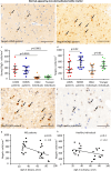
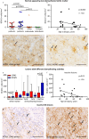
A reduced regenerative capacity may contribute to faster disease progression and poorer relapse recovery in multiple sclerosis patients with disease onset after the age of 50, a condition known as late-onset multiple sclerosis (LOMS). We hypothesized that lesions in LOMS patients show more pronounced axonal damage, less remyelination and an altered inflammatory composition, and performed a detailed histopathological analysis of MS biopsies in patients with early-stage LOMS. The number of T cells, B cells, plasma cells, microglia/macrophages, different oligodendrocyte populations as well as the axonal density and acute axonal damage were assessed in 31 LOMS and 30 normal-onset MS (NOMS, 20-40 years old) patients. No major differences in the inflammatory infiltrate or axonal damage were found. BCAS1-positive oligodendrocytes indicating early myelinating oligodendrocytes, and mature oligodendrocytes were significantly lower in the normal-appearing white matter of LOMS compared to NOMS patients (p = 0.05; p = 0.01), with a negative correlation with age (r = - 0.5, p = 0.01). In active demyelinating lesions, the number of BCAS1-positive oligodendrocytes did not differ between LOMS and NOMS, but NOMS lesions showed a higher proportion of ramified cells indicating active remyelination. In LOMS, BCAS1-positive oligodendrocytes decreased with increasing lesion age, with the lowest numbers found in inactive demyelinated lesions. In contrast, NOMS patients showed high numbers of BCAS1-positive cells with an activated morphology, even in inactive demyelinated lesions. At the last follow-up, LOMS patients had a significantly higher EDSS score (median 3.5) than NOMS patients (median 3.0, p = 0.05). A higher EDSS score correlated with fewer mature and oligodendrocyte precursor cells in active demyelinating lesions (r = - 0.4, p = 0.01 and r = - 0.6, p = 0.003). These findings suggest a clinically relevant impaired oligodendrocyte differentiation and remyelination in LOMS. Since remyelination is essential for axonal protection, it will be necessary to consider the complex and dynamic tissue environment when researching therapeutics aimed at fostering the differentiation of oligodendrocyte precursor cells into myelinating oligodendrocytes.

Keywords: Acute axonal damage; Axonal density; BCAS1; Inflammation; Late-onset multiple sclerosis; Oligodendrocytes; Remyelination.


Conflict of interest statement

Declarations. Conflict of interest: LS: reports funding from Sanofi outside the submitted work; SS has nothing to disclose; AK: has nothing to disclose; IM: reports funding from Sanofi for this study. IM reports personal fees from Biogen, Bayer Healthcare, TEVA, Serono, Novartis, Sanofi, Roche, BMS, Neuraxpharm as well as grants from Biogen outside the submitted work.

Figures

Fig. 1
Fig. 1
Histopathology of different demyelinating lesion activities. Panels a-c illustrate an early active demyelinating lesion, representing the initial stage of demyelination. At this stage, degradation products from both major (e.g., MBP) and minor (e.g., MAG) myelin proteins are present within phagocytes. a LFB/PAS staining reveals a demyelinated lesion containing multiple foamy macrophages. The inset shows an enlarged microscopic image of foamy macrophages with LFB-positive myelin debris; b Anti-myelin basic protein (MBP) staining of the early active lesion reflects myelin destruction and highlights numerous macrophages filled with myelin degradation products; c Anti-myelin-associated glycoprotein (MAG) staining also shows multiple macrophages containing MAG-positive degradation products, a minor myelin protein. Panels d-f depict a late active demyelinating lesion, representing the subsequent stage of demyelination, where only degradation products from major myelin proteins (e.g., MBP) can be identified within phagocytes. d LFB/PAS staining shows a completely demyelinated lesion with reactive astrogliosis and foamy macrophages; e Anti-MBP staining reflects an advanced stage of demyelination, with only a few macrophages containing MBP-positive degradation products (see inset for MBP-positive macrophages); f In contrast, no degradation products from minor myelin proteins, such as MAG, are detected within macrophages, as demonstrated by anti-MAG staining. Panels gi present an inactive demyelinated lesion, where no active demyelination processes are observed. g LFB/PAS staining reveals a demyelinated lesion accompanied by reactive astrogliosis and foamy macrophages; h Anti-MBP staining shows newly formed, thin myelinated fibers, indicating early remyelination. However, no myelin degradation products are observed within macrophages; i Anti-MAG staining also identifies early remyelination. LFB/PAS Luxol fast blue/periodic acid-Schiff, MBP myelin basic protein, MAG myelin-associated glycoprotein, Scale bars: a, d and g 200 µm; b, f and i 50 µm; e and h 100 µm; c 20 µm
Fig. 2
Fig. 2
Oligodendrocyte populations in the normal-appearing white matter of MS patients and white matter of healthy individuals a A median of 316.8 NogoA-positive mature oligodendrocytes per mm2 is present in the normal-appearing, non-demyelinated white matter of LOMS patients (anti-NogoA). b In contrast, significantly higher numbers (median of 618.7 NogoA-positive mature oligodendrocytes per mm2) are found in the normal-appearing white matter of NOMS patients (anti-NogoA). c The number of strongly Olig2-positive oligodendrocyte progenitor cells is increased in the normal-appearing white matter of MS patients compared to healthy individuals (p < 0. 0001), with no significant age-related differences. d In contrast, a significant reduction in the number of NogoA-positive mature oligodendrocytes is observed in the normal-appearing white matter of LOMS compared to NOMS patients (p = 0. 02), as well as in older compared to younger healthy individuals (p = 0.03). e Anti-Olig2 staining of the non-demyelinated white matter of an LOMS patient: Multiple strongly Olig2-positive nuclei (arrows), corresponding to oligodendrocyte precursors, as well as weakly positive nuclei are seen. f Anti-Olig2 staining of the white matter of a healthy older individual: Both strongly (arrows) and weakly Olig2-positive nuclei are identified. g A negative correlation between the number of NogoA-positive mature oligodendrocytes and patient age at the time of biopsy is observed (r =− 0.5, p = 0.01). h No significant correlation is found between the number of NogoA-positive mature oligodendrocytes and the age of healthy individuals (r = − 0.2, p = 0.3). a and b Scale bar = 100µm; c and d Error bars represent the median with interquartile range; e and f Scale bar = 50µm; g and h Non-parametric Spearman correlation index is shown. LOMS late-onset multiple sclerosis, NOMS normal-onset multiple sclerosis
Fig. 3
Fig. 3
BCAS1-positive early myelinating oligodendrocytes in MS patients and healthy individuals a The number of BCAS1-positive early myelinating oligodendrocytes is increased in the normal-appearing, non-demyelinated white matter of MS patients compared to age-matched controls (p < 0.0001 for the older cohort and p = 0.01 for the younger cohort), and is significantly lower in LOMS compared to NOMS patients (p = 0.02). b In MS patients, the number of BCAS1-positive early myelinating oligodendrocytes in the normal-appearing white matter shows a negative correlation with patient age at biopsy (r = − 0.6, p = 0.004). c A median of 3.4 BCAS1-positive cells/mm2 is present in the normal-appearing white matter of LOMS patients (arrows). d: In contrast, significantly higher numbers of BCAS1-positive cells (median 16.5 cells/mm2) are found in the normal-appearing white matter of NOMS patients (arrows). e The number of BCAS1-positive early myelinating oligodendrocytes within lesions with different demyelinating activities, indicating lesion evolution, is shown. BCAS1-positive cells are significantly lower in the normal-appearing white matter and in inactive lesions of LOMS compared to NOMS patients (p = 0.02 for both regions). A biphasic increase in BCAS1-positive early myelinating oligodendrocytes is observed in NOMS patients, with an initial rise in active lesions and a second increase in inactive lesions. In contrast, LOMS patients show a further decrease in inactive lesions. f A lower number of BCAS1-positive early myelinating oligodendrocytes in inactive MS lesions is associated with an older age of patients (r =− 0.6, p = 0.01). g Single BCAS1-positive cells are observed in an inactive lesion of a LOMS patient (median: 7.3 cells/mm2). h Multiple BCAS1-positive cells are present in an inactive lesion of a NOMS patient (median: 101.7 cells/mm2). a Error bars represent the median with interquartile range; b and f Non-parametric Spearman correlation index is shown; c, d, g and h: Scale bar: 50µm; e box plot showing the median number of cells with 10th–90th percentile; whiskers indicate minimum and maximum values. LOMS late-onset multiple sclerosis, NOMS normal-onset multiple sclerosis
Fig. 4
Fig. 4
Distribution of BCAS1-positive oligodendrocytes with resting and activated morphologies in MS lesions with different demyelinating activities a BCAS1-positive cells with strongly positive cell bodies and ramified processes are shown, referred to as activated BCAS1-positive oligodendrocytes. b Lesions with BCAS1-positive cells showing strongly positive cell bodies and ramified processes, as well as cells with BCAS1 expression only on the cell membrane and within the cytoplasm, referred to as activated and quiescent BCAS1-positive oligodendrocytes, are depicted. c BCAS1-positive cells with expression predominantly on the cell membrane and within the cytoplasm, but not showing ramified processes, are found in this case, referred to as quiescent BCAS1-positive oligodendrocytes. d Finally, some lesions show no BCAS1-positive cells. e 53% of early active demyelinating lesions in NOMS patients show mostly activated BCAS1-positive oligodendrocytes, whereas the proportion is lower in LOMS (36%). The percentage of lesions with mostly quiescent BCAS1-positive oligodendrocytes and lesions with both activated and quiescent BCAS1 + oligodendrocytes does not differ significantly between groups. However, a small percentage (14%) of lesions without any BCAS1-positive cells is observed only in LOMS. f In late active demyelinating lesions, the percentage of lesions with mostly activated BCAS1-positive cells increases in both cohorts, reaching 75% of lesions in NOMS and 47% in LOMS. The presence of lesions with mostly quiescent BCAS1-positive cells decreases in both cohorts, but the reduction is more pronounced in NOMS, from 35 to 8%, and less pronounced in LOMS, from 27 to 20%. Lesions without any BCAS1-positive cells are present only in LOMS (13%). g Inactive demyelinated lesions of NOMS patients show only lesions with activated BCAS1-positive cells (100%). In LOMS, the percentage of these lesions does not change compared to late active demyelinating lesions (44%). One in three lesions in LOMS patients have no BCAS-1 cells at all (33%). ad: Scale bar = 50µm. LOMS late-onset multiple sclerosis, NOMS normal-onset multiple sclerosis
Fig. 5
Fig. 5
Correlation of mature oligodendrocytes and oligodendrocyte precursor cells with the EDSS score at the last follow-up a The EDSS score at the last follow-up was significantly higher in LOMS patients compared to NOMS patients (median: LOMS 3.5 vs. NOMS 2.0, p = 0.05). b A lower number of NogoA-positive mature oligodendrocytes is associated with a higher disability score at the last follow-up, as measured by the EDSS (r =− 0.4, p = 0.01). c: A Lower number of strongly Olig2-positive oligodendrocyte precursor cells is associated with a higher EDSS score at the last follow-up (r = − 0.6, p = 0.003). a Error bars represent the median with interquartile range; b and c: Non-parametric Spearman correlation index is shown. LOMS late-onset multiple sclerosis, NOMS normal-onset multiple sclerosis, EDSS Expanded disability status scale

References

    1. Barateiro A, Fernandes A (2014) Temporal oligodendrocyte lineage progression: in vitro models of proliferation, differentiation and myelination. Biochem Biophys Acta 1843:1917–1929. 10.1016/j.bbamcr.2014.04.018 - PubMed
    1. Bergner CG, van der Meer F, Franz J, Vakrakou A, Würfel T, Nessler S et al (2024) BCAS1-positive oligodendrocytes enable efficient cortical remyelination in multiple sclerosis. Brain. 10.1093/brain/awae293 - PMC - PubMed
    1. Bodini B, Veronese M, García-Lorenzo D, Battaglini M, Poirion E, Chardain A et al (2016) Dynamic imaging of individual remyelination profiles in multiple sclerosis. Ann Neurol 79:726–738. 10.1002/ana.24620 - PMC - PubMed
    1. Bramow S, Frischer JM, Lassmann H, Koch-Henriksen N, Lucchinetti CF, Sørensen PS et al (2010) Demyelination versus remyelination in progressive multiple sclerosis. Brain 133:2983–2998. 10.1093/brain/awq250 - PubMed
    1. Bruck W, Popescu B, Lucchinetti CF, Markovic-Plese S, Gold R, Thal DR et al (2012) Neuromyelitis optica lesions may inform multiple sclerosis heterogeneity debate. Ann Neurol 72:385–394. 10.1002/ana.23621 - PubMed

Publication types

LinkOut - more resources