

Neurological disability and brain grey matter atrophy in primary progressive multiple sclerosis are determined by microstructural lesional changes, but not by lesion load
- PMID: 40167785
- PMCID: PMC11961454
- DOI: 10.1007/s00415-025-13043-x
Neurological disability and brain grey matter atrophy in primary progressive multiple sclerosis are determined by microstructural lesional changes, but not by lesion load
Abstract
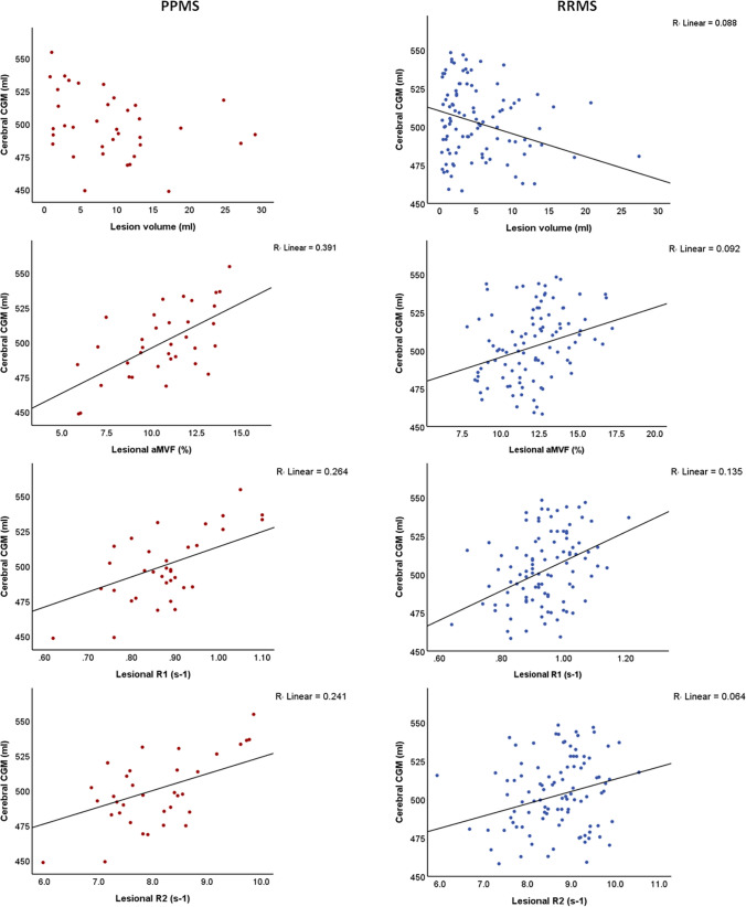
Background: Conventional MRI measures, such as the number and volume of MS lesions, are histologically non-specific and cannot sufficiently explain clinical disability or brain atrophy in MS. Nevertheless, demyelinating plaques exhibit distinct histopathological features in relapsing and progressive multiple sclerosis (MS) subtypes. The aim of this study was to assess microstructural characteristics of MS lesions using quantitative MRI and explore their associations with grey matter (GM) atrophy and clinical disability.
Methods: 56 control subjects (CS), 121 patients with relapsing-remitting (RRMS), and 38 patients with primary progressive MS (PPMS) underwent 1.5 T MRI scans and clinical examinations. Lesion and brain segmentation based on T1-weighted and FLAIR images were performed using SAMSEG. The MDME sequence and SyMRI software were used to estimate relaxation rates and myelin volume fraction in MS lesions and normal-appearing white matter (NAWM). Associations between quantitative lesional and NAWM MRI parameters with GM atrophy and clinical disability were investigated.
Results: Brain regional volumes and quantitative lesional and NAWM MRI parameters were significantly decreased in patients with PPMS compared to those with RRMS. Quantitative lesional MRI parameters demonstrated statistically significant associations with cortical and deep GM volumes as well as with disability scores in RRMS and especially in PPMS. In contrast to RRMS, lesion volume was not associated with either GM atrophy or clinical disability in the PPMS group.
Conclusions: Quantitative lesional MRI measures, but not lesion load, were strongly associated with clinical disability and GM atrophy in PPMS patients, likely reflecting differences in lesion pathology between MS subtypes.
Keywords: Advanced neuroimaging; EDSS; Grey matter atrophy; MDME; MRI; Multiple sclerosis.
© 2025. The Author(s).
Conflict of interest statement
Declarations. Conflicts of interest: B.K., B.B., Z.A., J.CJ., and J.B: nothing to disclose. R.S. has received speaker's honoraria from Bayer HealthCare, Alexion Pharma, Novartis Pharma, and Roche Pharma AG, congress travel support from Merck, Biogen Idec GmbH, and has received research scientific grant support from Novartis Pharma. R.G.: has received compensation for serving as a consultant or speaker from Bayer HealthCare, Biogen Idec, Merck Serono, Novartis, and Teva Neuroscience; he, or the institution he works for, has received research support from Bayer HealthCare, Biogen Idec, Merck Serono, Novartis, and Teva Neuroscience; he has also received honoraria as a Journal Editor from SAGE and Thieme Verlag. C.L.: received a research grant by the German Federal Ministry for Education and Research, BMBF, German Competence Network Multiple Sclerosis (KKNMS), Grant No. 01GI1601I, has received consulting and speaker's honoraria from Biogen Idec, Bayer Schering, Daiichi Sanykyo, Merck Serono, Novartis, Sanofi, Genzyme, and TEVA.e. T.L.: has received research scientific grant support from Novartis Pharma. Ethical approval: The study was approved by the local ethics committee of Ruhr University Bochum (Approval No. 20–7054-BR), and all patients provided written informed consent prior to participation in the study.
Figures


References
-
- Miller DH, Leary SM (2007) Primary-progressive multiple sclerosis. Lancet Neurol 6(10):903–912 - PubMed
-
- Thompson AJ, Banwell BL, Barkhof F, Carroll WM, Coetzee T, Comi G, Correale J, Fazekas F, Filippi M, Freedman MS et al (2018) Diagnosis of multiple sclerosis: 2017 revisions of the McDonald criteria. Lancet Neurol 17(2):162–173 - PubMed
-
- Nijeholt GJ, van Walderveen MA, Castelijns JA, van Waesberghe JH, Polman C, Scheltens P, Rosier PF, Jongen PJ, Barkhof F (1998) Brain and spinal cord abnormalities in multiple sclerosis. Correlation between MRI parameters, clinical subtypes and symptoms. Brain 121(4):687–697 - PubMed
-
- Barkhof F (2002) The clinico-radiological paradox in multiple sclerosis revisited. Curr Opin Neurol 15(3):239–245 - PubMed
MeSH terms
LinkOut - more resources
Full Text Sources
