Review
. 2025 Jun;130(6):934-964.
doi: 10.1007/s11547-025-02008-9. Epub 2025 Apr 1.

New insights into imaging of pulmonary metastases from extra-thoracic neoplasms

Affiliations
Review

New insights into imaging of pulmonary metastases from extra-thoracic neoplasms

Giuseppe Cicchetti et al. Radiol Med. 2025 Jun.

Abstract

The lung is one of the most common sites of metastases from extra-thoracic neoplasms. Lung metastases can show heterogeneous imaging appearance, thus mimicking a wide range of lung diseases, from benign lesions to primary lung cancer. The proper interpretation of pulmonary findings is crucial for prognostic assessment and treatment planning, even to avoid unnecessary procedures and patient anxiety. For this purpose, computed tomography (CT) is one of the most used imaging modalities. In the last decades, cancer patients' population has steadily increased and, due to the widespread application of CT for staging and surveillance, the detection of pulmonary nodules has raised, making their characterization and management an urgent and mostly unsolved problem for both radiologists and clinicians. This review will highlight the pathways of dissemination of extra-thoracic tumours to the lungs and the heterogeneous CT imaging appearance of pulmonary metastases, providing useful clues to properly address the diagnosis. Furthermore, we will deal with the promising applications of radiomics in this field. Finally, a focus on the hot-topic of pulmonary nodule management in patients with extra-thoracic neoplasms (ETNs) will be discussed.

Keywords: Cancer; Differential diagnoses; Management; Metastasis; Multimodal imaging; Pulmonary nodules.


Conflict of interest statement

Declarations. Ethics approval and consent to participate: Not applicable. Consent for publication: Not applicable. Competing interests: The authors declare that they have no competing interests.

Figures

Fig. 1
Fig. 1
Typical CT appearance and distribution of hematogenous pulmonary metastases, lymphangitic carcinomatosis and endobronchial metastasis. a, b Lung metastases in a 32 y.o. woman affected by Ewing sarcoma. Axial (a) and coronal (b) computed tomography (CT) images show multiple solid nodular and rounded lesions of variable size, preferentially located in the basal regions of the lungs. c, d Lymphangitic carcinomatosis in a 74 y.o. man affected by advanced stage colon cancer. Axial CT images show bilateral smooth thickening of the interlobular septa and peribronchovascular interstitium, predominant in the lung bases, associated with bilateral pleural effusion and atelectasis of the right lower lobe. Note the absence of architectural distortion of the secondary pulmonary lobules. e, f Endobronchial metastasis in a 57 y.o. patient with melanoma (e, f). Contrast-enhanced CT images show a well-defined, homogeneous and strongly enhancing solid nodule (white arrows in e), that obstructs the right middle lobe bronchus, determining distal lobar atelectasis (arrowheads in e, f)
Fig. 2
Fig. 2
Drug-induced organizing pneumonia in an oncologic patient, with complete resolution after treatment. Drug-induced organizing pneumonia in a 29 y.o. woman with melanoma treated with nivolumab. Axial computed tomography (CT) image a shows bilateral perilobular consolidations with peripheral distribution in the lung bases. Axial CT image b performed after corticosteroid treatment shows almost complete resolution of the abnormalities, with some residual hazy ground glass opacities and subtle bands in the lower lobes
Fig. 3
Fig. 3
Pulmonary macronodular sarcoidosis in a patient affected by an extra-thoracic neoplasm. Pulmonary sarcoidosis in a 61 y.o. man affected by follicular lymphoma of the scalp. Axial (a) and coronal (b) computed tomography (CT) images show bilateral clusters of confluent micronodules (“galaxy sign”), preferentially located in the middle and superior zones of both lungs, with peribronchovascular and subpleural distribution
Fig. 4
Fig. 4
DIPNECH in an oncologic female patient. Diffuse idiopathic pulmonary neuroendocrine cell hyperplasia (DIPNECH) in a 63 y.o. woman who underwent gastric resection due to gastric adenocarcinoma. Axial computed tomography (CT) maximum intensity projection (MIP) image (a), expiratory low dose image (b) with minimum intensity projection (MinIP) reconstruction (c) showing well-defined bilateral nodules of various size and diffuse inhomogeneous density of lung parenchyma due to air trapping, better depicted in the expiratory images
Fig. 5
Fig. 5
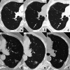
Organizing pneumonia versus lung adenocarcinoma, appearing as a solitary pulmonary nodule in two different cancer patients. ac Solitary pulmonary nodule in a 50 y.o. woman appeared during the follow-up for breast cancer treated with surgery and radiotherapy. At first computed tomography (CT) exam a no pulmonary nodules are detected. Six months later b a tiny micronodule (arrows) can be observed in the superior segment of the right lower lobe. One year later c the nodule significantly increased in size and patient was addressed to surgery. Histology revealed a focus of organizing pneumonia. df Solitary pulmonary part-solid nodule in the superior segment of the left lower lobe in a 68 y.o. man with bladder cancer. A progressive increase of the solid component within the nodule, with retraction of the adjacent fissure, is observed during the follow-up, after 3 months (b) and 9 months (c). At surgery, histology revealed a lung adenocarcinoma
Fig. 6
Fig. 6
Miliary distribution of lung metastases versus miliary tuberculosis. a Miliary metastases in a 72 y.o. man with gastric carcinoid tumour endoscopically resected. Axial computed tomography (CT) maximum intensity projection (MIP) image shows multiple metastatic micronodules with a miliary distribution and variable size. Note the feeding vessel sign in the two larger nodules located in the right lower lobe. b Miliary tuberculosis in a 33 y.o. man. Axial CT scan depicts multiple micronodules widely disseminated throughout the lungs
Fig. 7
Fig. 7
Cystic and cavitary pulmonary metastases. a, b Axial (a) and coronal (b) images in a 64 y.o. man with history of esophageal adenocarcinoma showing cystic lesions with thickened and lobulated wall, the majority with a solid and irregular component inside or at the periphery (arrowheads), histologically proved to be cystic metastases. cf Cavitary pulmonary metastases in a 61 y.o. man affected by ampullary cancer. Axial computed tomography (CT) scans show two indeterminate subcentimeter nodules in the anterior segment of the left upper lobe (arrows) (c) and in the posterior segment of the right lower lobe (circles) (d), respectively. An increase in size and the appearance of small cavitation within both lesions at CT performed after 18 months suggested the hypothesis of metastases (e, f)
Fig. 8
Fig. 8
Pulmonary calcified metastases versus pulmonary calcified hamartoma. a, b Pulmonary calcified metastases in a 23 y.o. man affected by osteosarcoma. Contrast-enhanced axial computed tomography (CT) images showing two adjacent solid nodules with inhomogeneous contrast enhancement and eccentric irregular calcifications. c, d Pulmonary calcified hamartoma in a 73 y.o. man with a history of pancreatic adenocarcinoma, treated with pancreatectomy and adjuvant radio-chemotherapy. Axial CT images depict a nodule showing central dense calcifications with popcorn-like appearance; note the associated fat tissue density (-46 HU) within the nodule
Fig. 9
Fig. 9
Calcified miliary metastases versus calcified nodules resulting from a previous infection. a, b Calcified metastases in a 33 y.o. man, who had undergone thyroidectomy due to a medullary thyroid cancer 10 months before. a Axial computed tomography (CT) maximum intensity projection (MIP) image depicts multiple sharply defined calcified micronodules with miliary distribution; note calcified hilar and mediastinal lymph nodes in b. Differential diagnosis with pneumoconiosis might be hard even though miliary distribution is less likely to be related to exposure, and patient’s age and history should always be taken into consideration. c, d Calcified nodules in a 38 y.o. man with previous varicella pneumonia. Axial CT MIP images show multiple calcified nodules and micronodules randomly distributed throughout the lungs, which are commonly found years after varicella infection
Fig. 10
Fig. 10
Uncommon CT features of pulmonary metastases. Metastases with less common morphological features. a, b Pulmonary metastasis in a 47 y.o. man with renal cell carcinoma. Axial computed tomography (CT) chest image a shows a pulmonary solid nodule surrounded by a peripheral ground-glass attenuation (halo sign) in the left upper lobe. Contrast-enhanced axial CT scan of the upper abdomen b depicts a large solid neoplastic mass of the right kidney, with marked heterogeneous contrast enhancement. c, d Pulmonary metastasis in a 78 y.o. man with renal cell carcinoma. Axial CT images depict a well-defined and contrast-enhancing pulmonary nodule; note the fat tissue density within the nodule in d. e, f Endoarterial tumoral embolization in a 61 y.o. man with gastric cancer. Axial CT images show vascular tree-in-bud pattern; note the irregular enlargement and occlusion of the arterial pulmonary vessels determined by intraluminal neoplastic tissue, evident until the centrilobular core
Fig. 11
Fig. 11
Proposal of diagnostic flowchart for IPN(s) management in patients with ETNs. Proposed diagnostic flowchart for indeterminate pulmonary nodule(s) (IPNs) in patients affected by extra-thoracic neoplasms (ETNs). This diagnostic algorithm should be regarded as a guide only. A tailored approach in a multidisciplinary context should be ensured, taking into account the risk factors related to both primary tumour (e.g., histology, stage) and patient (e.g., laboratory testing, symptoms, oncologic treatment). *See Table 1. †See text (Sect. “Introduction”) and Table 1. ‡Fever, productive cough, leucocytosis, C-reactive protein elevation, subsolid density, multiplicity and clustering, bronchial wall thickening/mucoid impaction, centrilobular nodularity, COPD (among the others). §OP, sarcoidosis, DIPNECH (among the others). ¶Increase in average diameter by at least 2 mm. ETN, Extra-thoracic neoplasm; CT, Computed Tomography; FDG-PET/CT, Fluorodeoxyglucose Positron Emission Tomography/Computed Tomography

References

    1. Gerull WD, Puri V, Kozower BD (2021) The epidemiology and biology of pulmonary metastases. J Thorac Dis 13:2585–2589. 10.21037/jtd.2020.04.28 - PMC - PubMed
    1. Gould MK, Tang T, Liu I-LA et al (2015) Recent trends in the identification of incidental pulmonary nodules. Am J Respir Crit Care Med 192:1208–1214. 10.1164/rccm.201505-0990OC - PubMed
    1. Araujo-Filho JAB, Halpenny D, McQuade C et al (2021) Management of pulmonary nodules in oncologic patients: AJR expert panel narrative review. Am J Roentgenol 216:1423–1431. 10.2214/AJR.20.24907 - PubMed
    1. Donin N, Filson C, Drakaki A et al (2016) Risk of second primary malignancies among cancer survivors in the United States, 1992 through 2008. Cancer 122:3075–3086. 10.1002/cncr.30164 - PMC - PubMed
    1. Zheng X, Li X, Wang M et al (2020) Second primary malignancies among cancer patients. Ann Transl Med 8:638–638. 10.21037/atm-20-2059 - PMC - PubMed

MeSH terms