. 2025 Apr 1;8(6):e202503243.
doi: 10.26508/lsa.202503243. Print 2025 Jun.

The RBR E3 ubiquitin ligase HOIL-1 can ubiquitinate diverse non-protein substrates in vitro

Affiliations

The RBR E3 ubiquitin ligase HOIL-1 can ubiquitinate diverse non-protein substrates in vitro

Xiangyi S Wang et al. Life Sci Alliance. .

Abstract

HOIL-1 is a RING-between-RING-family E3 ubiquitin ligase and a component of the linear ubiquitin chain assembly complex. Although most E3 ubiquitin ligases conjugate ubiquitin to protein lysine sidechains, HOIL-1 has also been reported to ubiquitinate hydroxyl groups in protein serine and threonine sidechains and glucosaccharides, such as glycogen and its building block maltose, in vitro. However, HOIL-1 substrate specificity is currently poorly defined. Here, we show that HOIL-1 is unable to ubiquitinate lysine but can efficiently ubiquitinate serine and a variety of model and physiologically relevant di- and monosaccharides in vitro. We identify a critical catalytic histidine residue, His510, in the flexible catalytic site of HOIL-1 that enables this O-linked ubiquitination and prohibits ubiquitin discharge onto lysine sidechains. We use HOIL-1's in vitro non-proteinaceous ubiquitination activity to produce preparative amounts of different ubiquitinated saccharides that can be used as tool compounds and standards in the rapidly emerging field of non-proteinaceous ubiquitination. Finally, we report an engineered, constitutively active HOIL-1 variant that simplifies in vitro generation of ubiquitinated saccharides.


Conflict of interest statement

The authors declare that they have no conflict of interest.

Figures

Figure 1.
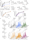
Figure 1.. HOIL-1 ubiquitination of Ser and Thr residues in vitro is dependent on His510.
(A) Peptide ubiquitination assay with 20 mM synthetic Ser- or Thr-containing peptides (Ac-EGxGN-NH2, where x is either Ser or Thr). (B) Peptide ubiquitination assays as in panel A with Lys- or Arg-containing peptides. Experiments in panels A and B were performed three times with consistent results. (C) Peptide ubiquitination assay with 20 mM (left) and 50 mM (right) synthetic Ser-, Thr-, or Lys-containing peptides and WT HOIL-1. Samples were taken at the indicated time points and analysed via SDS–PAGE under reducing conditions. Half of the samples taken at 30 min were treated with hydroxylamine (NH2OH) as indicated. (D) Peptide ubiquitination assay as in panel (C) but with the HOIL-1 H510A mutant. Experiments in panels (C, D) were performed at least twice with consistent results. Representative gels are shown. Ub: ubiquitin. The dotted lines in panels (C, D) are added for readability and do not indicate gel splicing. Source data are available for this figure.
Figure 2.
Figure 2.. Crystal structure of the HOIL-1 RING2 domain bound by ubiquitin.
(A) Overview of the crystallographic asymmetric unit shows two HOIL-1 RING2 domains (green, grey) with one of them (green) bound to ubiquitin (pink). Bound zinc ions are shown as purple spheres with zinc ion–coordinating amino acid sidechains shown as sticks. (B) Detailed view of the interactions between ubiquitin (pink) and the HOIL-1 RING2 domain (green). Key interacting residues are shown as sticks and labelled. (C) Structural superposition of the HOIL-1 RING2 domains in the free state (grey) and bound to ubiquitin (green with ubiquitin in pink) highlights the conformational change between the core RING2 domain and the N-terminal helix upon ubiquitin binding. (D) Structural superposition of the HOIL-RING2 domains in the free state (grey) and bound to ubiquitin (green with ubiquitin in pink) highlights the conformational changes in the active site loop, W462 and H510. The active site Cys460 in the structure is mutated to Ala (C460A). The N-terminal part of the HOIL-1 RING2 domain is omitted for clarity.
Figure S1.
Figure S1.. Crystal structure of HOIL-1 RING2/ubiquitin and comparison with other HOIL-1 RING2 structures.
(A) Asymmetric unit of the HOIL-1 RING2 (green, grey) structure with ubiquitin (pink) with the 2Fo-Fc electron density (blue mesh) shown contoured at 1σ. (B) Comparison of the UbcH7-ubiquitin conjugate–bound HOIL-1 RING2 domain (left, UbcH7 in wheat, HOIL-1 RING2 in green, ubiquitin in pink, PDB ID 8EAZ [Wang et al, 2023]) and the HOIL-1 RING2/ubiquitin complex described here. (C) Overlays of the HOIL-1 RING2/ubiquitin complex described here (HOIL-1 in dark green, ubiquitin in pink) with two different free HOIL-1 RING2 domain structures. Left: PDB ID 7YUJ (light green) (Xu et al, 2023), right: PDB ID 8BVL (cyan) (Wu et al, 2022). (D) Overlay of the HOIL-1 RING2/ubiquitin complex (HOIL-1 in dark green, ubiquitin in pink) and E2-ubiquitin conjugate–bound HOIL-1 RING2 (PDB ID 8EAZ (Wang et al, 2023), HOIP in light green, ubiquitin in bright pink, UbcH7 in wheat). (E) Overlay of the free HOIL-1 RING2 from this work (grey) and HOIL-1 RING2 structures previously published (PDB ID 7YUJ [Xu et al, 2023], chain A in pink; 8BVL [Wu et al, 2022], chain A in blue). Only the helix-RING2 regions of all structures are shown for simplicity.
Figure 3.
Figure 3.. HOIL-1 ubiquitinates multiple disaccharides and physiological monosaccharides in vitro.
(A) Quantification of HOIL-1–mediated in vitro substrate ubiquitination comparing Ser and Thr peptides with maltose. The proportion of ubiquitinated substrate over total ubiquitin as quantified from Coomassie-stained SDS–PAGE gels is plotted over time. Individual data points and connecting lines of the mean are shown (n = 3). (B) Chemical structures of disaccharide substrates tested. Differences to maltose are highlighted in red font. (C) Quantification of HOIL-1–mediated in vitro substrate ubiquitination reactions comparing the disaccharides maltose and cellobiose. Individual data points and connecting lines of the mean are shown (n = 3–4). (D) Quantification of HOIL-1–mediated in vitro substrate ubiquitination reactions comparing disaccharides as indicated. Individual data points and connecting lines of the mean are shown (n = 2–4). Maltose data in panels (C, D) are from the same experiment. (E) Chemical structures of the physiological monosaccharide substrates tested. (F) MALDI-TOF analysis of HOIL-1–mediated in vitro substrate ubiquitination reactions of the substrates shown in panel (E). Experiments were performed twice with consistent results. Exemplary data are shown. Ub: ubiquitin. Source data are available for this figure.
Figure S2.
Figure S2.. In vitro ubiquitination of peptides and disaccharides by HOIL-1.
(A) Coomassie-stained SDS–PAGE gels showing HOIL-1 ubiquitination of different substrates as indicated. Ubiquitin (Ub) and Ub-substrate bands were quantified for the graph in Fig 3A. Exemplary gels of three experiments are shown. (B) Extended MALDI-TOF mass spectrometry trace of ubiquitinated maltose shows the expected mass of ubiquitin-maltose (8,889 D) with no evidence for dual-modified maltose. These data are from the same experiment as those presented in Fig 3F (maltose, 4 h). (C) Coomassie-stained SDS–PAGE gels showing HOIL-1 ubiquitination of different disaccharides as indicated. Ub-substrate and Ub bands were quantified for the graph in Fig 3C and D. Exemplary gels of two to four experiments are shown. Source data are available for this figure.
Figure 4.
Figure 4.. Large-scale preparation and purification of ubiquitinated maltose.
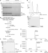
(A) Time course showing HOIL-1–mediated ubiquitination of maltose using low (10 μM) and higher (100 μM) ubiquitin concentrations. To enable comparison of ubiquitin (Ub) and ubiquitin-maltose bands between the different samples, the samples containing 100 μM were diluted 10-fold before SDS–PAGE analysis. o/n, overnight. (B) Workflow for large-scale ubiquitin-maltose preparation and purification. (C) Coomassie-stained SDS–PAGE gel showing successful large-scale ubiquitin-maltose generation (Reaction) and His-tag affinity purification step (HisTrap). Frac: fractions. (D) Size-exclusion chromatography purification of His-ubiquitin-maltose after HisTrap purification. (E) Intact mass spectrometry of His-ubiquitin (top) and purified His-ubiquitin-maltose (bottom). The mass difference of 325 D confirms mono-ubiquitination of a single maltose molecule. The slightly heavier minor species in the two spectra likely represent α-N-6-phosphogluconoylation of the His-tag, often identified in His-tagged proteins expressed in E. coli (Geoghegan et al, 1999). Source data are available for this figure.
Figure S3.
Figure S3.. Examples of other ubiquitinated non-proteinaceous biomolecules generated by HOIL-1 in vitro.
(A) Ubiquitination of N-acetyllactosamine (LacNAc) and maltose control. The chemical structure of LacNAc is shown. (B) Ubiquitination of GalNAc and GlcNAc. (C) Ubiquitination of glycosylated serine bearing the Tn antigen, T antigen, and sTn antigen after 18 h. Chemical structures of the different substrates are shown on the right. Coomassie-stained SDS–PAGE gels are shown. Source data are available for this figure.
Figure S4.
Figure S4.. Crystal structure of HOIL-1 RING2/ubiquitin-maltose.
(A) Asymmetric unit of the HOIL-1 RING2 (green, blue) structure with ubiquitin-maltose (pink) with the 2Fo-Fc electron density (grey mesh) shown contoured at 1σ. (A, B) Structural comparison of HOIL-1 RING2/ubiquitin-maltose (coloured as in (A)) and HOIL-1 RING2/ubiquitin (grey). (A, B, C) Electron density maps of the ubiquitin C terminus (pink sticks; Gly76 modelled in two conformations, (A, B)) in the HOIL-1 (green sticks) active site lack clear density for the maltose molecule. 2Fo-Fc map (grey mesh) shown contoured at 1σ, and Fo-Fc difference density map (green and red mesh) shown contoured at ±2.5σ. Key residues are labelled. Water molecules are shown as red spheres. (D) Intact mass spectrometry analysis of redissolved crystals taken from two different crystallisation drops shows that most (>75%) of ubiquitin present in the crystals is conjugated to maltose. Source data are available for this figure.
Figure 5.
Figure 5.. Proof-of-principle DUB assay towards ubiquitin-maltose hydrolysis.
(A) Table summarising known activities of DUBs and hydroxylamine (NH2OH) against different amino acid ubiquitin linkages (Lys, M1, Thr) from the literature and results from our analysis with Ub-maltose. Grey cells indicate activity with the indicated species. For Lys chain type–specific DUBs, the cleaved linkages are indicated. (B) Ubiquitin-maltose DUB assay. His-ubiquitin-maltose was incubated with different DUBs as indicated and analysed via SDS–PAGE. The first lane contains untreated sample, and the sample in the last lane was treated with hydroxylamine (NH2OH) to fully cleave oxyester-linked His-ubiquitin-maltose. DUBs were used at different, optimal concentrations (Hospenthal et al, 2015); hence, only DUBs used at high concentrations (USP2, ATXN3) are resolved on the gel. Ub: ubiquitin. Source data are available for this figure.
Figure S5.
Figure S5.. Quality control of the DUBs used for the ubiquitin-maltose DUB assay.
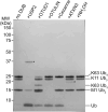
To verify that DUBs used in Fig 5 are active and specific at the concentrations used, we tested their activity against a mixture of M1 di-ubiquitin, K11 tri-ubiquitin, and K63 tetra-ubiquitin. As expected, USP2 cleaves all linkages, OTUD1-only K63 tetra-ubiquitin, OTULIN-only M1 di-ubiquitin, and Cezanne-only K11 tri-ubiquitin. The DUB ATXN3 and the chemical hydroxylamine (NH2OH) do not cleave any of these (iso)peptide-linked ubiquitin oligomers. Ub: ubiquitin. DUB bands are marked with an asterisk (*). Source data are available for this figure.
Figure 6.
Figure 6.. Design of a constitutively active HOIL-1 variant.
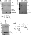
(A) Domain organisation of human HOIL-1 (top) and the constitutively active M1-di-Ub-HOIL-1 fusion protein that contains an N-terminal M1-linked di-ubiquitin in which glycine 76 in both protomers is mutated to valine (G76V). (B) Maltose-ubiquitination assay comparing HOIL-1 with the M1-di-Ub(G76V)-HOIL-1 fusion protein in the presence and absence of additional allosteric M1-linked di-ubiquitin. Ub: ubiquitin. Source data are available for this figure.
Figure S6.
Figure S6.. Design of a constitutively active HOIL-1 variant.
(A) Initial purification (HisTrap) comparing WT human HOIL-1 and the first generation of the M1-di-Ub-HOIL-1 fusion protein. Although both proteins express well in E. coli as indicated by the relevant bands in the whole-cell lysate, M1-di-Ub-HOIL-1 is not stable during HisTrap purification indicated by a large amount of di-ubiquitin in the HisTrap elution sample. (B) HisTrap purification of linker-less M1-di-Ub(G76V)-HOIL-1 showing that removal of the linker between the di-ubiquitin and HOIL-1 and mutation of Gly76 to Val (G76V) in both copies of ubiquitin yield a more stable fusion protein. For the assay shown in Fig 6B, the protein was further purified via size-exclusion chromatography (see the Materials and Methods section). Sup.: supernatant after centrifugation of the whole-cell lysate; LFT: load flow-through of applying supernatant to a HisTrap column; W1: wash 1; W2: wash 2. Source data are available for this figure.

References

    1. Abdul Rehman SA, Cazzaniga C, Di Nisio E, Antico O, Knebel A, Johnson C, Şahin AT, Ibrahim P, Lamoliatte F, Negri R, et al. (2024) Discovery and characterization of noncanonical E2-conjugating enzymes. Sci Adv 10: eadh0123. 10.1126/sciadv.adh0123 - DOI - PMC - PubMed
    1. Afonine PV, Moriarty NW, Mustyakimov M, Sobolev OV, Terwilliger TC, Turk D, Urzhumtsev A, Adams PD (2015) FEM: Feature-enhanced map. Acta Crystallogr D Biol Crystallogr 71: 646–666. 10.1107/S1399004714028132 - DOI - PMC - PubMed
    1. Akimov V, Barrio-Hernandez I, Hansen SVF, Hallenborg P, Pedersen AK, Bekker-Jensen DB, Puglia M, Christensen SDK, Vanselow JT, Nielsen MM, et al. (2018) UbiSite approach for comprehensive mapping of lysine and N-terminal ubiquitination sites. Nat Struct Mol Biol 25: 631–640. 10.1038/s41594-018-0084-y - DOI - PubMed
    1. Aragão D, Aishima J, Cherukuvada H, Clarken R, Clift M, Cowieson NP, Ericsson DJ, Gee CL, Macedo S, Mudie N, et al. (2018) MX2: A high-flux undulator microfocus beamline serving both the chemical and macromolecular crystallography communities at the Australian Synchrotron. J Synchrotron Radiat 25: 885–891. 10.1107/S1600577518003120 - DOI - PMC - PubMed
    1. Corcilius L, Payne RJ (2013) Stereoselective synthesis of sialylated tumor-associated glycosylamino acids. Org Lett 15: 5794–5797. 10.1021/ol402845e - DOI - PubMed

LinkOut - more resources