. 2025 Jun 2;64(23):e202505717.
doi: 10.1002/anie.202505717. Epub 2025 Apr 10.

Fast and Autonomous Mannosylated Nanomotors for Dynamic Cancer Cell Targeting

Affiliations

Fast and Autonomous Mannosylated Nanomotors for Dynamic Cancer Cell Targeting

Yuechi Liu et al. Angew Chem Int Ed Engl. .

Abstract

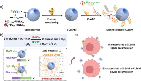
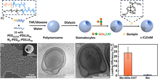
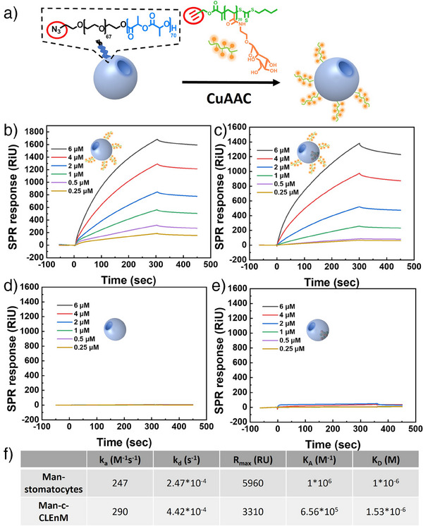
An attractive strategy in cancer cell therapy is to employ motile nanoparticles that can actively search for their target. Herein, we introduce mannosylated compartmentalized cross-linked enzyme-driven nanomotors (c-CLEnM), which exhibit specific and efficient targeting of Hep G2 cells through elevated autonomous motion. In this design, we constructed biodegradable bowl-shaped stomatocytes encapsulating the enzymes glucose oxidase (GOx) and catalase (CAT) within their nanocavity. A subsequent enzyme crosslinking reaction was performed to guarantee their stability. Furthermore, the c-CLEnM were surface modified with a mannose-functional glycopolymer, enabling binding with receptors expressed on Hep G2 cells. Interestingly, the targeting ligands on the nanomotors not only improved their specificity toward cancer cells but also enhanced motility. Compared to the non-mannosylated nanomotors, mannosylated c-CLEnM exhibited enhanced motion and higher targeting efficiency to cells in glucose-containing ionic environments. The unexpected acceleration in speed resulted from the surface modification of these nanomotors with a glycopolymer layer, which increased the zeta potential and created a shielding effect that mitigated the influence of the surrounding ions. This nanomotor design highlights the synergistic effect of functional glycopolymer modification on cellular uptake, adding an additional level of control to nanomotors for application in cancer therapy.

Keywords: Cancer cell targeting; Enhanced autonomous motion; Mannosylated enzymatic nanomotors; Stomatocytes.


Conflict of interest statement

The authors declare no conflict of interest.

Figures

Scheme 1
Scheme 1
Schematic illustration of mannosylated compartmentalized cross‐linked enzymatic nanomotors (Man‐c‐CLEnM). a) Poly(ethylene glycol)‐b‐poly(D,L‐lactide) (PEG‐PDLLA) with two different PEG lengths and functionalized with an azide moiety was co‐assembled into stomatocytes, encapsulating catalase (CAT) and glucose oxidase (GOx); the enzymes were subsequently crosslinked in the stomatocyte cavity. Mannose glycopolymer was introduced on the nanoparticle surface via Cu‐catalyzed azide‐alkyne click chemistry. b) The addition of D‐glucose leads to a cascade reaction that produces oxygen, leading to motility. The glycopolymer modified on the nanomotors can increase the zeta potential and potentially generate a shielding effect that mitigates the influence of surrounding ions, thus enhancing motility. c) The mannosylated nanomotors show effective accumulation on the Hep G2 cell membrane compared to galactosylated nanomotors and non‐decorated nanomotors. Schematic illustrations of cancer cells were created with BioRender.com.
Figure 1
Figure 1
Preparation and characterization of crosslinked enzyme‐driven nanomotors (c‐CLEnM). a) Schematic illustration of the supramolecular assembly of the c‐CLEnM. b) SEM image of empty stomatocytes and zoom in of stomatocytes, scale bar = 4 µm, 400 nm (insert). c) Morphological characterization of empty stomatocytes using cryo‐TEM, scale bar = 200 nm. d) Cryo‐TEM image revealing the structure of the c‐CLEnM, scale bar = 200 nm. e) Quantification of GOx and CAT loading efficiency of enzyme‐loaded stomatocytes and empty stomatocytes by using a bicinchoninic acid (BCA) protein assay.
Figure 2
Figure 2
Characterization of mannosylated stomatocytes. a) Schematic illustration of conjugating mannose glycopolymer to the surface of mannosylated stomatocytes. b) Surface plasmon resonance (SPR) analysis illustrating the binding of mannosylated stomatocytes (Man‐stomatocytes) with mannose‐binding lectin (MBL). c) SPR analysis demonstrating the binding of mannosylated c‐CLEnM (Man‐c‐CLEnM) with MBL. d) SPR analysis of the binding of empty stomatocytes with MBL. e) SPR analysis of the binding of c‐CLEnM with MBL. f) Kinetic values (k a, k d, R max, K A, K D) obtained from fitting experimental SPR curves with a 1:1 Langmuir binding model.
Figure 3
Figure 3
Motion behavior of galactosylated c‐CLEnM, mannosylated c‐CLEnM, c‐CLEnM, and empty stomatocytes. a) MSD curves of c‐CLEnM as a function of a range of glucose concentrations (0, 5, 10, 30, 50 mM). b) Average speed of c‐CLEnM 6.40 ± 0.38 (5 mM), 8.04 ± 0.80 (10 mM), 12.23 ± 0.50 (30 mM), 15.53 ± 0.75 (50 mM). c) MSDs of galactosylated c‐CLEnM, mannosylated c‐CLEnM, c‐CLEnM, and empty stomatocytes in the presence of glucose (+, 25 mM) and absence of glucose (−) in MilliQ water. d) MSDs of galactosylated c‐CLEnM, mannosylated c‐CLEnM, c‐CLEnM, and empty stomatocytes in the presence (+, 25 mM) and absence of glucose (−) in live cell imaging solution. e) Average speed of galactosylated c‐CLEnM, mannosylated c‐CLEnM, and c‐CLEnM in the presence of glucose (+, 25 mM) in MilliQ water and live cell imaging solution. f) Zeta potential of galactosylated c‐CLEnM, mannosylated c‐CLEnM, c‐CLEnM, and empty stomatocytes in the presence (+, 25 mM) and absence of glucose (−), in live cell imaging solution. **p < 0.01; NA, not significant, calculated by t‐test.
Figure 4
Figure 4
Evaluation of active cell targeting of mannosylated c‐CLEnM toward Hep G2 cells. a) Schematic illustration of mannosylated c‐CLEnM targeting cancer cells. b) Flow cytometry mean fluorescence intensities of Hep G2 cells treated with representative nanoparticles were measured in the presence (25 mM glucose, DMEM) and absence of glucose (n = 2); ***p < 0.001; NA, not significant, calculated by t‐test. c) Flow cytometry histograms of Hep G2 cells treated with mannosylated c‐CLEnM, c‐CLEnM, and empty stomatocytes in the presence (25 mM glucose, DMEM) and absence of glucose. d) Magnified confocal laser scanning microscopy (CLSM) images of Hep G2 cells incubated with mannosylated c‐CLEnM, galactosylated c‐CLEnM, and c‐CLEnM for 2 h in the presence (25 mM glucose, DMEM) and absence of glucose, scale bar = 10 µm. (+): in the presence of glucose, (−): in the absence of glucose. Schematic illustrations of cancer cells were created with BioRender.com.

References

    1. Dreyfus R., Baudry J., Roper M. L., Fermigier M., Stone H. A., Bibette J., Nature 2005, 437, 862–865. - PubMed
    1. Palagi S., Fischer P., Nat. Rev. Mater. 2018, 3, 113–124.
    1. Sengupta S., Ibele M. E., Sen A., Angew. Chem. Int. Ed. 2012, 51, 8434–8445. - PubMed
    1. Wang W., J. Am. Chem. Soc. 2023, 145, 27185–27197. - PubMed
    1. Ye Z., Wang Y., Liu S., Xu D., Wang W., Ma X., J. Am. Chem. Soc. 2021, 143, 15063–15072. - PubMed

LinkOut - more resources