. 2025 Apr 1;16(1):234.
doi: 10.1038/s41419-025-07593-1.

PLA2G7 promotes immune evasion of bladder cancer through the JAK-STAT-PDL1 axis

Affiliations

PLA2G7 promotes immune evasion of bladder cancer through the JAK-STAT-PDL1 axis

Ding Peng et al. Cell Death Dis. .

Abstract

Targeting immune checkpoints such as Programmed death ligand-1 (PD-L1) and Programmed cell death 1 (PD-1) has been approved for treating bladder cancer and shows promising clinical benefits. However, the relatively low response rate highlights the need to seek an alternative strategy to traditional PD-1/PD-L1 targeting immunotherapy. In this study, we found that PLA2G7 is significantly elevated in bladder cancer and correlates with worse prognosis. In vitro experiments demonstrated that knockdown of PLA2G7 does not significantly affect the proliferation, migration, and invasion of bladder cancer cells. Flow cytometry detection, as well as protein and RNA detection, showed that knockdown of PLA2G7 significantly inhibits PD-L1 expression and suppresses the growth of transplanted tumors by promoting CD8 + T-cell infiltration. Further experiments showed that PLA2G7 regulates the JAK-STAT pathway to promote PD-L1 expression by upregulating the phosphorylation of STAT1 and STAT3. Meanwhile, results from syngeneic mouse models indicated that PLA2G7 suppression and anti-CTLA4 therapy have synergistic effects on tumor burden and mouse survival. In addition, we found that ETS1 promotes PLA2G7 overexpression in bladder cancer cells. In summary, our findings provide a novel immunotherapeutic strategy against bladder cancer through targeting the ETS1-PLA2G7-STAT1/STAT3-PD-L1 axis.


Conflict of interest statement

Competing interests: The authors declare no competing interests. Ethics approval and consent to participate: This study was approved by the Biomedical Research Ethics Committee of The First Affiliated Hospital School of Medicine, Zhejiang University (Number: IIT20230241A). All methods were performed in accordance with the relevant guidelines and regulations. Informed consent was obtained from all participants.

Figures

Fig. 1
Fig. 1. Elevated PLA2G7 expression is correlated with poor clinical outcome of bladder cancer.
A The level of PLA2G7 mRNA in bladder cancer tumors compared to matched normal tissues (TCGA cohort). B The level of PLA2G7 mRNA in bladder cancer tumors compared to matched normal tissues (qRT-PCR result). C Western blot analysis showing PLA2G7 protein levels in normal bladder cell line compared bladder cancer cell lines, and bladder cancer tissues compared to matched normal tissues. D IHC results showing the protein level of PLA2G7 in bladder cancer tumors compared to matched normal tissues. E The level of PLA2G7 protein in different stage bladder cancer tissues. F Kaplan–Meier survival analysis of bladder cancer patients by different levels of PLA2G7 protein in tumors.
Fig. 2
Fig. 2. PLA2G7 does not affect bladder cancer cell proliferation, migration, and invasion.
A Western blot analysis demonstrating the quality of PLA2G7 knockdown in 5637 and T24 cells. B MTS assays of sh-NC and sh-PLA2G7 cell viability. C Clone formation assays for sh-NC and sh-PLA2G7 cells. D Wound healing assays for sh-NC and sh-PLA2G7 cells. E Transwell assays for sh-NC and sh-PLA2G7 cells.
Fig. 3
Fig. 3. PLA2G7 depletion inhibits PD-L1 expression and tumor immune evasion.
A Surface PD-L1 protein levels analyzed by FACS in sh-NC and sh-PLA2G7 cells. B Western blot analysis showing the levels of PD-L1 in sh-NC and sh-PLA2G7 cells with or without IFN-γ. C RT-qPCR analysis of the mRNA levels of PD-L1 in sh-NC and sh-PLA2G7 cells with or without IFN-γ. D Results from the T cell killing assays of sh-NC and sh-PLA2G7 cells. E Results from the expression of T cell activity factors including PRF1, GAMB, GNLY and IFNG of T cells that cocultured with sh-NC and sh-PLA2G7 cells.
Fig. 4
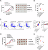
Fig. 4. In vivo experiments in mice confirmed that PLA2G7 knockdown inhibited tumor immune escape.
A Tumor growth curves for immunodeficient NOD-SCID mice injected with sh-NC and sh-PLA2G7 cells. B Tumor weights for immunodeficient NOD-SCID mice injected with sh-NC and sh-PLA2G7 cells. C Tumor growth curves for immunocompetent BALB/C mice injected with sh-NC and sh-PLA2G7 cells. D Tumor weights for immunocompetent BALB/C mice injected with sh-NC and sh-PLA2G7 cells. E Flow cytometry analysis and statistical diagram showing the proportion of CD4+ and CD8+ T cells in tumors.
Fig. 5
Fig. 5. PLA2G7 regulates the JAK–STAT pathway through the phosphorylations of STAT1 and STAT3.
A Volcano plot of genes differentially expressed in sh-NC and sh-PLA2G7 cells following stimulation with IFN-γ. B KEGG enrichment analysis for significant down-regulated genes. C The correlation of PLA2G7 with dysfunction score in TCGA bladder cancer cohort. D The correlation of PLA2G7 with exhausted score in TCGA bladder cancer cohort. E The correlation of PLA2G7 with PD-L1 mRNA level in TCGA bladder cancer cohort. F The correlation of PLA2G7 with PD-L1 expression in bladder cancer tissues analyzed by RT-qPCR. G Western blot analysis showing PLA2G7, JAK2, STAT1, p-STAT1, STAT3, p-STAT3, IRF1 and PD-L1 levels in sh-NC and sh-PLA2G7 cells.
Fig. 6
Fig. 6. PLA2G7 inhibitor darapladib suppresses bladder cancer immune evasion.
A MTS assays of 5637 cell viability treated with placebo and darapladib. B RT-qPCR analysis of the mRNA levels of PD-L1 in bladder cancer cells treated with placebo and darapladib. C Western blot analysis showing p-STAT1, p-STAT3 and PD-L1 levels in bladder cancer cells treated with placebo and darapladib. D Tumor growth curves for immunocompetent BALB/C mice injected with bladder cancer cells treated with placebo and darapladib. E Tumor weights for immunocompetent BALB/C mice injected with bladder cancer cells treated with placebo and darapladib.
Fig. 7
Fig. 7. PLA2G7 overexpression promotes PD-L1 expression and tumor immune evasion.
A MTS cell viability assay of OE-NC and OE-PLA2G7 cells. B Surface PD-L1 protein levels analyzed by FACS in OE-NC and OE-PLA2G7 cells. C Results from the T cell killing assays of OE-NC and OE-PLA2G7 cells. D Western blot analysis of PLA2G7, p-STAT1, p-STAT3 and PD-L1 levels in OE-NC and OE-PLA2G7 cells with or without STAT1 and STAT3 knockdown.
Fig. 8
Fig. 8. PLA2G7 inhibition sensitizes the efficacy of CTLA4 blockade.
A Tumor growth of MBT-2 cells with stable PLA2G7 depletion in BALB/C mice with or without anti-CTLA4 antibody treatment. B Comparative analysis of tumor weight in Fig. 8A. C Flow cytometry analysis and statistical plot showing the proportion of CD4+ and CD8+ T-cells in the tumors. D Tumor growth of MBT-2 cells in BALB/C mice treated with darapladib and anti-CTLA4 antibody. E Comparative analysis of tumor weight in Fig. 8D.
Fig. 9
Fig. 9. Transcription factor ETS1 regulates the expression of PLA2G7.
A In silico analysis of potential transcription factors in PLA2G7 promoter region. B The correlation of PLA2G7 with ETS1 expression in bladder cancer tissues from the TCGA database. C The correlation of PD-L1 with ETS1 expression in bladder cancer tissues from the TCGA database. D RT-qPCR analysis of ETS1 mRNA levels in sh-NC and sh-ETS1 cells. E RT-qPCR analysis of PLA2G7 mRNA levels in sh-NC and sh-ETS1 cells. F RT-qPCR analysis of PD-L1 mRNA levels in sh-NC and sh-ETS1 cells. G Surface PD-L1 levels were analyzed by FACS in sh-NC cells and sh-ETS1 cells. H Western blot analysis showing ETS1, PLA2G7, and PD-L1 levels in sh-NC and sh-ETS1 cells. I Luciferase reporter assays of PLA2G7 promoter in sh-NC and sh-ETS1 cells.

References

    1. Siegel RL, Miller KD, Wagle NS, Jemal A. Cancer statistics, 2023. CA Cancer J Clinicians. 2023;73:17–48. - PubMed
    1. Babjuk M, Burger M, Capoun O, Cohen D, Compérat EM, Dominguez Escrig JL, et al. European Association of Urology Guidelines on Non-muscle-invasive Bladder Cancer (Ta, T1, and Carcinoma in Situ). European Urol. 2022;81:75–94. - PubMed
    1. Witjes JA, Bruins HM, Cathomas R, Compérat EM, Cowan NC, Gakis G, et al. European Association of Urology Guidelines on Muscle-invasive and Metastatic Bladder Cancer: Summary of the 2020 Guidelines. European Urol. 2021;79:82–104. - PubMed
    1. Powles T, Bellmunt J, Comperat E, De Santis M, Huddart R, Loriot Y, et al. Bladder cancer: ESMO Clinical Practice Guideline for diagnosis, treatment and follow-up. Ann Oncol. 2022;33:244–58. - PubMed
    1. Chen X, Zhang J, Ruan W, Huang M, Wang C, Wang H, et al. Urine DNA methylation assay enables early detection and recurrence monitoring for bladder cancer. J Clin Invest. 2020;130:6278–89. - PMC - PubMed