. 2025 Apr 1;16(1):231.
doi: 10.1038/s41419-025-07577-1.

Mechanical sensing protein PIEZO1 controls osteoarthritis via glycolysis mediated mesenchymal stem cells-Th17 cells crosstalk

Affiliations

Mechanical sensing protein PIEZO1 controls osteoarthritis via glycolysis mediated mesenchymal stem cells-Th17 cells crosstalk

Yikun Zhou et al. Cell Death Dis. .

Abstract

Aberrant mechanical stimuli can cause tissue attrition and activate mechanosensitive intracellular signaling, impacting the progression of osteoarthritis (OA). However, the precise relationship between mechanical loading and bone metabolism remains unclear. Here, we present evidence that Piezo1 senses the mechanical stimuli to coordinate the crosstalk between mesenchymal stem cells (MSCs) and T helper 17 (Th17) cells, leading to the deterioration of bone and cartilage in osteoarthritis (OA). Mechanical loading impaired the property of MSCs by inhibiting their osteo-chondrogenic differentiation and promoting inflammatory signaling to enhance Th17 cells. Mechanistically, mechanical stimuli activated Piezo1, thereby facilitating Ca2+ influx which upregulated the activity of Hexokinase 2(HK2), the rate-limiting enzyme of glycolysis. The resultant increase in glycolytic activity enhanced communication between MSCs and T cells, thus promoting Th17 cell polarization in a macrophage migration inhibitory factor (MIF) dependent manner. Functionally, Wnt1cre; Piezo1fl/fl mice reduced bone and cartilage erosion in the temporomandibular joint condyle following mechanical loading compared to control groups. Additionally, we observed activated Piezo1 and HK2-mediated glycolysis in patients with temporomandibular joint OA, thereby confirming the clinical relevance of our findings. Overall, our results provide insights into how Piezo1 in MSCs coordinates with mechano-inflammatory signaling to regulate bone metabolism. The schema shows that mechanical sensing protein PIEZO1 in MSCs controls osteoarthritis via glycolysis mediated MSCs and Th17 cells crosstalk in a MIF dependent manner.


Conflict of interest statement

Competing interests: The authors declare no competing interests. Ethics approval: The study was approved by the Animal Use and Care Committee of Peking University approval (LA2021488). All institutional and national guidelines for the care and use of laboratory animals were followed. Patient sample collection was approved by the ethics committee of Hospital & School of Stomatology, Peking University (2022-12-83-07).

Figures

None
The schema shows that mechanical sensing protein PIEZO1 in MSCs controls osteoarthritis via glycolysis mediated MSCs and Th17 cells crosstalk in a MIF dependent manner.
Fig. 1
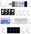
Fig. 1. Excessive mechanical stimulus leads to temporomandibular joint OA.
A The morphology of control and force-treated condyle tissues showed by micro-CT. Quantitative analysis of (B) BV/TV, (C) Tb.Th, (D) Tb.Sp and (E) Tb.N in subchondral bone of TMJ condyle determined by micro-CT analysis. F The morphology of control and force-treated condyle tissues showed by HE staining and Masson staining. G The expression of osteoblast and osteoclasts markers in control and force-treated TMJ condyles, assessed by ALP staining and TRAP staining. H The expression of IL-17 and IFN-γ in control and force-treated TMJ condyles. I The cell proliferation in control and force treated MSCs determined by flow cytometry. J Alizarin red S staining in control and force treated MSCs after osteogenesis induction for 21 days. K The expression levels of ALP and RUNX2 in control and force-treated MSCs analyzed by western blot. L Alcian blue staining in control and force-treated MSCs undergo chondrogenic differentiation for 21 days. M The expression levels of SOX9 and ACAN in control and force treated MSCs, as assessed by western blot. TMJ: temporomandibular joint, Scale bar: 100 μm (F, H, J and L); 50 μm (G); data are presented as the mean ± SEM, P < 0.05; ∗∗P < 0.01; ∗∗∗P < 0.005.
Fig. 2
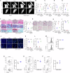
Fig. 2. The mechanosensory channel Piezo1 in MSCs controls temporomandibular joint OA development.
A The gene expression levels of mechanically activated ion channel in control and force treated groups determined by RT-PCR. B The MIF protein expression levels of Piezo1 in control and force treated MSCs. C The morphology of condyles in control and TMJOA human specimens were shown by CBCT analysis. D The histological characteristics of TMJ condyles in control and temporomandibular joint OA human specimens analyzed by HE staining and safranin O staining. E The expression of Piezo1 in control and TMJOA patients samples analyzed by immunofluorescence staining. F The morphology of TMJ condyle tissues in control and Wnt1cre; Piezo1fl/fl mice with or without force treated groups was shown by micro-CT analysis. Quantitative analysis of (G) BV/TV, (H) Tb.Th, (I) Tb.Sp and (J) Tb.N in subchondral bone of TMJ condylar heads determined by micro-CT. K The histological characteristics of condyles in control and Wnt1cre; Piezo1fl/fl mice with or without force treated groups was shown by HE staining, Masson staining and safranin O staining. LN The ALP positive and TRAP-positive cells in control and Wnt1cre; Piezo1fl/fl mice with or without force-treated groups were assessed by ALP staining and TRAP staining. OQ The expression of IL-17 and IFN-γ in control and Wnt1cre; Piezo1fl/fl mice with or without force treated condyle tissues. TMJ temporomandibular joint, Scale bar: 100 μm; data are presented as the mean ± SEM, P < 0.05; ∗∗P < 0.01; ∗∗∗P < 0.005.
Fig. 3
Fig. 3. Piezo1 in CD4+ T cells failed to control temporomandibular joint OA development.
A The morphology of TMJ condyle tissues in control, force treated and CD4cre; Piezo1fl/fl mice and CD4cre; Piezo1fl/fl mice with force treated groups was shown by micro-CT analysis. Quantitative analysis of (B) BV/TV, (C) Tb.Th, (D) Tb.Sp and (E) Tb.N in subchondral bone of TMJ condylar heads determined by micro-CT. F The histological characteristics of condyles in control, force treated and CD4cre; Piezo1fl/fl mice and CD4cre; Piezo1fl/fl mice with force treated groups were shown by HE staining, Masson staining, and safranin O staining. GI The ALP positive and TRAP-positive cells in control, force treated and CD4cre; Piezo1fl/fl mice and CD4cre; Piezo1fl/fl mice with force treated groups were assessed by ALP staining and TRAP staining. JL The expression of IL-17 and IFN-γin control, force treated and CD4cre; Piezo1fl/fl mice and CD4cre; Piezo1fl/fl mice with force treated groups condyle tissues. M The Ki-67 positive cells ratio in native CD4+ T cells from control and force treated groups. N Foxp3 positive cells ratio in native CD4+ T cells from control and force treated groups. O IFN-γ positive cells ratio in native CD4+ T cells from control and force treated groups. P IL-17 positive cells ratio in native CD4+ T cells from control and force treated groups. TMJ temporomandibular joint, Scale bar: 100 μm; data are presented as the mean ± SEM, P < 0.05; ∗∗P < 0.01; ∗∗∗P < 0.005.
Fig. 4
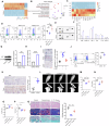
Fig. 4. Mechanical stimulus promoted the crosstalk between MSCs with T cells via MIF.
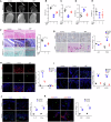
A A heatmap depicting the upregulated and downregulated genes between control and force-treated MSCs. B The KEGG analysis showed that IL-17 signaling pathway was one of the top ten different enriched clusters between control and force-treated MSCs. C A heatmap depicting the different gene expression related with IL-17 signaling pathway. D Th17 cell differentiation in control, co-cultured with MSCs, and co-cultured with mechanical stimulus pretreated MSCs groups was analyzed by flow cytometry. E Cytokine secretion profiles of MSCs from control and force-treated groups. F Semi-quantification of cytokine array results. G The protein expression levels of MIF in control and force-treated groups. H The gene expression levels of MIF in control and force-treated groups. I The recruitment of T cells by MSCs in control, force with or without Anti-MIF groups. J Th17 cell differentiation in control and MIF-treated groups was analyzed by flow cytometry. K The expression of MIF in control and force-treated condyle tissues, was assessed by immunohistochemistry staining. L The morphology of TMJ condyle tissues in control, force-treated and IL-17-/- mice with force-treated groups was shown by micro-CT analysis. Quantitative analysis of (M) BV/TV, (N) Tb.Th, (O) Tb.Sp and (P) Tb.N in subchondral bone of TMJ condylar heads determined by micro-CT. Q The histological characteristics of condyles in control, force-treated, and IL-17-/- mice with force-treated groups were shown by HE staining, Masson staining, and safranin O staining. TMJ temporomandibular joint, Scale bar: 100 μm; data are presented as the mean ± SEM, P < 0.05; ∗∗P < 0.01; ∗∗∗P < 0.005.
Fig. 5
Fig. 5. Mechanical stimulus promoted MIF secretion via HK2 mediated glycolysis.
A A heatmap depicting the different gene expressions related with the glycolysis pathway. B The lactate level produced by control and force-treated MSCs. (C)The glucose consumption rate in control and mechanical force treated MSCs. D Extracellular acidification rate (ECAR) in control and force-treated MSCs. E The expression of glycolytic enzymes HK2 and LDHA in control and force-treated MSCs, assessed by western blot. F The expression of HK2 and LDHA in control and force-treated TMJ condyle, was assessed by immunohistochemistry staining. G The protein expression levels of MIF in control, force with or without SiHK2 pre-treated MSCs groups. H Semi-quantification of the protein expression levels of MIF. I The gene expression levels of MIF in control, force with or without SiHK2 pre-treated MSCs groups. J The recruitment of T cells by MSCs in control, force with or without SiHK2 groups. K Th17 cell differentiation in control, force with or without SiHK2 groups was analyzed by flow cytometry. L The osteogenic differentiation of MSCs in control, force with or without SiHK2 pre-treated groups after osteogenic induction for 21 days, assessed by Alizarin red S staining. M The protein expression levels of ALP and RUNX2 in control, force with or without SiHK2 pre-treated groups. N The chondrogenic differentiation of MSCs in in control, force with or without SiHK2 pre-treated groups, as assessed by Alcian blue staining. O The protein expression levels of SOX9 and ACAN in control, force with or without SiHK2 pre-treated groups. TMJ temporomandibular joint, Scale bar: 100 μm; data are presented as the mean ± SEM, P < 0.05; ∗∗P < 0.01; ∗∗∗P < 0.005.
Fig. 6
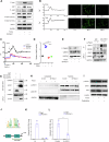
Fig. 6. Mechanical stimulus activated glycolysis via STAT1 signaling.
A The protein expression levels of p-JAK1/JAK1, p-JAK2/JAK2, and p-STAT1/STAT1 in control, the force with or without Piezo1 siRNA treated MSCs. B The Ca2+ influx induced by force and Yoda treatment in MSCs. C The Ca2+ influx induced by force treatment in MSCs was inhibited with Piezo1 inhibitor GsMTx4 or Piezo1 siRNA treatment. D, E The enzymatic activity of CaMKII and expression of phosphorylated CaMKII in control, force with or without Piezo1 siRNA treated for 24 h MSCs. F Co-immunoprecipitation assay showing that CaMKII interact STAT1 directly, which was attenuated by Piezo1 siRNA treatment. G GST pull-down assays were performed with GST-fusion proteins containing the portions of CaMKII and in vitro translated STAT1. The bound proteins were separated by SDS/PAGE and visualized by autoradiography. H The localization of p-STAT1 in the nuclei and cytoplasm fraction in control, force with or without Piezo1 siRNA treated MSCs. I The expression of p-STAT1 and HK2 in control, the force with or without fludarabine pre-treated MSCs analyzed by western blot. J It’s predicted that STAT1 could target on the promoter region of the HK2 gene hinted by JASPAR database. K, L The enrichment of STAT1 on the element 1 and element 2 of HK2 promoter was determined by ChIP-qPCR. Scale bar: 100 μm; data are presented as the mean ± SEM, P < 0.05; ∗∗P < 0.01; ∗∗∗P < 0.005.
Fig. 7
Fig. 7. Temporomandibular joint OA could be alleviated by inhibiting glycolysis.
A The morphology of TMJ joint in control, force with or without 3-BP treated groups was shown by micro-CT analysis. Quantitative analysis of (B) BV/TV, (C) Tb.Th, (D) Tb.Sp and (E) Tb.N in subchondral bone of temporomandibular joint determined by micro-CT measurements. F The histological characteristics of TMJ subcondral bone in control, force with or without 3-BP treated groups, showed by HE staining, Masson staining and safranin O staining. G The ALP and TRAP-positive cells in control, force with or without 3-BP treated TMJ subchondral bone were shown by ALP staining and TRAP staining. The expression of HK2, MIF (H) and IL-17, IFN-γ (I) in control, force with or without 3-BP treated groups, assessed by immunofluorescence staining. J The expression of IL-17 and IFN-γ in control and TMJOA human condyle specimens, assessed by immunofluorescence staining. K The expression of HK2 and LDHA in control and TMJOA human condyle specimens, was assessed by immunofluorescence staining. TMJ temporomandibular joint, Scale bar: 100 μm; data are presented as the mean ± SEM, P < 0.05; ∗∗P < 0.01; ∗∗∗P < 0.005.

References

    1. Feng X, McDonald JM. Disorders of bone remodeling. Annu Rev Pathol Mech Dis. 2011;6:121–45. - PMC - PubMed
    1. Vico L, Collet P, Guignandon A, Lafage-Proust M-H, Thomas T, Rehailia M, et al. Effects of long-term microgravity exposure on cancellous and cortical weight-bearing bones of cosmonauts. Lancet. 2000;355:1607–11. - PubMed
    1. Wang L, You X, Zhang L, Zhang C, Zou W. Mechanical regulation of bone remodeling. Bone Res. 2022;10:16. - PMC - PubMed
    1. Findlay DM, Kuliwaba JS. Bone–cartilage crosstalk: a conversation for understanding osteoarthritis. Bone Res. 2016;4:16028. - PMC - PubMed
    1. Hosseini SM, Wilson W, Ito K, van Donkelaar CC. A numerical model to study mechanically induced initiation and progression of damage in articular cartilage. Osteoarthr Cartil. 2014;22:95–103. - PubMed

LinkOut - more resources