. 2025 Apr 1;13(1):42.
doi: 10.1038/s41413-025-00412-5.

M2 macrophages-derived exosomes for osteonecrosis of femoral head treatment: modulating neutrophil extracellular traps formation and endothelial phenotype transition

Affiliations

M2 macrophages-derived exosomes for osteonecrosis of femoral head treatment: modulating neutrophil extracellular traps formation and endothelial phenotype transition

Guanzhi Liu et al. Bone Res. .

Abstract

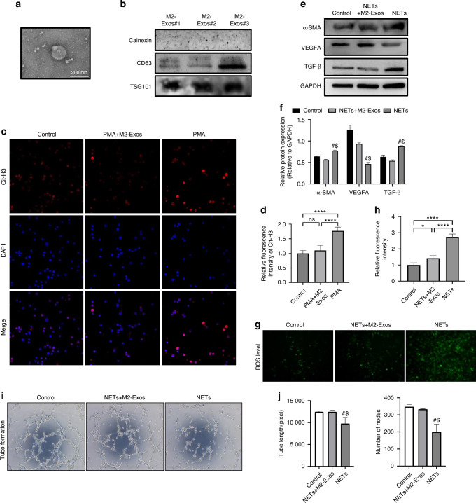
Exosomes have shown good potential in ischemic injury disease treatments. However, evidence about their effect and molecular mechanisms in osteonecrosis of femoral head (ONFH) treatment is still limited. Here, we revealed the cell biology characters of ONFH osteonecrosis area bone tissue in single cell scale and thus identified a novel ONFH treatment approach based on M2 macrophages-derived exosomes (M2-Exos). We further show that M2-Exos are highly effective in the treatment of ONFH by modulating the phenotypes communication between neutrophil and endothelium including neutrophil extracellular traps formation and endothelial phenotype transition. Additionally, we identified that M2-Exos' therapeutic effect is attributed to the high content of miR-93-5p and constructed miR-93-5p overexpression model in vitro and in vivo based on lentivirus and adeno-associated virus respectively. Then we found miR-93-5p can not only reduce neutrophil extracellular traps formation but also improve angiogenic ability of endothelial cells. These results provided a new theoretical basis for the clinical application of ONFH therapeutic exosomes.


Conflict of interest statement

Competing interests: The authors declare no competing interests.

Figures

Fig. 1
Fig. 1
Single-cell transcriptome analysis of ONFH subchondral bone tissue. a ScRNA sequencing cell cluster and annotation analysis of total samples (ONFH n = 2; FHF n = 2; hip osteoarthritis n = 3). b HOA group cell cluster and cell-cell communication analysis. c FHF group cell cluster and cell-cell communication analysis. d ONFH group cell cluster and cell-cell communication analysis
Fig. 2
Fig. 2
ONFH rat model construction and NETs formation in femoral head. a TONFH rat models and SONFH rat models establishment pipelines. b HE stanning of rat femoral heads in control group, TONFH group and SONFH group. c, d MicroCT analysis of rat femoral heads in control group, TONFH group and SONFH group. e, f Immunofluorescent staining of NETs (MPO: red; Cit-H3: green; DAPI: blue). All data were presented as means ± SD, n ≥ 3 per group; ns P ≥ 0.05. *P < 0.05. **P < 0.01. ***P < 0.001. ****P < 0.000 1; Statistical significance was determined by two-tailed Student’s t test (d, f).Triangle marker: area with empty osteocytic lacunae
Fig. 3
Fig. 3
M2-Exos enhance HUVECs angiogenesis by reducing NETs formation and endothelial phenotype transition in vitro. a Transmission electron microscopy (TEM) image of M2-Exos. b Western blotting assay for specific markers of exosomes like CD63, Tsg101 and endoplasmic reticulum protein Calnexin. c, d In vitro immunofluorescent staining for NETs formation after the treatment of PMA and M2-Exos (Cit-H3: red; DAPI: blue). e, f Western blotting assay for angiogenesis markers and mesenchymal markers in HUVECs after the treatment of NETs and M2-Exos. gj HUVECs ROS levels and tube formation ability after the treatment of NETs and M2-Exos. All data were presented as means ± SD, n ≥ 3 per group; ns P ≥ 0.05. *P < 0.05. **P < 0.01. ***P < 0.001. ****P < 0.000 1; In (f, j), # represents the differences between NETs group and Control group have statistical significance (P < 0.05); $ represents the differences between NETs+M2-Exos group and NETs group have statistical significance (P < 0.05). Statistical significance was determined by two-tailed Student’s t test (d, f, j, h)
Fig. 4
Fig. 4
M2-Exos improve ONFH by reducing NETs formation in vivo. a SONFH mice model establishment and M2-Exos injection pipeline. b Representative fluorescence images after intravenous injections of DIR (control group) and DIR-labeled M2-Exos for 48 h and 72 h. c Representative fluorescence images of the distribution of M2-Exos in the femur after 72 h. d Representative fluorescence images of the distribution of M2-Exos in the main organs after 72 h. e HE stanning of SONFH mice femoral heads in control group, SONFH + M2-Exos group and SONFH group. f, g Immunofluorescent staining for NETs formation in SONFH mice femoral head (MPO: red; Cit-H3: green; DAPI: blue). All data were presented as means ± SD, n ≥ 3 per group; ns P ≥ 0.05. *P < 0.05. **P < 0.01. ***P < 0.001. ****P < 0.000 1; Statistical significance was determined by two-tailed Student’s t test (g)
Fig. 5
Fig. 5
M2-Exos miR-93-5p regulate NETs-HUVECs interaction and endothelial phenotype transition activation in vitro. a ONFH associated M2-Exos miRNA analysis (GSE97467: M2-Exos miRNA dataset 1; GSE209957: M2-Exos miRNA dataset 2; GSE89587: ONFH patients serum deferential expressed miRNAs compared to control group). be ROS levels and tube formation ability after the treatment of NETs in HUVECs group, HUVECs overexpression control group and HUVECs miR-93-5p overexpression group. f, g HUVEC transwell migration assays. h, i Western blotting assay for angiogenesis markers and mesenchymal markers in each HUVEC group. All data were presented as means ± SD, n ≥ 3 per group; ns P ≥ 0.05. *P < 0.05. **P < 0.01. ***P < 0.001. ****P < 0.000 1; In (e, i), # represents the differences between NETs+oemiR-93-5p group and NETs group have statistical significance (P < 0.05); $ represents the differences between NETs+oemiR-93-5p group and NETs+oeNC group have statistical significance (P < 0.05). Statistical significance was determined by two-tailed Student’s t test (c, e, f, gi)
Fig. 6
Fig. 6
MiR-93-5p is involved with the therapeutic effects of M2-Exos for ONFH by prohibiting NETs formation. a ONFH rat model adeno-associated virus (AAV) treatment pipeline. b HE stanning of ONFH rat femoral heads in ONFH group, AAV negative control group and AAV-miR-93-5p group. c, d MicroCT images and structure analyses of rat femoral heads. e, f Immunofluorescent staining for NETs formation in rats’ femoral head (Cit-H3: red; DAPI: blue). All data were presented as means ± SD, n ≥ 3 per group; ns P ≥ 0.05. *P < 0.05. **P < 0.01. ***P < 0.001. ****P < 0.000 1; Statistical significance was determined by two-tailed Student’s t test (d, f)

References

    1. Chen, C. Y. et al. Glucocorticoid-induced loss of beneficial gut bacterial extracellular vesicles is associated with the pathogenesis of osteonecrosis. Sci. Adv.8, eabg8335 (2022). - PMC - PubMed
    1. Liu, G. et al. Osteogenesis-related long noncoding RNA GAS5 as a novel biomarker for osteonecrosis of femoral head. Front. Cell Dev. Biol.10, 857612 (2022). - PMC - PubMed
    1. Tan, H. et al. Extracorporeal shock wave therapy with imaging examination for early osteonecrosis of the femoral head: A systematic review. Int. J. Surg.111, 1144–1153 (2024). - PMC - PubMed
    1. Yoon, B. H. et al. The 2019 revised version of association research circulation osseous staging system of osteonecrosis of the femoral head. J. Arthroplast.35, 933–940 (2020). - PubMed
    1. Nakamura, J. et al. Time elapsed from definitive diagnosis to surgery for osteonecrosis of the femoral head: A nationwide observational study in Japan. BMJ Open14, e082342 (2024). - PMC - PubMed

LinkOut - more resources