. 2025 Apr 1;16(1):3133.
doi: 10.1038/s41467-025-58504-z.

Intranasal influenza virus-vectored vaccine offers protection against clade 2.3.4.4b H5N1 infection in small animal models

Affiliations

Intranasal influenza virus-vectored vaccine offers protection against clade 2.3.4.4b H5N1 infection in small animal models

Ying Liu et al. Nat Commun. .

Abstract

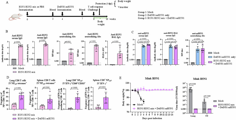
The highly pathogenic avian influenza (HPAI) H5N1 virus has been endemic in aquatic birds since 1997, causing outbreaks in domestic poultry and occasional human infections worldwide. Recently, the cross-species transmission of a new reassortant variant from clade 2.3.4.4b of H5N1 to cattle in the US has heightened concerns regarding the expansion of host range and potential human infection. As eradicating the H5N1 virus from its reservoir is impossible, it is essential to prepare for a potential pandemic caused by an H5N1 derivative. Utilizing a deleted-NS1 live attenuated influenza viral vector vaccine system (DelNS1 LAIV), a system we have previously used in the development of a COVID-19 vaccine, we have rapidly developed an intranasal vaccine for cattle H5N1 and related clade 2.3.4.4b strains, based on publicly available sequences. Our research demonstrates that a single intranasal immunization can provide effective protection against lethal challenges from HPAI cattle or mink H5N1 variants, offering strong, sustained immunity after two months in female mouse and male hamster models. Immunogenicity analysis reveals that intranasal vaccination with DelNS1 LAIV induces robust neutralizing antibody, mucosal IgA and T cell responses in mice. It is crucial to further evaluate the DelNS1-H5N1 LAIV system to prepare for potential future H5N1 outbreaks in humans.


Conflict of interest statement

Competing interests: The authors declare that the University of Hong Kong owns patents on work related to the generation and application of DelNS1 live attenuated influenza vaccines and the associated platform, with H.C., P.W. and K-Y.Y. included as co-inventors. There is no restriction on the publication of data. The other authors declare that they have no competing interests.

Figures

Fig. 1
Fig. 1. Generation and characterization of DelNS1-H5N1 vaccines.
A Illustration of construction and generation of DelNS1-H5N1 vaccines. NCR, non-coding region. DCP: deletion of polybasic cleavage site. B Growth kinetics of DelNS1-cH5N1 and DelNS1-mH5N1 at 33 °C were analyzed in MDCK cells inoculated at a multiplicity of infection (MOI) of 0.01 (n = 3), and viral titers of DelNS1-cH5N1 and DelNS1-mH5N1 were measured in day 8 embryonated chicken eggs 48 h after inoculation with different initial doses (n = 3). Viral supernatants or allantoic fluids were collected and titrated by plaque assay in MDCK cells. C Virulence of DelNS1-cH5N1 and DelNS1-mH5N1 was examined in mice. Mice were inoculated intranasally with DelNS1-cH5N1 (106 pfu, n = 8) or cattle H5N1 (5000 pfu, n = 8), DelNS1-mH5N1 (106 pfu, n = 8) or mink H5N1 (104 pfu, n = 8). Body weights were observed for 14 days. D Mice were inoculated intranasally with DelNS1-cH5N1 (106 pfu, n = 3) or cattle H5N1 (5000 pfu, n = 4), with virus titers in lungs, nasal turbinates (NT) and brain tissues of mice being examined at day 2, day 4 and day 6 post infection. LOD limit of detection. Data represents mean values ± standard deviation (SD) of results. N/A not available. Statistical comparisons between means were performed by Student’s t-test (two-tailed). Cartoons were created in BioRender. Wang, P. (2025) https://BioRender.com/n81t049. Source data are provided as a Source Data file.
Fig. 2
Fig. 2. Immunogenicity evaluation of DelNS1-H5N1 vaccines.
A Schedule of immunization, blood collection, and bronchoalveolar lavage (BAL) fluid and nasal wash collection from BALB/c mice. Mice (n = 8 for each group) were intranasally immunized with 106 pfu DelNS1-cH5N1 or 106 pfu DelNS1-mH5N1, or mock-immunized (PBS). B At week 3, serum were collected and tested for IgG titers against the cH5N1 (DelNS1-cH5N1) virus, mH5N1 (DelNS1-mH5N1) virus, or H5N1-HA1 protein. C Microneutralization (MN) titers were measured against live viruses. At week 4 post immunization, mice from all groups were sacrificed and BAL and nasal wash samples collected. D BAL IgA levels and E nasal wash IgA levels against cH5N1, mH5N1, or H5N1-HA1 were measured. LOD: limit of detection. Data represents the mean values ± SD of results. Statistical analysis was performed for (B) and (C) using one-way ANOVA followed by Dunn’s multiple comparisons test. Statistical analysis was performed for (D) and (E) using Student’s t-test (two-tailed). The mouse cartoon was created in BioRender. Wang, P. (2025) https://BioRender.com/n83i107. Source data are provided as a Source Data file.
Fig. 3
Fig. 3. DelNS1-H5N1 vaccines induce CD4+ and CD8+ T cell responses.
A Schedule for immunization of BALB/c mice. At week 4 after immunization, 2 μg of PE-Cy5 conjugated CD45-specific antibody was injected intravenously via the tail vein 5 min before sacrifice (n = 8 for each group). B Lung cells and splenocytes were obtained and stained with Zombie, anti-CD8 and NP147 tetramer. Flow cytometry gating was performed to assess the frequency of live DelNS1-cH5N1-induced non-circulating (IV CD45-PE-Cy5 negative) NP-specific CD8+ T cells among lung cells and splenocytes (percentage IV-CD8 + NP147 tetramer+ out of all live IV-CD8+ T cells). C Lung or spleen cells were stimulated with NP147 or NP55 peptide overnight. Surface markers (CD69, CD103, CD4, CD8 and Zombie) were stained, followed by fixation, permeabilization, and intracellular staining for IFN-γ. DelNS1-cH5N1 induced tissue-resident memory cells in lungs and spleens were displayed (IV-IFNγ + CD69 + CD103 + CD8 + T cells and IV-IFNγ+CD69+CD4+ T cells out a percentage of all live IV- CD8+ and CD4+ T cells, respectively). D DelNS1-mH5N1 induced non-circulating T cells in lung cells and spleens (IV-CD8 + NP147 tetramer out of all live IV-CD8 T cells). E DelNS1-mH5N1 induced tissue-resident memory cells in lungs and spleen were displayed (IV-IFNγ+CD69+ CD103+CD8+ T cells and IV-IFNγ + CD69+ CD4+ T cells out a percentage of all live IV- CD8+ and CD4 + T cells, respectively). Data represents the mean values ± SD of results. Statistical comparisons between means were performed by Student’s t-test (two-tailed). The mouse cartoon was created in BioRender. Wang, P. (2025) https://BioRender.com/n83i107. Source data are provided as a Source Data file.
Fig. 4
Fig. 4. DelNS1-H5N1 vaccines provide protection against different 2.3.4.4b clade viruses.
A Illustration of schedule of immunization, virus challenge and sacrifice for BALB/c mice. Mice were intranasally vaccinated with DelNS1-cH5N1 (106 pfu, n = 32, two independent experiments), DelNS1-mH5N1 (106 pfu, n = 32, two independent experiments) or mock-vaccinated (n = 16, two independent experiments), then challenged with either cattle H5N1 virus (5000 pfu) or mink H5N1 virus (104 pfu) 4 weeks after immunization. B Mice immunized with DelNS1-cH5N1 and challenged with cattle H5N1 virus; body weights were monitored for 14 days and viral titers in the lungs, NT and brain were measured at 4 dpi. C Mice immunized with DelNS1-mH5N1 and challenged with mink H5N1 virus; body weights were monitored for 14 days and viral titers in the lungs, NT and brain were measured at 4 dpi. D DelNS1-cH5N1-vaccinated mice were challenged with mink H5N1 virus, and body weights monitored for 14 days and viral titers in the lungs, NT and brain measured at 4 dpi. E DelNS1-mH5N1-vaccinated mice were challenged with cattle H5N1, and body weights monitored for 14 days and viral titers in the lungs, NT and brain measured at 4 dpi. LOD: limit of detection. Data represents mean values ± SD of results. Statistical comparisons between means were performed by Student’s t-test (two-tailed). Mouse cartoon was created in BioRender. Wang, P. (2025) https://BioRender.com/n83i107. Source data are provided as a Source Data file.
Fig. 5
Fig. 5. Immunization reduces expression of pro-inflammatory genes in mice post-challenge.
A Illustration of schedule of immunization, virus challenge and sacrifice for BALB/c mice. B Mice were intranasally vaccinated with DelNS1-cH5N1 (106 pfu), or PBS (n = 4 for each group), then challenged with cattle H5N1 virus (5000 pfu) 4 weeks after immunization. Pro-inflammatory genes were measured by RT-qPCR after isolation of RNA from lungs at 4 dpi. C Mice were intranasally vaccinated with DelNS1-mH5N1 (106 pfu), or PBS (n = 4 for each group), then challenged with mink H5N1 virus (104 pfu) 4 weeks after immunization. Pro-inflammatory genes were measured by RT-qPCR after isolation of RNA from lungs at 4 dpi. Data represent mean values ± SD of results. Statistical analysis was performed using Student’s t-test (two-tailed). The mouse cartoon was created in BioRender. Wang, P. (2025) https://BioRender.com/n83i107. Source data are provided as a Source Data file.
Fig. 6
Fig. 6. Effect of pre-existing immunity on the immunogenicity and protective ability of DelNS1-H5N1 vaccine.
A Schedule of immunization, serum collection, T cell response, virus challenge and sacrifice for BALB/c mice. Mice were intranasally vaccinated with H1N1/H3N2 mix (106 pfu mixture of H3N2 and H1N1 DelNS1 LAIVs (1:1), n = 23), or mock-vaccinated (n = 8). 4 weeks after immunization, 15 mice from H1N1/H3N2 mix group and 8 mice from mock group were vaccinated with DelNS1-mH5N1 (106 pfu). Mice were challenged with mink H5N1 virus (104 pfu) 4 weeks later. B Serum was collected at week 3 post H1N1/H3N2 mix immunization and tested for IgG titers against H3N2 and H1N1 DelNS1 LAIVs and micro-neutralization titers against live viruses (n = 8 each group). BAL IgA level against H1N1 DelNS1 LAIVs were measured 8 weeks after H1N1/H3N2 mix vaccination (n = 8 each group). C Serum were collected 3 weeks after DelNS1-mH5N1 immunization and tested for IgG titers against live virus or H5N1-HA1 protein and micro-neutralization titers against live virus (n = 8 each group). D 8 weeks after prime immunization, for the H1N1/H3N2 mix group and H1N1/H3N2 mix + DelNS1-mH5N1 group (n = 6), PE-Cy5 CD45 antibody was injected intravenously 5 min before sacrifice. Lung cells and splenocytes were stained with Zombie, anti-CD8 and NP147 tetramer. The frequency of IV-CD8 + NP147 tetramer out of all live IV-CD8 T cells was displayed. Lung cells and splenocytes were stimulated with NP147 and stained with surface markers, followed by fixation, permeabilization, and intracellular staining for IFN-γ. The frequency of tissue-resident memory cells in lungs (IV-IFNγ+CD69+CD103+ CD8+ T cells out of all live IV-CD8 T cells) and spleens (IV-IFNγ+CD8+ T cells out of all live IV-CD8 T cells) was displayed. E 8 weeks after prime immunization, mice in H1N1/H3N2 mix + DelNS1-mH5N1 group (n = 9) and mock group (n = 8, two independent experiments) were challenged with mink H5N1 virus (104 pfu); body weights were monitored for 14 days, and viral titers in lungs and NT were measured at 4 dpi. LOD: limit of detection. Data represent mean values ± SD of results. Statistical analysis was performed using Student’s t-test (two-tailed) (B, D, E) and one-way ANOVA (C). The mouse cartoon was created in BioRender. Wang, P. (2025) https://BioRender.com/n83i107. Source data are provided as a Source Data file.
Fig. 7
Fig. 7. Effective protection after two-month DelNS1-H5N1 immunization.
A Illustration of schedule of immunization, virus challenge and sacrifice for BALB/c mice. Mice were intranasally vaccinated with DelNS1-cH5N1 (106 pfu, n = 16) or mock-vaccinated (n = 12). B Serum was collected at week 8 and tested for IgG titers against cH5N1 (DelNS1-cH5N1) virus or H5N1-HA1 protein and micro-neutralization titers against live viruses. Levels of IgA against H5N1-HA1 in BAL were measured at week 8 following DelNS1-mH5N1 vaccination. C At 8 weeks after immunization, mice were challenged with cattle H5N1 virus (5000 pfu); body weights were monitored for 14 days and viral titers in the lungs, NT and brain were measured at 4 dpi (n = 4 for mock group; n = 8 for DelNS1-cH5N1 two-month group). D Mice were intranasally vaccinated with DelNS1-mH5N1 (106 pfu, n = 16) or mock-vaccinated (n = 12), and serum collected at week 8 tested for IgG titers against mH5N1 (DelNS1-mH5N1) virus or H5N1-HA1 protein and micro-neutralization titers against live virus. BAL levels of IgA recognizing H5N1-HA1 were measured at week 8 after DelNS1-mH5N1 vaccination. E Eight weeks after immunization, mice were challenged with mink H5N1 virus (104 pfu); body weights were monitored for 14 days and viral titers in the lungs and NT were measured at 4 dpi (n = 4 for mock group; n = 8 for DelNS1-mH5N1 two-month group). LOD limit of detection. Data represent mean values ± SD of results. Statistical analysis was performed using Student’s t-test (two-tailed). Mouse cartoon was created in BioRender. Wang, P. (2025) https://BioRender.com/n83i107. Source data are provided as a Source Data file.

References

    1. Yuen, K. Y. et al. Clinical features and rapid viral diagnosis of human disease associated with avian influenza A H5N1 virus. Lancet351, 467–471 (1998). - PubMed
    1. Li, K. S. et al. Genesis of a highly pathogenic and potentially pandemic H5N1 influenza virus in eastern Asia. Nature430, 209–213 (2004). - PubMed
    1. Chen, H. et al. Establishment of multiple sublineages of H5N1 influenza virus in Asia: implications for pandemic control. Proc. Natl Acad. Sci. USA103, 2845–2850 (2006). - PMC - PubMed
    1. Chen, H. et al. Avian flu: H5N1 virus outbreak in migratory waterfowl. Nature436, 191–192 (2005). - PubMed
    1. Lee, D. H., Bertran, K., Kwon, J. H. & Swayne, D. E. Evolution, global spread, and pathogenicity of highly pathogenic avian influenza H5Nx clade 2.3.4.4. J. Vet. Sci.18, 269–280 (2017). - PMC - PubMed

MeSH terms