. 2025 Apr 1;8(1):98.
doi: 10.1038/s42004-025-01497-z.

Development of potential immunomodulatory ligands targeting natural killer T cells inspired by gut symbiont-derived glycolipids

Affiliations

Development of potential immunomodulatory ligands targeting natural killer T cells inspired by gut symbiont-derived glycolipids

Jesang Lee et al. Commun Chem. .

Abstract

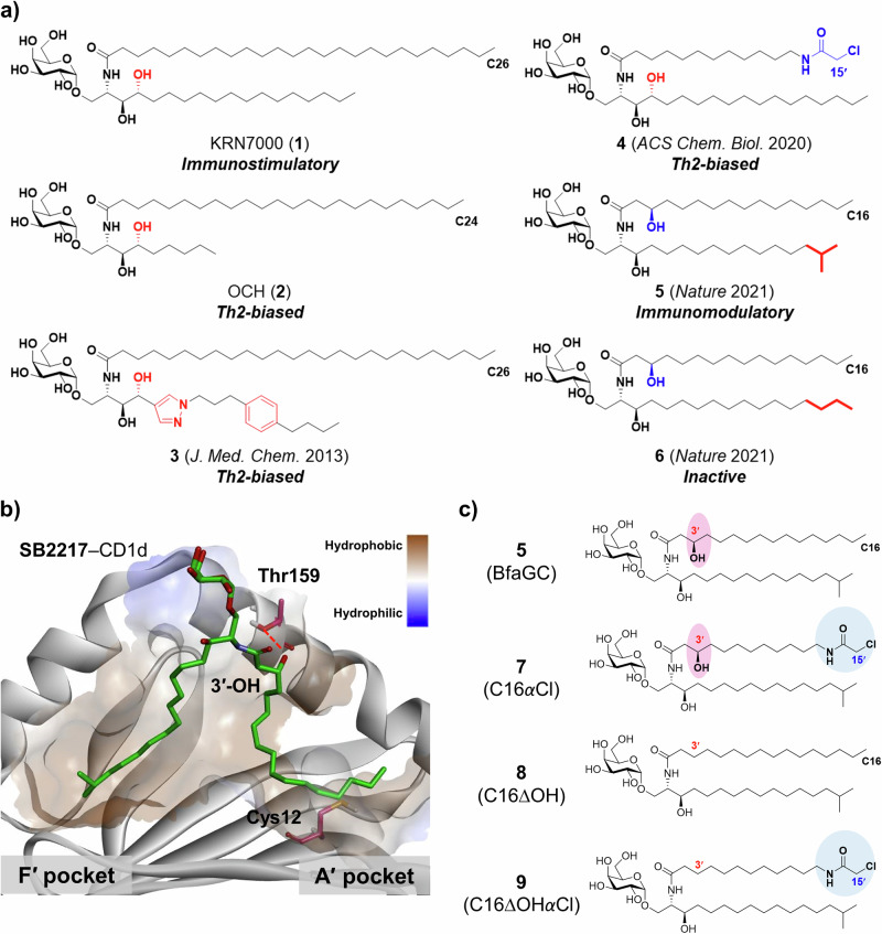
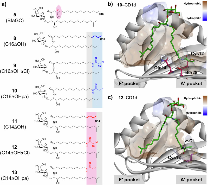
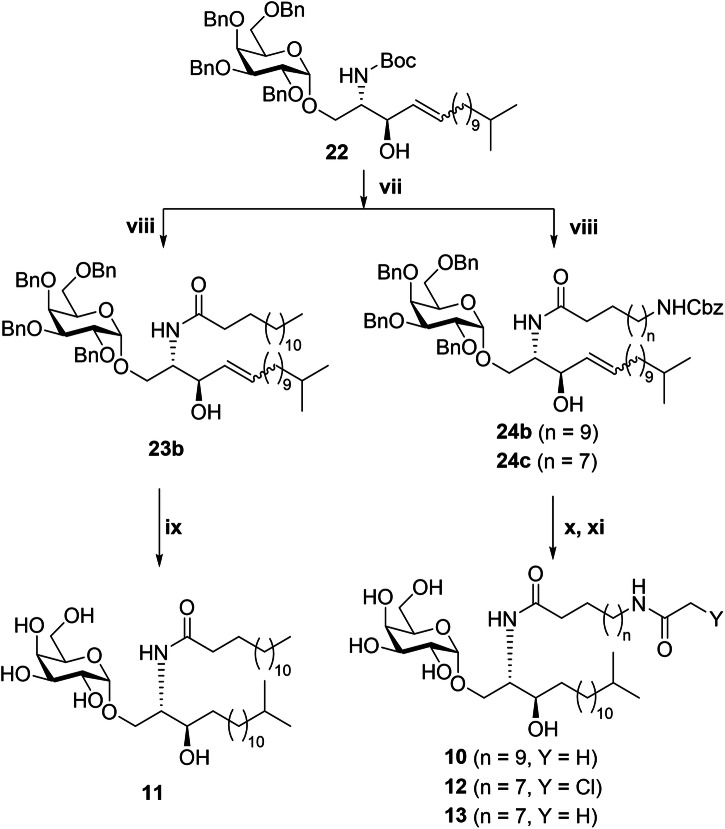
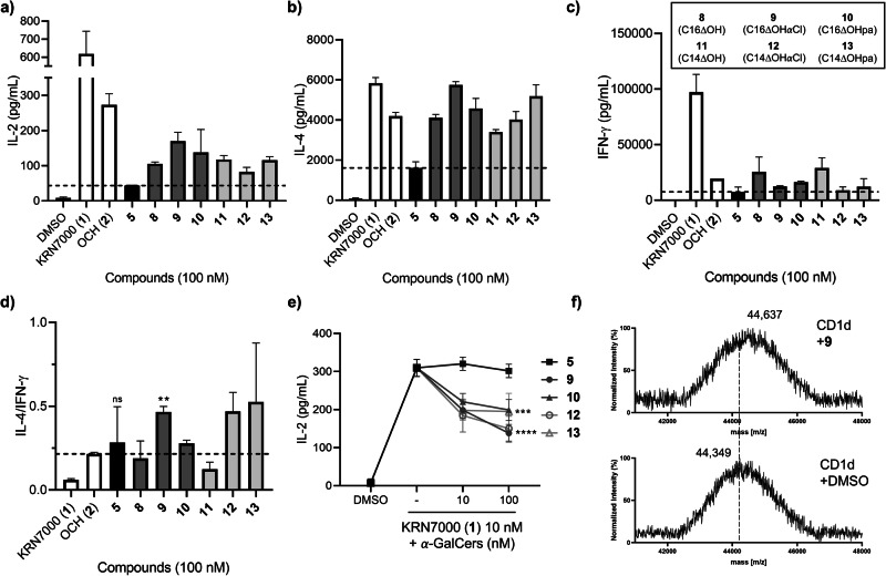
α-Galactosylceramide (α-GalCer) is a prototypical antigen recognized by natural killer T (NKT) cells, a subset of T cells crucial for immune regulation. Despite its significance, the complex structure-activity relationship of α-GalCer and its analogs remains poorly understood, particularly in defining the structural determinants of NKT cell responses. In this study, we designed and synthesized potential immunomodulatory ligands targeting NKT cells, inspired by glycolipids derived from the gut symbiont Bacteroides fragilis. A series of α-GalCer analogs with terminal iso-branched sphinganine backbones was developed through rational modification of the acyl chain. Our results identified the C3' hydroxyl group as a structural element that impairs glycolipid presentation by CD1d, as evidenced by reduced IL-2 secretion and weak competition with a potent CD1d ligand. Notably, among C3'-deoxy α-GalCer analogs, those containing an α-chloroacetamide group exhibited robust NKT cell activation with Th2 selectivity. Computational docking and mass spectrometry analyses further confirmed the substantial interaction of α-chloroacetamide analogs to CD1d. These findings underscore the potential of leveraging microbiota-derived glycolipid structures to selectively modulate NKT cell functions for therapeutic purposes.


Conflict of interest statement

Competing interests: The authors declare no competing interests.

Figures

Fig. 1
Fig. 1. Structures of α-GalCer analogs.
a Chemical structures of α-GalCer analogs inducing different NKT cell responses–KRN7000 (1), OCH (2), α-GalCer with pyrazole sphingosine backbone (3), α-GalCer with chloroacetamide acyl chain (4), and two representative Bacteroides fragilis-derived α-GalCers (BfaGCs, 56). b Crystal structure of representative immunomodulatory BfaGC with mCD1d (PDB: 7M72). Two hydrophobic pockets (A′ pocket and F′ pocket) of CD1d are shaded in brown. The ligand is shown in green sticks, and Cys12 and Thr159 are shown in pink. Hydrogen bonding is indicated as a red dotted line. c Chemical structures of BfaGC (5) and three α-GalCer analogs (79) with iso-branched sphinganine backbone and rationally designed acyl chains. C3′ hydroxyl groups were highlighted in pink, and chloroacetamide moieties were shown in blue.
Scheme 1
Scheme 1. General synthetic scheme for α-GalCer analogs (79)a.
aReagents, conditions, and yields; (i) ref. []; (ii) benzoic acid, DCC, DMAP, DCM, 0 °C to r.t., 16 h, 97% as a crude; (iii) FeCl3.6H2O, DCM, r.t., 1 h, 80% over 2 steps; (iv) 13-methyltetradec-1-ene, Grubbs catalyst 2nd generation, DCM, 40 °C, 8 h, 58%; (v) 2,3,4,6-tetra-O-benzyl-α-d-galactopyranosyl iodide, TBAI, DIPEA, 4 Å molecular sieves, benzene, 70 °C, 5 h; (vi) NaOMe, THF, 0 °C to r.t., 3 h, 41% over 2 steps; (vii) HCl, EtOH, 70 °C, 3 h; (viii) fatty acid (hexadecenoic acid, S13, or S14), EDC.HCl, HOBt.H2O, DIPEA, DCM, 0 °C to r.t., 16–18 h, yields over 2 steps (76% for 23a; 45% for 24a; 83% for 24b); (ix) H2 (g, 1 atm), Pd(OH)2/C, DCM/MeOH (1:3, v/v), r.t., 18 h, 73%; (x) H2 (g, 10 atm), Pd(OH)2/C, DCM/MeOH (1:3, v/v), r.t., 17–23 h; and (xi) N-hydroxysuccinimide ester (S20), DMF, r.t., 4–5 h, yields over 2 steps (50% for 7; 43% for 9).
Fig. 2
Fig. 2. Immunological evaluations of α-GalCer analogs (79).
a IL-2 secretion from murine hepatic mononuclear cells (HMNCs) was measured after 48 h upon treatment of α-GalCer analogs (n = 2, biological replicates. *p < 0.05, **p < 0.01, ***p < 0.005, and ns: not significant by one-way ANOVA). Data are shown as mean ± SD; b IL-2 production of NKT cells was modulated by α-GalCer analogs. Each analog was pre-treated to HMNCs before 30 min of KRN7000 (1) treatment, and IL-2 secretion was measured after 72 h (n = 3, biological replicates. *p < 0.05, **p < 0.01, ****p < 0.0001, and ns: not significant by two-way ANOVA). Data are shown as mean ± SD; c overview of the binding mode of 7 and its location of the A′ pocket and F′ pocket in mCD1d (PDB: 7M72) using computational docking analysis. The structure of 7 is shown as green sticks. Thr159 and Cys12 in the A′ pocket of mCD1d are shown in pink.
Fig. 3
Fig. 3. Structures of C3′-deoxy α-GalCer analogs with B.fragilis-derived sphinganine backbone and functionalized acyl chains.
a Chemical structures of α-GalCer analogs (813) with acyl chains equivalent to C16:0 and C14:0; b docking mode of 10 in the binding pocket of mCD1d (PDB: 7M72). The structure of 10 is shown as green sticks. Hydrophilic residues (Cys12, Gln14, Ser28) are shown in pink; c docking mode of 12 in the binding pocket of mCD1d (PDB: 7M72). The structure of 12 is shown as green sticks, while Cys12 is demonstrated in pink.
Scheme 2
Scheme 2. General synthetic scheme for α-GalCer analogs (1013)a.
aReagents, conditions, and yields; (vii) HCl, EtOH, 70 °C, 5 h; (viii) fatty acid (tetradecanoic acid, S14, or S15), EDC.HCl, HOBt.H2O, DIPEA, DCM, 0 °C to r.t., 16–18 h, yields over 2 steps (92% for 23b; 77% for 24b; and 73% for 24c); (ix) H2 (g, 1 atm), Pd(OH)2/C, DCM/MeOH (1:3, v/v), r.t., 17 h, 77%; (x) H2 (g, 10 atm), Pd(OH)2/C, DCM/MeOH (1:3, v/v), r.t., overnight; and (xi) N-hydroxysuccinimide ester (S20 or S21*), DMF, r.t., 4 h, yields over 2 steps (62% for 10; 50% for 12; 56% for 13). *K2CO3 was used as a base with S21.
Fig. 4
Fig. 4. Immunological evaluations of α-GalCer analogues (813).
a IL-2 secretion from murine hepatic mononuclear cells (HMNCs) was measured after 48 h upon treatment of α-GalCer analogs (n = 2, biological replicates); b IL-4 and c IFN-γ secretion from HMNCs was measured after 72 h upon treatment of α-GalCer analogs (n = 2, biological replicates); d IL-4/IFN-γ ratio of α-GalCer analogs (n = 2, biological replicates. **p < 0.01 and ns: not significant by Student’s t-test); e IL-2 production of NKT cells were modulated by α-GalCer analogs. Each analog was pre-treated to HMNCs before 30 min of KRN7000 (1) treatment, and IL-2 secretion was measured after 72 h (n = 3, biological replicates. ***p < 0.005 and ****p < 0.0001 by two-way ANOVA). Data are shown as mean ± SD; and f MALDI-TOF mass analysis of mCD1d with and without incubating 9.

References

    1. Brennan, P. J., Brigl, M. & Brenner, M. B. Invariant natural killer T cells: an innate activation scheme linked to diverse effector functions. Nat. Rev. Immunol.13, 101–117 (2013). - PubMed
    1. Bendelac, A., Savage, P. B. & Teyton, L. The biology of NKT cells. Annu. Rev. Immunol.25, 297–336 (2007). - PubMed
    1. Rossjohn, J., Pellicci, D. G., Patel, O., Gapin, L. & Godfrey, D. I. Recognition of CD1d-restricted antigens by natural killer T cells. Nat. Rev. Immunol.12, 845–857 (2012). - PMC - PubMed
    1. Borg, N. A. et al. CD1d-lipid-antigen recognition by the semi-invariant NKT T-cell receptor. Nature448, 44–49 (2007). - PubMed
    1. Zhou, D. et al. Lysosomal glycosphingolipid recognition by NKT cells. Science306, 1786–1789 (2004). - PubMed

LinkOut - more resources