. 2025 Apr 1;20(1):44.
doi: 10.1186/s13020-025-01092-3.

Astragaloside IV accelerates hematopoietic reconstruction by improving the AMPK/PGC1α-mediated mitochondrial function in hematopoietic stem cells

Affiliations

Astragaloside IV accelerates hematopoietic reconstruction by improving the AMPK/PGC1α-mediated mitochondrial function in hematopoietic stem cells

Ling Zhang et al. Chin Med. .

Abstract

Background: Radiotherapy can damage hematopoietic stem cells (HSC) in bone marrow, leading to impaired hematopoietic function. Current treatments mainly target differentiated hematopoietic progenitor cells, which may accelerate their depletion. Astragaloside IV (AS-IV), derived from Astragalus membranaceus, shows potential in hematopoiesis, but its direct effects on HSC remain unclear.

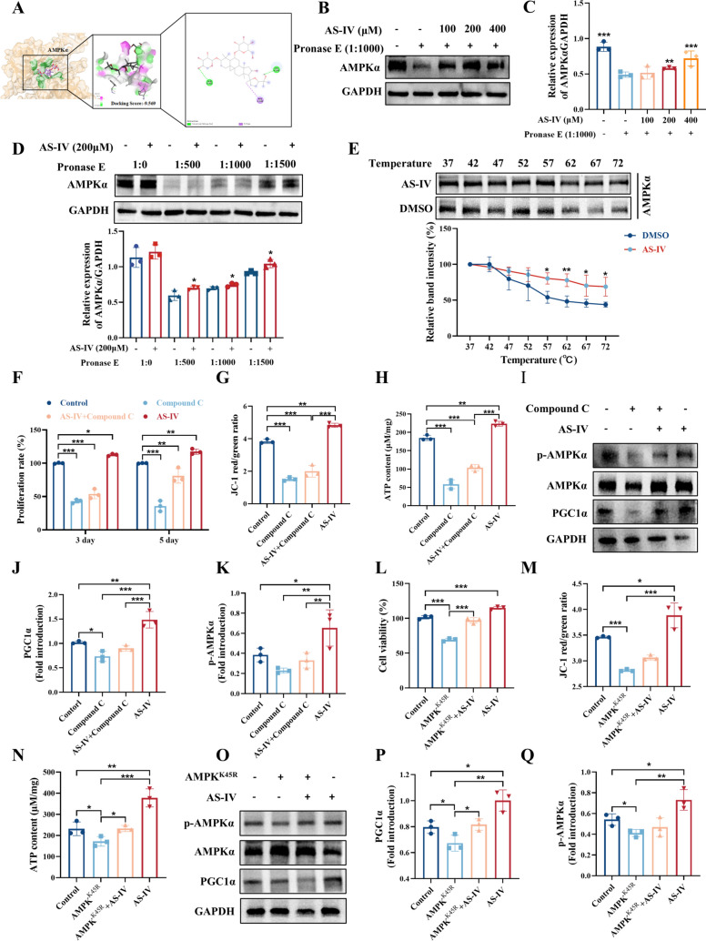
Methods: The study employed both in vitro and in vivo approaches. In vitro experiments utilized K562 cells and mouse bone marrow nucleated cells (BMNCs) to evaluate AS-IV's effects on cell proliferation and mitochondrial function. In vivo studies involved a 4.0 Gy total body irradiation mouse model treated with different doses of AS-IV (50 mg/kg and 100 mg/kg). The mechanism of action was investigated through Western blot, flow cytometry, and metabolomics analyses. The AMPK/PGC1α pathway regulation was verified using AMPK inhibitors and mutant plasmid, with molecular docking confirming AS-IV's direct binding to AMPK.

Results: In vitro studies demonstrated that AS-IV significantly promoted the proliferation of K562 cells and BMNC while enhancing their mitochondrial membrane potential, mitochondrial mass, and ATP production. In the irradiated mouse model, AS-IV treatment led to significant improvements in peripheral blood cell counts, including white blood cells, red blood cells, and hemoglobin levels. Further investigation revealed that AS-IV increased the proportion of HSC in both bone marrow and spleen while improving their mitochondrial function. Transcriptomic sequencing and Western blot analysis identified the AMPK/PGC1α signaling pathway as the key mechanism underlying AS-IV-mediated mitochondrial enhancement. These findings were validated through pharmacological inhibition of AMPK and AMPKK45R mutation experiments.

Conclusion: AS-IV accelerates hematopoietic reconstruction following radiation injury via activation of the AMPK/PGC1α signaling pathway, which enhances HSC mitochondrial function.

Keywords: AMPK/PGC1α pathway; Astragaloside IV; Hematopoietic stem cells; Mitochondrial function.


Conflict of interest statement

Declarations. Ethics approval and consent to participate: The animal study protocol was approved by the laboratory animal ethics committee of Southwest Medical University (License NO.20220812-029). Consent for publication: All authors agree to publish the manuscript. Competing interests: No potential competing interests were reported by the author(s).

Figures

Fig. 1
Fig. 1
AS-IV enhances mitochondrial function and proliferation in K562 cells in vitro. A Effect of AS-IV on K562 cell viability. Effects of different time points and dosages on K562 cell viability (%); B Effect of AS-IV on K562 cells proliferation. Effect of different time points and dosages on the proliferation rate (%) of K562 cells; C, E K562 cells treated with the fluorescent probe JC-1 were labelled with AS-IV (2.5, 5, 10 μM) to assess mitochondrial membrane potential changes by inverted fluorescence microscopy. JC-1 polymer appears as red, whereas JC-1 monomer appears as green. Scale bar: 100 μm; D, F Mito-Tracker green fluorescent probe for detection of mitochondrial mass after drug administration. Nuclei were stained with Hoechst 33258 (Scale bar: 100 μm); G ATP Assay Kit detected the concentration of ATP produced by cells; HI Representative immunoblot images and biochemical quantification of mitochondrial metabolism-associated pathway proteins (AMPK/PGC1α pathway) in K562 cells after 5 days treatment with AS-IV (2.5, 5, 10 μM). JN. The mitochondrial function-related genes (TFAM, NRF1, SDH, COXII, SOD2) were verified by qRT-PCR. Data represent the mean ± standard deviation of three independent experiments. *p < 0.05, **p < 0.01, ***p < 0.001 compared with control
Fig. 2
Fig. 2
AS-IV promotes mitochondrial function and proliferation of HSC in vitro. A BMNCs were isolated from KM mice and cultured in vitro. The cell viability of bone marrow nucleated cells was assessed using the CCK-8 assay on day 5 and 9 after administration of AS-IV intervention; B Bone marrow nucleated cells from eGFP-fluorescent mice were extracted and cultured in vitro, and their proliferation was counted by fluorescence photography and fluorescence intensity analysis after the intervention of AS-IV administration on day 5 and 9; C Cell colony formation assay to detect CFU-E, BFU-E, CFU-GM proliferation after AS-IV intervention on bone marrow nucleated cells; DE Bone marrow nucleated cells treated with AS-IV-labelled fluorescent probe JC-1 (2.5, 5, 10 μM) to assess changes in mitochondrial membrane potential by inverted fluorescence microscopy. JC-1 polymer appears as red, whereas JC-1 monomer appears as green. Scale bar: 100 μm; FG Mito-Tracker green fluorescent probe for detection of mitochondrial mass after drug administration. Nuclei were stained with Hoechst 33,258 (scale bar: 100 μm); H ATP Assay Kit detected the concentration of ATP produced by cells; I After 9 days of in vitro culture of bone marrow nucleated cells, the expression of c-kit+ HSC in each group was evaluated using flow cytometry; J Histograms showing the percentage of c-kit+ HSC in each group; KL Mitochondrial membrane potential changes of c-kit+ HSC detected by flow cytometry. Data represent the mean ± standard deviation of three independent experiments. *p < 0.05, **p < 0.01, ***p < 0.001 compared with control
Fig. 3
Fig. 3
AS-IV promotes hematopoietic recovery in irradiated mice. A The strategies of establishing the irradiated mice and treatment of each group; BE Blood counts showing (B) WBC, (C) RBC, (D) HGB, (E) LYMPH, and (F) PLT (day 0, 7, 10 and 13 after IR exposure). Data are expressed as mean ± SD (n = 8). *p < 0.05, **p < 0.01, ***p < 0.001 compared with model
Fig. 4
Fig. 4
AS-IV promotes HSC proliferation after radiation injury. The expression of HSC in the BM and SP was assessed using flow cytometry 10 days post-treatment. AB The frequencies of HSC (LSK, Lin sca-1+ c-kit+) in the BM cells of each group (n = 3); CD The frequencies of common lymphoid progenitor cell (CLP, Lin CD127+ sca-1+ c-kit +) in the BM cells of each group (n = 3); (EF) The frequencies of HSC in the SP cells of each group (n = 3); GH The frequencies of CLP in the SP cells of each group (n = 3). Data represent the mean ± standard deviation of three independent experiments. * p < 0.05, ** p < 0.01, *** p < 0.001 compared with model
Fig. 5
Fig. 5
AS-IV enhances HSC mitochondrial function after radiation injury. A Flow cytometry was utilized to assess the proportion of c-kit+ HSC in the bone marrow of each group 10 days post-treatment; B Histograms showing the percentage of c-kit+ HSC in the bone marrow of each group; C Histograms showing the changes in mitochondrial membrane potential of c-kit+ HSC in the bone marrow of each group; D An ATP assay kit was used to detect the concentration of ATP produced by BM cells; EK Targeted metabolomics analysis was utilized to analyze changes in the concentrations of citric acid, malic acid, fumaric acid, succinic acid, α-ketoglutaric acid, cis-aconitic acid, and oxaloacetic acid in bone marrow. Data represent the mean ± standard deviation of three independent experiments. *p < 0.05, **p < 0.01, ***p < 0.001 compared with model
Fig. 6
Fig. 6
AS-IV upregulates genes express associated with hematopoietic lineage and mitochondrial function. A Volcano plots depicting the DEGs in both the model and AS-IV treated groups. Red dots denote upregulated genes, while blue dots indicate downregulated genes (|log2FC| > 1 and p-value < 0.05); B Hierarchical clustering analysis of AS-IV regulated DEGs; C Cellular radar map; D GO enrichment analysis of DEGs; E Enrichment analysis of DEGs in KEGG pathways. The positive or negative log2FC value, the greater the fold change in gene expression; FG Representative immunoblot images and biochemical quantification of mitochondrial energy metabolism-related pathway proteins (AMPK/PGC1α pathway) in bone marrow and spleen of different groups of mice after irradiation injury. Data represent the mean ± standard deviation of three independent experiments. *p < 0.05, **p < 0.01, ***p < 0.001 compared with model
Fig. 7
Fig. 7
AS-IV enhances HSC proliferation via the AMPK/PGC1α signaling pathway. K562 cells were subjected to treatment with Compound C (5 μM), Compound C (5 μM) combined with AS-IV (10 μM), or AS-IV (10 μM) for a duration of 5 days. A Molecular docking shows the binding ability between AS-IV and its core target (AMPKα); BC DARTS assays demonstrated dose-dependent binding of AS-IV to AMPKα in K562 cells. Treatment with Streptomyces protease (1:1000) at 40 °C for 10 min; D The lysates of K562 cells were treated with AS-IV (200 μM) for 1 h, followed by the addition of streptomycin (Pronase E) at various concentrations (1:500, 1:1000 or 1:1500), and incubated at 40 °C for 10 min. AMPKα content was assessed using western blot analysis; E CETSA analysis of AMPKα degradation damage under different temperatures. The histogram shows the relative density of AMPKα to GAPDH; F CCK-8 assay for K562 cell proliferation; G Flow cytometry analysis was conducted to assess the mitochondrial membrane potential in K562 cells; H ATP assay kit to detect the concentration of ATP produced by K562 cells; IK The expression of PGC1α and p-AMPKα was detected by WB. L The bar graph shows the viability of K562 cells after AMPKK45R gene mutation, treated with 10 μM AS-IV; M Flow cytometry was used to detect the changes of mitochondrial membrane potential in K562 cells with AMPKK45R gene mutation. Loading or not loading AS-IV intervention; N ATP assay kit was used to detect the level of ATP production in K562 cells with AMPKK45R mutation; OQ Western blot was used to detect the changes of p-AMPKα and PGC1α in K562 cells with AMPKK45R mutation. Data represent the mean ± standard deviation of three independent experiments. *p < 0.05, **p < 0.01, ***p < 0.001 versus the corresponding control groups
Fig. 8
Fig. 8
This diagram elucidates the mechanism by which AS-IV enhances the mitochondrial functionality of HSC to facilitate hematopoietic reconstruction. AS-IV activates the AMPK/PGC1α signaling pathway, thereby augmenting HSC mitochondrial function through the promotion of the TCA cycle, ATP synthesis, and an increase in mitochondrial membrane potential. The enhancement of mitochondrial function in HSC fosters their proliferation, ultimately expediting hematopoietic reconstruction

References

    1. Chen W, Zheng R, Zhang S, et al. Cancer incidence and mortality in China in 2013: an analysis based on urbanization level. Chin J Cancer Res. 2017;29(1):1–10. 10.1147/j.issn.1000-9604.2017.01.01. - PMC - PubMed
    1. Dainiak N, Waselenko JK, Armitage JO, et al. The hematologist and radiation casualties. Hematology Am Soc Hematol Educ Program. 2003;(1):473–496. 10.1182/asheducation-2003.1.473. - PubMed
    1. Shao L, Luo Y, Zhou D. Hematopoietic stem cell injury induced by ionizing radiation. Antioxid Redox Signal. 2014;20(9):1447–62. 10.1089/ars.2013.5635. - PMC - PubMed
    1. Zhou BO, Yu H, Yue R, et al. Bone marrow adipocytes promote the regeneration of stem cells and haematopoiesis by secreting scf. Nat Cell Biol. 2017;19(8):891–903. 10.1038/ncb3570. - PMC - PubMed
    1. De Ruysscher D, Niedermann G, Burnet NG, et al. Radiotherapy toxicity. Nat Rev Dis Primers. 2019;5(1):13. 10.1038/s41572-019-0064-5. - PubMed

LinkOut - more resources