. 2025 Mar 31:24:e20240075.
doi: 10.1590/1677-5449.202400752. eCollection 2025.

Variations in the branching pattern of the internal iliac artery and its implications in trauma and surgery - a South Indian cadaveric study

Affiliations

Variations in the branching pattern of the internal iliac artery and its implications in trauma and surgery - a South Indian cadaveric study

Satheesha Nayak Badagabettu et al. J Vasc Bras. .

Abstract

Background: The internal iliac artery (IIA) frequently shows variations in its branching pattern. Knowledge of its variations is helpful during gynecological and orthopedic surgical procedures.

Objectives: To observe the branching pattern of IIA in the human pelvises and discuss its clinical implications.

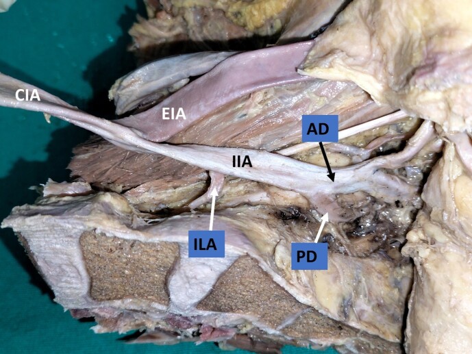
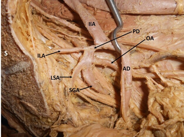
Methods: The study was conducted on 80 male hemipelvises (40 left halves and 40 right halves). The pelvic halves were obtained by making mid-line saw cuts through formalin embalmed adult human cadavers aged approximately 50-80 years. The IIA were dissected and cleaned. Variations of the internal iliac artery and its branches were noted. Relevant photographs were taken. Results were expressed as percentages.

Results: Variations in the branching pattern were observed in 49 (61%) hemipelvises (right: 21, left: 28). Variations were more common (48%) in the branching pattern of the anterior division of IIA than the posterior division (20%). Variations of the main trunk were observed in 29% of cases. In 3% of cases, the IIA did not divide into two divisions. Among the individual branches, the iliolumbar artery showed variations in 29% of cases and the obturator artery in 25%. A common trunk of the internal pudendal and middle rectal arteries was found in 24% of cases and variations of the inferior gluteal artery were seen in 18% of cases.

Conclusions: The study showed a high rate of occurrence of variant IIA branching patterns. Understanding the anatomical variations of the IIA and its branches is essential to minimize intraoperative blood loss and other complications during pelvic surgeries.

Contexto: A artéria ilíaca interna (AII) frequentemente apresenta variações em seus padrões de ramificação. Conhecer suas variações é útil durante procedimentos cirúrgicos ginecológicos e ortopédicos.

Objetivos: Observar o padrão de ramificação da IIA na pelve humana e discutir suas implicações clínicas.

Métodos: O estudo foi realizado em 80 hemipelves masculinas (40 hemipelves esquerdas e 40 hemipelves direitas). As hemipelves foram obtidas através de cortes com serra na linha média em cadáveres humanos adultos de aproximadamente 50-80 anos de idade embalsamados em formaldeído. As AIIs foram dissecadas e limpas. Foram observadas as variações da AII e seus ramos. Obtiveram-se fotografias relevantes. Os resultados foram expressos em porcentagem.

Resultados: Foram encontradas variações no padrão de ramificação em 49 (61%) das hemipelves (direita: 21, esquerda: 28). As variações foram mais comuns (48%) no padrão de ramificação da divisão anterior da AII do que na sua divisão posterior (20%). Variações no tronco principal foram observadas em 29% dos casos. Em 3% dos casos, a AII não estava dividida em duas divisões. Com relação aos ramos individuais, a artéria ileolombar apresentou variações em 29% dos casos, e a artéria obturatória em 25%. Foi encontrado um tronco comum entre as artérias pudenda interna e retal média em 24% dos casos, e variações na artéria glútea inferior foram observadas em 18% dos casos.

Conclusões: O estudo demonstrou uma alta ocorrência de variações no padrão de ramificação da AII. Entender as variações anatômicas da AII e seus ramos é essencial para minimizar a perda sanguínea intraoperatória e outras complicações durante cirurgias pélvicas.

Keywords: iliolumbar artery; internal iliac artery; obturator artery; pelvis; vascular variation.


Conflict of interest statement

Conflicts of interest: NIL.

Figures

Figure 1
Figure 1. Dissected left hemipelvis showing the internal iliac artery giving off all its branches directly, without dividing into anterior and posterior trunks. IIA = internal iliac artery; EIA = external iliac artery; MUL = medial umbilical ligament; SVA = superior vesical artery; IVA = inferior vesical artery; MRA = middle rectal artery; IGA = inferior gluteal artery; IPA = internal pudendal artery; SGA = superior gluteal artery; LSA = lateral sacral artery; ILA = iliolumbar artery; S = sacrum.
Figure 2
Figure 2. Dissected left hemipelvis showing origin of the iliolumbar artery from the main trunk of the internal iliac artery. CIA = common iliac artery; IIA = internal iliac artery; EIA = external iliac artery; AD = anterior division; PD = posterior division; ILA = iliolumbar artery.
Figure 3
Figure 3. Dissected left hemipelvis, showing the abnormal obturator artery arising from the inferior epigastric artery. LIF = left iliac fossa; EIA = external iliac artery; EIV = external iliac vein; IEA = inferior epigastric artery; OA = obturator artery; ON = obturator nerve; OV = obturator vein; OF = obturator foramen.
Figure 4
Figure 4. Dissected left hemipelvis showing origin of the obturator artery from the posterior division of the internal iliac artery. IIA = internal iliac artery; AD = anterior division; PD = posterior division; SGA = superior gluteal artery; LSA = lateral sacral artery; ILA = iliolumbar artery; OA = obturator artery; S = sacrum.
Figure 5
Figure 5. Dissected left hemipelvis showing origin of the obturator artery from the inferior gluteal artery (inferior gluteal artery is a branch of the posterior division of the internal iliac artery here) forming the corona mortis. IIA = internal iliac artery; EIA = external iliac artery; AD = anterior division; PD = posterior division; SRA = Sub-branch middle rectal artery; IGA = inferior gluteal artery; OA = obturator artery; ON = obturator nerve; IPA = internal pudendal artery; SGA = superior gluteal artery; ILA = iliolumbar artery; S = sacrum.
Figure 6
Figure 6. Dissected left hemipelvis showing a common trunk of the internal pudendal artery and middle rectal artery. IIA = anterior division of internal iliac artery; IIV = internal iliac vein; MUL = medial umbilical ligament; SVA = superior vesical artery; IVA = inferior vesical artery; MRA = middle rectal artery; IGA = inferior gluteal artery; IPA = internal pudendal artery; CT = common trunk of origin of internal pudendal and middle rectal arteries; EIV = external iliac vein; UB = urinary bladder; R = rectum; S = sacrum.
Figure 7
Figure 7. Dissected left hemipelvis showing a common trunk of the inferior gluteal artery and middle rectal artery. AD = anterior division of internal iliac artery; MUL = medial umbilical ligament; SVA = superior vesical artery; IVA = inferior vesical artery; IPA = internal pudendal artery; CT = common trunk of origin of the inferior gluteal and middle rectal arteries; IGA = inferior gluteal artery; MRA = middle rectal artery; UB = urinary bladder; R = rectum; S = sacrum.
Figure 8
Figure 8. Dissected left hemipelvis showing the origin of inferior gluteal artery from the posterior division of the internal iliac artery. CIA = common iliac artery; IIA = internal iliac artery; EIA = external iliac artery; AD = anterior division; PD = posterior division; SGA = superior gluteal artery; IGA = inferior gluteal artery; CT = common trunk of origin of the middle rectal and internal pudendal arteries; MRA = middle rectal artery; IPA = internal pudendal artery; ON = obturator nerve; ILA = iliolumbar artery; LSA = lateral sacral artery; S = sacrum.
Figure 9
Figure 9. Dissected right gluteal region, showing a common trunk of the superior and inferior gluteal arteries. GMX = gluteus maximus; GMD = gluteus medius; GT = greater trochanter; SN = sciatic nerve; PI = piriformis; IGA = inferior gluteal artery; SGA = superior gluteal artery; CT = common trunk.
Figure 10
Figure 10. Graph showing the incidence of types of branching pattern of the internal iliac artery (IIA) (based on Adachi classification), n=80.

Similar articles

References

    1. Williams PL, Bannister LH, Berry MM. Gray’s anatomy: the anatomical basis of medicine and surgery. 40th. London: Churchill Livingstone; 2008. 1036
    1. Bilhim T, Casal D, Furtado A, Pais D, O’Neill JEG, Pisco JM. Branching patterns of the male internal iliac artery: imaging findings. Surg Radiol Anat. 2011;33(2):151–159. doi: 10.1007/s00276-010-0716-3. - DOI - PubMed
    1. Bangal V, Kwatra A, Raghav S. Role of internal iliac artery ligation in control of pelvic hemorrhage. Pravara Med Rev. 2009;1(2):23–24.
    1. Li L, Wu K, Liu Y, Lai H, Zeng Z, Zhang B. Angiographic evaluation of the internal iliac artery branch in pelvic tumour patients: diagnostic performance of multislice computed tomography angiography. Oncol Lett. 2019;17(5):4305–4312. doi: 10.3892/ol.2019.10084. - DOI - PMC - PubMed
    1. Joshi VM, Otiv SR, Majumder R, Nikam YA, Shrivastava M. Internal iliac artery ligation for arresting postpartum haemorrhage. BJOG. 2007;114(3):356–361. doi: 10.1111/j.1471-0528.2006.01235.x. - DOI - PubMed

LinkOut - more resources