. 2025 Apr 1:8:0663.
doi: 10.34133/research.0663. eCollection 2025.

A Self-Assembled Metabolic Regulator Reprograms Macrophages to Combat Cytokine Storm and Boost Sepsis Immunotherapy

Affiliations

A Self-Assembled Metabolic Regulator Reprograms Macrophages to Combat Cytokine Storm and Boost Sepsis Immunotherapy

Junyan Zhuang et al. Research (Wash D C). .

Abstract

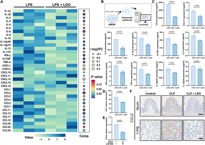
Sepsis, a life-threatening inflammatory disorder characterized by multiorgan failure, arises from a dysregulated immune response to infection. Modulating macrophage polarization has emerged as a promising strategy to control sepsis-associated inflammation. The endogenous metabolite itaconate has shown anti-inflammatory potential by suppressing the stimulator of interferon genes (STING) pathway, but its efficacy is inhibited by hyperactive glycolysis, which sustains macrophage overactivation. Here, we revealed a critical crosstalk between the itaconate-STING axis and glycolysis in macrophage-mediated inflammation. Building on this interplay, we developed a novel nanoparticle LDO (lonidamine disulfide 4-octyl-itaconate), a self-assembled metabolic regulator integrating an itaconate derivative with the glycolysis inhibitor Lonidamine. By concurrently targeting glycolysis and STING pathways, LDO reprograms macrophages to restore balanced polarization. In sepsis models, LDO effectively attenuates CCL2-driven cytokine storms, alleviates acute lung injury, and significantly enhances survival via metabolic reprogramming. This study offers a cytokine-regulatory strategy rooted in immunometabolism, providing a foundation for the translational development of immune metabolite-based sepsis therapies.


Conflict of interest statement

Competing interests: The authors declare that they have no competing interests.

Figures

Fig. 1.
Fig. 1.
The structure and mechanism of nanoparticle LDO. The schematic illustration of LDO recalibrates macrophage polarization from M1 to M2, inhibiting CCL2-driven cytokine storm, ultimately alleviating sepsis.
Fig. 2.
Fig. 2.
Therapeutic potential of HK2 and STING-mediated macrophages metabolic reprogramming in sepsis. (A) Schematic overview of the experimental design of RNA-seq. (B) Heatmap of differentially expressed genes in sepsis or healthy mice. (C) KEGG and GO enrichment analysis of differentially expressed genes in sepsis or healthy mice. (D) A network diagram depicts the relationships between differentially expressed pathways and their associated genes. (E and F) GSEA of genes in glycolytic process (E) and type I interferon signaling pathway (F). (G) Experimental establishment for investigating HK2 and STING in sepsis or healthy condition. (H) hk2 and sting expression in PBMCs from patients. (I and J) Bioinformatic analyses of hk2 and sting expression (I) and their correlation (J) in GEO datasets (GSE26378). (K) Western blot for HK2 and STING in RAW264.7 cells and BMDMs with or without LPS treatment. (L) Western blot for HK2 and STING in RAW264.7 cells after shRNA-mediated knockdown. (M) Western blot for CD86 and CD206 in hk2, sting, or co-knockdown RAW264.7 cells with or without LPS treatment.
Fig. 3.
Fig. 3.
LDO’s self-assembling potential and superior efficacy in alleviating sepsis. (A) Chemical structure and composition of LDO. (B) The nanoparticles were characterized based on their morphology and size distribution; scale bars: 200 nm; PDI, polydispersity index. (C) Experimental design: sepsis or healthy C57BL/6 mice were treated with PBS, 4-OI, LND, and LDO. (D to F and J to L) Survival rate (n = 10), blood routine (n = 3), and liver function examinations (n = 3) were measured after treatment with PBS, 4-OI, LND, and LDO. (G and H, and M and N) The concentrations of cytokines in serum samples, including TNF-α, IL-1β, IL-6, IL-10, IFN-γ, and IFN-β (n = 3), were quantified using ELISA. (I and O) Representative H&E-stained sections of ileum tissue. Morphology was examined using light microscopy. Scale bar: 120 μm.
Fig. 4.
Fig. 4.
LDO alleviates sepsis-induced lung injury. (A and I) Schematic overview of the experimental design of CLP- or LPS-induced lung injury. (B and J) Representative images of H&E staining of lung tissue. Scale bar: 80 μm. Representative images of TUNEL staining of lung tissue. Scale bar: 65 μm. Morphology was examined using light microscopy. (C to E and K to M) Lung wet/dry ratio (C and K), LDH activity (D and L), and MDA level (E and M) of lung tissues were measured after treatment with PBS, 4-OI, LND, and LDO (n = 7). (F and N) The total cell and protein content in the BALF were measured (n = 7). (G and H, and O and P) ELISA was used to measure the levels of cytokines (TNF-α, IL-1β, IL-6, IL-10, IFN-γ, and IFN-β) in BALF. (n = 6).
Fig. 5.
Fig. 5.
LDO inhibit HK2-mediated glycolysis and cGAS-STING pathway. (A and H) Western blot for HK2 was performed, after treatment of 4-OI, LND, or LDO in RAW264.7 cells and BMDMs. (B and I) Immunofluorescence of HK2 in RAW264.7 cells and BMDMs. Scale bars: 20 μm. (C and J) Lactate level in RAW264.7 cells and BMDMs after treatment of 4-OI, LND, or LDO. (D and K) Real-time OCR and ECAR of RAW264.7 cells and BMDMs. (E and L) Western blot results for STING signaling cascades. (F and M) Immunofluorescence of p-STING in RAW264.7 cells and BMDMs. Scale bars: 30 μm. (G and N) ELISA was used to measure type I interferons (IFN-γ and IFN-β) in medium. (O to R) Immunofluorescence of HK2 and p-STING in ileum and lung tissue. Ileum scale bars: 50 μm. Lung scale bars: 25 μm.
Fig. 6.
Fig. 6.
LDO modulates cytokine signaling and promotes M2 macrophage polarization to alleviate septic inflammation. (A and B) KEGG (A) and GO (B) enrichment analysis of differentially expressed genes in BMDMs after LPS or LPS + LDO treatment. (C) Heatmap of main genes in the cytokine–cytokine receptor interaction pathway (n = 3). (D to G) GSEA of genes in chemokine-mediated signaling pathway (D), chemokine activity (E), cell chemotaxis (F), and CCR chemokine receptor binding (G) pathway. (H) R–L network in macrophages during sepsis. (I) Western blot for NLRP3 and ASC in RAW264.7 cells and BMDMs. (J) Immunofluorescence of NLRP3 in RAW264.7 cell and BMDMs. Scale bars: 30 μm. (K) Scheme of LDO’s therapeutic mechanism.
Fig. 7.
Fig. 7.
LDO-induced macrophage polarization: shifting from pro-inflammatory M1 to anti-inflammatory M2 phenotypes. (A) Experimental establishment for macrophage phenotype characterization in RAW264.7 and BMDM model. (B) Western blot for CD86, CD206, and INOS was performed after treatment of 4-OI, LND, or LDO in RAW264.7 cells and BMDMs. (C) Immunofluorescence of CD86 and CD206 in RAW264.7 cells and BMDMs. Scale bars: 30 and 50 μm. (D) CD86 and CD206 expression in RAW264.7 cells and BMDMs (n = 3). (E) Experimental establishment for macrophage phenotype characterization in PBMCs, PMs, ileum tissues, lung tissues, and spleen tissues. (F) Western blot for CD86, CD206, and INOS was performed in PBMCs and PMs. (G) Immunofluorescence of CD86 and CD206 in ileum and lung tissue. Ileum scale bars: 50 μm. Lung scale bars: 25 μm. (H) CD86 and CD206 expression in ileum and lung tissue (n = 3).
Fig. 8.
Fig. 8.
LDO inhibits CCL2-driven cytokine storm in sepsis. (A) Heatmap of BMDMs’ cytokines after treatment of LPS or LPS + LDO. (B) Luminex analysis for macrophage cytokines in BMDM supernatants. (C) The expression of 10 cytokines changed most significantly. (D) Relative expression of ccl2 in the transcriptome level. (E) CCL2 expression of RAW 264.7 or co-knockdown RAW264.7 after LPS treatment. (F) Immunohistochemistry of CCL2 in ileum and lung tissue. Ileum scale bars: 40 μm. Lung scale bars: 80 μm.
Fig. 9.
Fig. 9.
LDO alleviates sepsis and pulmonary injury by modulating CCL2-mediated macrophage infiltration and polarization. (A) Schematic overview of in vivo and in vitro experimental design. (B) Macrophage migration of RAW 264.7 cells and Calcein AM/PI staining assay of MLE-12 cells. Scale bars: 120 μm. (C to F) Survival rate (n = 7), blood routine (n = 3), BALF (n = 3), wet/dry ratio (n = 7), and LDH activity (n = 3) were measured. (G) Representative images of H&E staining of ileum and lung tissue, and TUNEL staining of ileum tissue. Scale bars: 120, 80, and 65 μm. (H) CD86 and CD206 expression in ileum and lung tissue (n = 3). (I) Immunofluorescence of CD86 and CD206 in ileum and lung tissue. Ileum scale bars: 50 μm. Lung scale bars: 25 μm. (J) Schematic overview of in vivo experimental design after treatment of LDO or Bindarit. (K to N) Survival rate (n = 10), blood routine (n = 3), BALF (n = 3), wet/dry ratio (n = 6), and LDH activity (n = 3) were measured. (O) Representative images of H&E staining of ileum and lung tissue. Scale bars: 120 and 80 μm. (P) Immunofluorescence of CD86 and CD206 in ileum and lung tissue. Ileum scale bars: 50 μm. Lung scale bars: 25 μm.

References

    1. Beane A, Shankar-Hari M. Long-term ill health in sepsis survivors: An ignored health-care challenge? Lancet. 2024;404(10459):1178–1180. - PubMed
    1. Garcia Lopez A, Schäuble S, Sae-Ong T, Seelbinder B, Bauer M, Giamarellos-Bourboulis EJ, Singer M, Lukaszewski R, Panagiotou G. Risk assessment with gene expression markers in sepsis development. Cell Rep Med. 2024;5(9):Article 101712. - PMC - PubMed
    1. Kabat AM, Pearce EJ. Inflammation by way of macrophage metabolism. Science. 2017;356(6337):488–489. - PubMed
    1. Hegarty LM, Jones G-R, Bain CC. Macrophages in intestinal homeostasis and inflammatory bowel disease. Nat Rev Gastroenterol Hepatol. 2023;20(8):538–553. - PubMed
    1. Zou R, Tao J, Qiu J, Lu H, Wu J, Zhu H, Li R, Mui D, Toan S, Chang X, et al. . DNA-PKcs promotes sepsis-induced multiple organ failure by triggering mitochondrial dysfunction. J Adv Res. 2022;41:39–48. - PMC - PubMed

LinkOut - more resources