. 2025 Apr 1;15(13):9952-9967.
doi: 10.1039/d5ra00063g. eCollection 2025 Mar 28.

PDMS biointerfaces featuring honeycomb-like well microtextures designed for a pro-healing environment

Affiliations

PDMS biointerfaces featuring honeycomb-like well microtextures designed for a pro-healing environment

Andreea Mariana Negrescu et al. RSC Adv. .

Abstract

Even today, the reduction of complications following breast implant surgery together with the enhancement of implant integration and performance through the modulation of the foreign body response (FBR), remains a fundamental challenge in the field of plastic surgery. Therefore, tailoring the material's physical characteristics to modulate FBR can represent an effective approach in implantology. While polydimethylsiloxane (PDMS) patterning on 2D substrates is a relatively established and available procedure, micropatterning multiscaled biointerfaces on a controlled large area has been more challenging. Therefore, in the present work, a specific designed honeycomb-like well biointerface was designed and obtained by replication in PDMS at large scale and its effectiveness towards creating a pro-healing environment was investigated. The grayscale masks assisted laser-based 3D texturing method was used for creating the required moulds in Polycarbonate for large area replication. By comparison to the smooth substrate, the honeycomb topography altered the fibroblasts' behaviour in terms of adhesion and morphology and reduced the macrophages' inflammatory response. Additionally, the microstructured surface effectively inhibited macrophage fusion, significantly limiting the colonization of both Gram-positive and Gram-negative microbial strains on the tested surfaces. Overall, this study introduces an innovative approach to mitigate the in vitro FBR to silicone, achieved through the creation of a honeycomb-inspired topography for prosthetic interfaces.


Conflict of interest statement

There are no conflicts to declare.

Figures
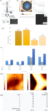
Fig. 1
Fig. 1. The experimental set-up used to process the moulds and to obtain the replicated textured PDMS (a). Surface characterization by AFM, SEM and wettability measurements: (b) profile of hexagonal unit forming matrices; (c) SEM images of a larger area obtained by PDMS replication. Scale bar 300 μm; (d) contact angle measurements of both treated, structured and untreated surfaces and their corresponding SE (e). The AFM image examples including the distribution of points where adhesion force was measured (f). The values of the adhesion force (in nN) measured on ridges, plateau and flat surfaces (g).
Fig. 2
Fig. 2. Viability/proliferation status of the CCD-1070Sk cells grown for 1- and 3-days onto the surface of the honeycomb microtextured and smooth PDMS, as assessed by (a) the CCK-8 test. Results are presented as means ± SD (n = 3; ****p < 0.0001, *p < 0.05 vs. smooth PDMS); (b) the live/dead assay (live and dead cells – green and red fluorescence, respectively). Scale bar represents 200 μm.
Fig. 3
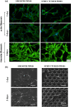
Fig. 3. (a) Fluorescence images of CCD-1070Sk fibroblasts grown for 1- and 3-days in contact with the PDMS surfaces highlighting the actin cytoskeleton (F-actin – green fluorescence), the nuclei (blue fluorescence) and vimentin filaments (green fluorescence). Scale bar represents 50 μm; (b) SEM micrographs of CCD-1070Sk fibroblasts grown onto the surface of the analysed PDMS-based samples after 1- and 3-days of culture. Scale bar represents 50 μm.
Fig. 4
Fig. 4. (a) Immunofluorescent labelling of the fibronectin network and type I pro-collagen synthesized by the CCD-1070SK cells cultured on the analysed surfaces (green fluorescence – positive signals for fibronectin and type I pro-collagen; blue fluorescence – nuclei). The white arrows indicate the better compacted lines of fibronectin fibres developed between adjacent cells on the smooth PDMS surface. Scale bar represents 50 μm; (b) fluorescence intensity measurement (n = 12, mean ± SD, ****p < 0.0001, **p < 0.01 vs. smooth PDMS).
Fig. 5
Fig. 5. The viability and proliferation rates of the RAW 264.7 cells cultured directly onto the smooth and honeycomb PDMS surfaces, under standard (−LPS) and LPS-stimulated (+LPS) conditions, after 1- and 3-days of culture. (a) live/dead assay: viable and dead cells – green and red fluorescence, respectively. Scale bar is 200 μm; (b) CCK-8 test. Results are presented as means ± SD (n = 3; *p < 0.05 vs. smooth PDMS).
Fig. 6
Fig. 6. The morphological characteristics of RAW 264.7 macrophages cultured on the smooth and honeycomb PDMS-based surfaces at 1- and 3-days post-seeding under standard (−LPS) and pro-inflammatory (+LPS) conditions: (a) fluorescence microscopy (actin cytoskeleton – green fluorescence). Scale bar represents 50 μm; (b) SEM microscopy. Scale bar represents 10 μm.
Fig. 7
Fig. 7. (a) The quantification of the inflammatory mediators secreted from the cells treated with LPS (100 ng mL−1) as assessed by ELISA: TNF-α: **p < 0.01 vs. smooth PDMS; IL-1β: *p < 0.05 vs. smooth PDMS; IL-10: ****p < 0.0001 vs. smooth PDMS; NO quantified by Griess diazotization reaction: *p < 0.05 vs. smooth PDMS. Results are expressed as means ± SD (n = 3); (b) fluorescence images showing RAW 264.7 cells' fusion and FBGCs formation on the smooth and honeycomb PDMS surfaces (actin cytoskeleton – green fluorescence; nuclei – blue fluorescence). Scale bar represents 50 μm. The values of the “multinuclear index” as determined by examining 6–10 microscopic fields for each sample.
Fig. 8
Fig. 8. (a) SEM micrographs after 1 day of incubation with microbial suspensions on the honeycomb PDMS support; (b) colony forming unit (CFU) analysis of S. aureus; E. coli and C. albicans after 1 day. Results are presented as means ± SD (n = 3) (****p < 0.0001, ***p < 0.001, *p < 0.05 vs. control).

References

    1. di Pompeo F. S. Paolini G. Firmani G. Sorotos M. JPRAS Open. 2022;32:166–177. - PMC - PubMed
    1. Chao A. H. Garza R. Povoski S. P. Expert Rev. Med. Devices. 2016;13:143–156. - PubMed
    1. Plastic Surgery Statistics, American Society of Plastic Surgeons, https://www.plasticsurgery.org/news/plastic-surgery-statistics, accessed 13 October 2023
    1. Łukasiewicz S. Czeczelewski M. Forma A. Baj J. Sitarz R. Stanisławek A. Cancers. 2021;13:4287. - PMC - PubMed
    1. Albornoz C. R. Bach P. B. Mehrara B. J. Disa J. J. Pusic A. L. McCarthy C. M. Cordeiro P. G. Matros E. Plast. Reconstr. Surg. 2013;131:15–23. - PubMed

LinkOut - more resources