. 2025 Jan 25;36(1):102464.
doi: 10.1016/j.omtn.2025.102464. eCollection 2025 Mar 11.

Effective gene therapy for metachromatic leukodystrophy achieved with minimal lentiviral genomic integrations

Affiliations

Effective gene therapy for metachromatic leukodystrophy achieved with minimal lentiviral genomic integrations

Lucas Tricoli et al. Mol Ther Nucleic Acids. .

Abstract

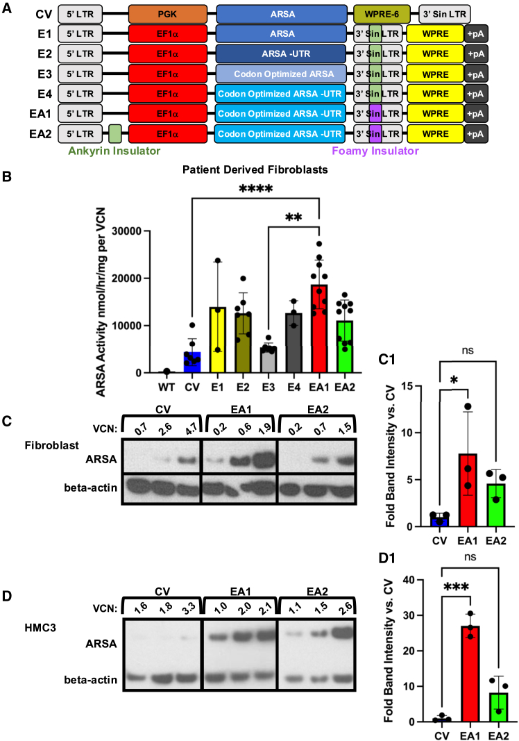
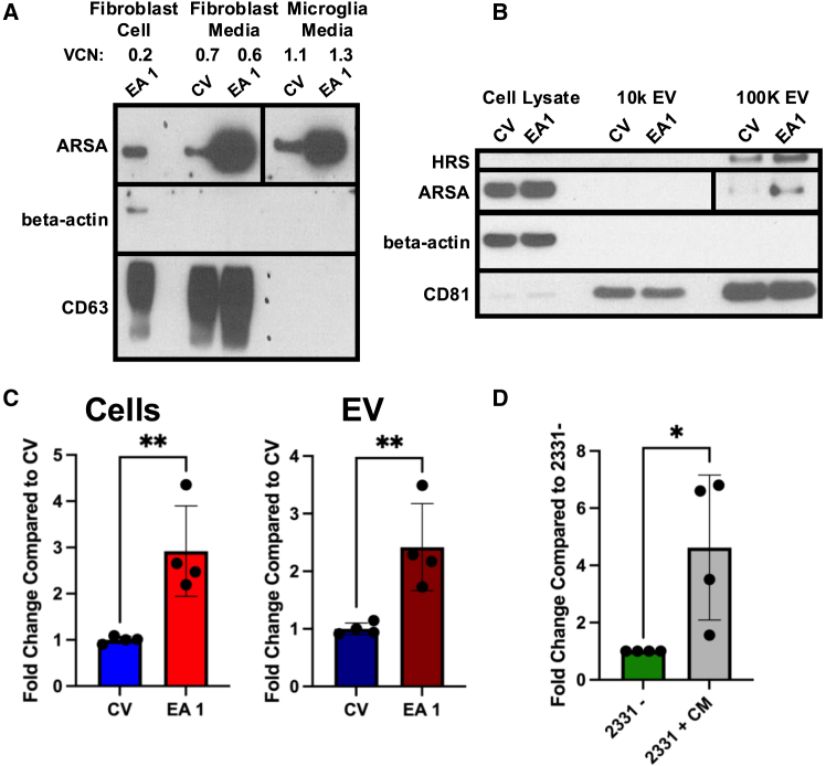
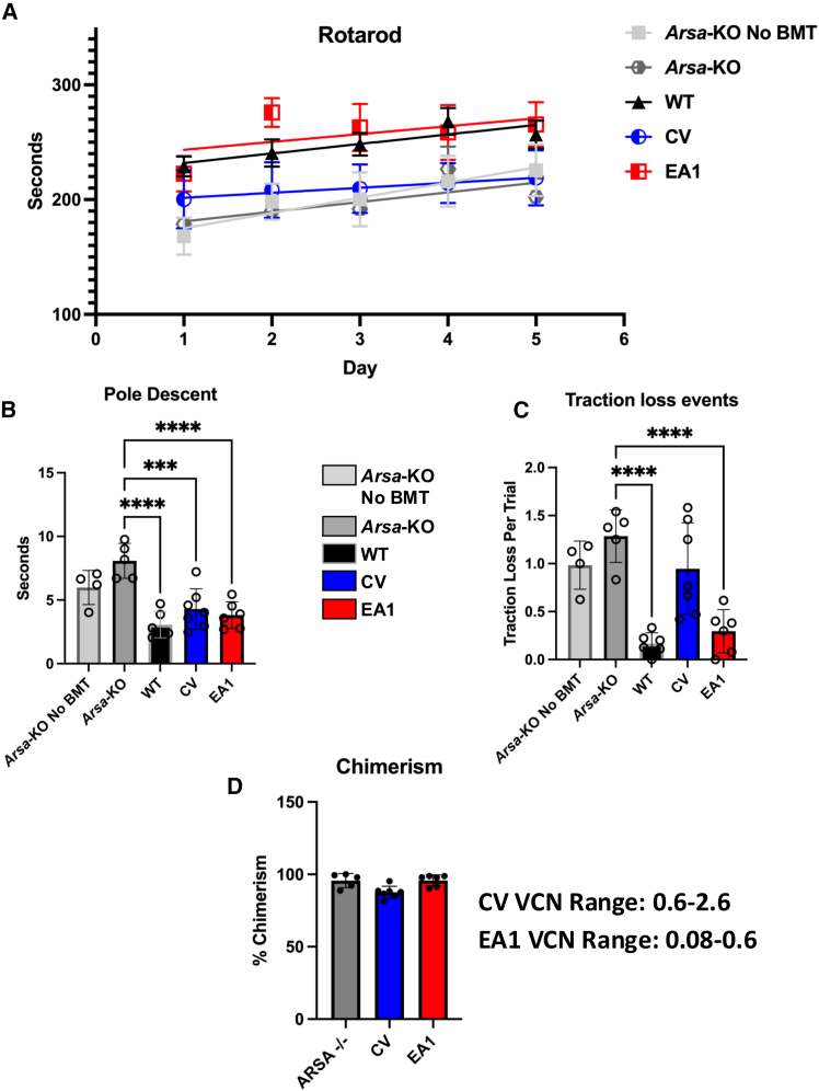
Metachromatic leukodystrophy (MLD) is a fatal lysosomal storage disease characterized by the deficient enzymatic activity of arylsulfatase A (ARSA). Combined autologous hematopoietic stem cell transplantion (HSCT) with lentiviral (LV)-based gene therapy has great potential to treat MLD. Achieving the optimal balance between high enzyme production for therapeutic efficacy and maintaining a low vector copy number (VCN) is crucial. Insufficient enzyme levels can lead to the progression of motor symptoms, undermining treatment goals. Conversely, elevated VCN increases the risk of genotoxicity, which poses safety concerns, and contributes to higher production costs, making the therapy less accessible. Striking this balance is essential to maximize clinical benefit while minimizing risks and costs. To address this need, we increased the expression of ARSA cDNA at single integration by generating novel LVs, optimizing ARSA expression and enhancing safety. In addition, our vectors achieved optimal transduction in mouse and human hematopoietic stem cells (HSCs) with minimal multiplicity of infection (MOI). Our top-performing vector (EA1) showed at least 4× more ARSA activity than the currently US and European Union (EU)-approved vector and a superior ability to secrete vesicle-associated ARSA, a critical modality to transfer functional enzymes from microglia to oligodendrocytes. Three-month-old Arsa-knockout (KO) MLD mice transplanted with Arsa-KO bone marrow (BM) cells transduced with 0.6 VCN of EA1 demonstrated behavior and CNS histology matching wild-type (WT) mice. Our novel vector boosts efficacy while improving safety as a robust approach for treating MLD patients.

Keywords: MLD; MT: Delivery Strategies; gene complementation; gene therapy; lysosomal storage disorder; metachromatic leukodystrophy; novel lentiviral vector; toxicity; translational research.


Conflict of interest statement

S.R. is a scientific advisory board member of Ionis Pharmaceuticals, Vifor, and Disc Medicine. Present to last 5 years: S.R. has been or is a consultant for GSK, BMS, Incyte, Cambridge Healthcare Res, Celgene Corporation, Catenion, First Manhattan Co., FORMA Therapeutics, Ghost Tree Capital, Keros Therapeutics, Noble insight, Protagonist Therapeutics, Sanofi Aventis U.S., Slingshot Insight, Spexis AG, Techspert.io, BVF Partners L.P., Rallybio LLC, venBio Select LLC, ExpertConnect LLC, and LifeSci Capital. The vector EA1 is protected in the patent-CHOP 2022-023: “Novel lentiviral vectors for the treatment of Multiple Sulfatase Deficiency (MSD), Metachromatic Leukodystrophy (MLD) and related disorder of sulfatase deficiency” (CHOP, L.T. and S.R.).

Figures

None
Graphical abstract
Figure 1
Figure 1
Optimal arrangement of vector components to maximize eGFP expression (A) Maps of the multiple vector arrangement used to determine the optimal configuration of EF1α promoter with ankyrin or foamy insulators and WPRE (W) with poly(A) tail (pA) region for optimal eGFP MFI. (B) MFI for all EF1α vector arrangements shown above (n = 3) in addition to select PGK arrangement compared to their EF1α counterparts to demonstrate the key expressive benefit of using Ef1α in these arrangements. LTR, long terminal repeat; Sin-LTR, self-inactivating long terminal repeat; WPRE, woodchuck hepatitis virus posttranscriptional regulatory element; WPRE6, mutated WPRE; PGK, phosphoglycerate kinase promoter; EF1α, elongation factor 1a (EF1α) promoter; pA, bovine growth hormone poly(A) signal. ∗∗∗∗p < 0.0001 Note: significance shown for key comparisons. Expanded significance is shown in Table S1. #PGK-G-W demonstrated the same MFI as the CVG construct and were not statistically different (not shown).
Figure 2
Figure 2
Improved activity and protein expression of ARSA in MLD patient-derived fibroblasts and ARSA-KO human microglia cells (A) Vector schematics comparing the CV and our six Ef1α vectors used to test ARSA expression based on the best MFIs from the eGFP pilot vectors. (B) Fibroblast ARSA enzyme activity normalized to VCN for our six test vectors, including the CV (n = 3–10). (C) A western blot for ARSA from the same transduced patient-derived fibroblast in (B). (D) A western blot using a human microglia cell line (HMC3-ARSA-KO) transduced with vectors CV, EA1, and EA2. Fold change in band intensity was normalized to beta-actin. (C1 and D1) Quantification of ARSA from western blots shown in (C) and (D) as average from independent experiments; n = 3. LTR, long terminal repeat; Sin-LTR, self-inactivating long terminal repeat; WPRE, woodchuck hepatitis virus posttranscriptional regulatory element; PGK, phosphoglycerate kinase promoter; EF1α, elongation factor 1 alpha (EF1α) promoter. ∗p < 0.05, ∗∗p < 0.01, ∗∗∗p < 0.001, ∗∗∗∗p < 0.0001.
Figure 3
Figure 3
Increased ARSA protein expression and activity in EVs secreted from transduced cell lines (A) Protein-precipitated medium harvested from vector-transduced patient fibroblasts and ARSA-KO microglia cells demonstrated much greater ARSA expression in EA1 than CV by western blot. The VCN of each cell line is noted above the treatment (top left). (B) EVs were then isolated via ultracentrifugation, yielding more expression of EV-related ARSA for EA1 at a VCN of 1.2 compared to CV VCN of 3.5. The cells the medium was derived from (cell lysate) are also shown for comparison in addition to the 10,000 × g large-vesicle fraction. (C) Fold change in ARSA activity assay compared to CV for harvested cells and EVs demonstrated higher ARSA activity in cells for EA1 at a VCN range of 0.6–2.4 compared to CV VCN in a range of 3.5–6.8 (n = 4). (D) Fold change in ARSA activity of the untransduced patient cell line alone (2331−) compared to when CM is added to the untransduced cells (2331+) demonstrates an increase in ARSA activity to the transferred cell line (n = 4). ∗p < 0.05, ∗∗p < 0.01.
Figure 4
Figure 4
Genotoxicity assay (A) A table of VCN and percentage of wells transformed in multi-well plate format of vector-transduced mouse HSCs. The positive-control pMSCV-eGFP vector-treated wells are the only plates with cell transformation and medium depletion (yellowing) due to cell immortalization. All other vector-transduced plates after 9 weeks show cellular senescence and absence of medium depletion. (B) A graphical representation of absence in cell transformation in all cell lines aside from the pMSCV-eGFP, indicating the safety of our vector at higher (>4) and clinically relevant (1.5>) VCN.
Figure 5
Figure 5
Transduction of eGFP CV, EA1, and EA2 vectors into WT mice demonstrates vector safety (A) The numbers of weeks post BMT and VCNs are indicated. (B) Erythropoiesis analysis (population I–V, from immature to enucleated red cells) by flow cytometry in WT controls, eGFP mice transduced with normal marrow or mice treated with CVG, EA1, or EA2, from top to bottom. (C) In the same animals, the lymphocyte B and T cell immunoprofiles are shown in addition to (D) the macrophage population. (E) Myeloid progenitor colony formation assay from transduced eGFP mouse mock control, CVG-, EA1-, and EA2-treated mice. CFU-GM, granulocyte-macrophage progenitor.
Figure 6
Figure 6
Rotarod and pole descent motor control assays of untreated Arsa-KO disease mice compared to vector-treated mice (A) Rotarod assessment over a 5-day time course for WT, untreated, or CV or EA1 vector-treated mice (n = 4–7). (B) Descent time duration for a predetermined pole segment in vector-treated groups compared to Arsa-KO mice (n = 4–7). (C) Report of times mice slipped or lost grip during the pole-descent assay (n = 4–7). (D) Table of mice assessed and corresponding VCN and chimerism. ∗∗∗p < 0.001, ∗∗∗∗p < 0.0001.
Figure 7
Figure 7
Neuropathological deficits of untreated Arsa-KO diseased mice relative to vector-treated mice Treatment groups include WT, untreated, CV, and EA1 vector treated at lower VCN. (A) Representative images of sulfatide accumulation in the corpus callosum across treatment groups. (A1) Quantification of sulfatide accumulation across groups. (B) Representative myelin staining (Eri-C) images of the corpus callosum in treatment groups. (B1) Graphical presentation of # of demyelinated vacuoles as shown by white arrows in (B). (C) Representative immunofluorescent images of GFAP (astrocytes) in different brain areas. (C1) Astrocytes (GFAP) counts/mm2 in the corpus callosum. (D) Representative immunofluorescent images of Iba1 (microglia) in different brain areas. (D1) Microglia (Iba1) counts/mm2 in the corpus callosum (n = 3–6 per group). ∗p < 0.05, ∗∗p < 0.01, ∗∗∗p < 0.001, ∗∗∗∗p < 0.0001. Extended significance is shown in Tables S2–S5.

Update of

References

    1. Rosenberg J.B., Kaminsky S.M., Aubourg P., Crystal R.G., Sondhi D. Gene therapy for metachromatic leukodystrophy. J. Neurosci. Res. 2016;94:1169–1179. - PMC - PubMed
    1. Brimley C.J., Lopez J., van Haren K., Wilkes J., Sheng X., Nelson C., Korgenski E.K., Srivastava R., Bonkowsky J.L. National variation in costs and mortality for leukodystrophy patients in US children's hospitals. Pediatr. Neurol. 2013;49:156–162.e1. - PMC - PubMed
    1. Papaioannou I., Owen J.S., Yáñez-Muñoz R.J. Clinical applications of gene therapy for rare diseases: A review. Int. J. Exp. Pathol. 2023;104:154–176. - PMC - PubMed
    1. Matzner U., Breiden B., Schwarzmann G., Yaghootfam A., Fluharty A.L., Hasilik A., Sandhoff K., Gieselmann V. Saposin B-dependent reconstitution of arylsulfatase A activity in vitro and in cell culture models of metachromatic leukodystrophy. J. Biol. Chem. 2009;284:9372–9381. - PMC - PubMed
    1. Schmidt J.L., Pizzino A., Nicholl J., Foley A., Wang Y., Rosenfeld J.A., Mighion L., Bean L., da Silva C., Cho M.T., et al. Estimating the relative frequency of leukodystrophies and recommendations for carrier screening in the era of next-generation sequencing. Am. J. Med. Genet. 2020;182:1906–1912. - PMC - PubMed

LinkOut - more resources