. 2025 Jun;56(6):1569-1580.
doi: 10.1161/STROKEAHA.124.048446. Epub 2025 Apr 2.

Small Extracellular Vesicles From Hypoxia-Neuron Maintain Blood-Brain Barrier Integrity

Affiliations

Small Extracellular Vesicles From Hypoxia-Neuron Maintain Blood-Brain Barrier Integrity

Wei Chen et al. Stroke. 2025 Jun.

Abstract

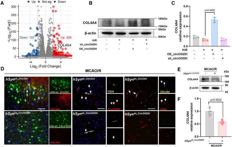
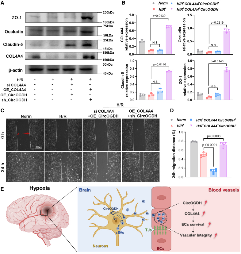
Background: Acute ischemic stroke disrupts communication between neurons and blood vessels in penumbral areas. How neurons and blood vessels cooperate to achieve blood-brain barrier repair remains unclear. Here, we reveal crosstalk between ischemic penumbral neurons and endothelial cells (ECs) mediated by circular RNA originating from oxoglutarate dehydrogenase (CircOGDH).

Methods: We analyzed clinical data from patients with acute ischemic stroke to explore the relationship between CircOGDH levels and hemorrhagic transformation events. In addition, a middle cerebral artery occlusion and reperfusion mouse model with neuronal CircOGDH suppression was used to assess endothelial permeability. ECs with increased CircOGDH expression were analyzed for changes in COL4A4 (collagen type IV alpha 4) levels, and in vitro coculture experiments were conducted to examine small extracellular vesicle-mediated CircOGDH transfer between neurons and ECs.

Results: Clinical data indicated that reduced CircOGDH levels were correlated with increased hemorrhagic transformation in patients with acute ischemic stroke. In the middle cerebral artery occlusion and reperfusion model, neuronal CircOGDH suppression impaired the restoration of endothelial permeability. ECs with increased CircOGDH expression exhibited higher COL4A4 levels, which helped maintain vascular stability. In vitro, hypoxic neurons transferred CircOGDH to ECs via small extracellular vesicles, leading to elevated COL4A4 expression and enhanced endothelial integrity.

Conclusions: Our findings highlight the significance of CircOGDH in neuron-EC crosstalk via small extracellular vesicles in the ischemic penumbra, emphasizing the need for balanced intervention strategies in acute ischemic stroke management.

Keywords: blood-brain barrier; circular RNA; endothelial cells; ischemic stroke; neurovascular coupling.


Conflict of interest statement

None.

Figures

Figure 1.
Figure 1.
Lower circular RNA originating from oxoglutarate dehydrogenase (CircOGDH) expression in patients with acute ischemic stroke (AIS) with hemorrhage compared with those without hemorrhage. A, Experimental scheme for identifying CircOGDH in the plasma of patients. Patients with AIS with hemorrhagic transformation (HT) and patients with AIS without HT were used to identify the relationship between CircOGDH and HT. Created in BioRender. B, Representative data from a patient with AIS with HT showing computed tomography (CT) images and relative CircOGDH expression levels (fold change). High-density areas (highlighted by yellow dashed lines) in CT images indicate the presence of hemorrhagic regions, with hematoma volume calculated using 3D Slicer software. The histogram depicts relative CircOGDH expression in patients with AIS with HT on the day of CT diagnosis of HT (day 1 of onset) compared with patients with AIS without HT (day 1) and those with noncerebrovascular disease (NCD; n=1). Data are presented as means±SD, nonparametric tests with the Mann-Whitney U test. C, Quantitative reverse transcription polymerase chain reaction of CircOGDH expression levels in the plasma of patients with AIS with or without HT (n=18) on the day of CT diagnosis of HT (within 24 hours). Data are presented as means±SEM; the Student t test was used. D, Spearman correlation between hematoma volume and CircOGDH expression levels in 15 patients with AIS with HT, controlled for age, sex, and TOAST criteria (Trial of ORG 10172 in Acute Stroke Treatment), partial correlation coefficient (r=−0.629; P=0.014). E and F, Images of CircOGDH (red) and their quantification (integrated optical density) in spontaneous intracerebral hemorrhage (sICH) and patients with AIS+HT (n=4). Nuclei were stained with 4′,6-diamidino-2-phenylindole (DAPI). Scale bar, 20 µm. Data are presented as means±SD (F); the Mann-Whitney U test was used.
Figure 2.
Figure 2.
Deficiency of neuronal circular RNA originating from oxoglutarate dehydrogenase (CircOGDH) exacerbates blood-brain barrier (BBB) permeability following middle cerebral artery occlusion and reperfusion (MCAO/R). A, Schematic illustration of the experimental procedure in mice. Neuron-specific knockdown of CircOGDH was achieved by administering adeno-associated virus (AAV)-hSynsh_CircOGDH into the lateral ventricles of the mice, with expression in the brain tissue after 21 days. Subsequently, the mice underwent middle cerebral artery occlusion (MCAO) for 1 hour, followed by a 24-hour reperfusion. The brains were then harvested after various treatments. Created in BioRender. B and C, Fluorescence in situ hybridization (FISH) showing CircOGDH localization (red) in the penumbra tissue (PEN; n=6). Scale bar, 20 µm. D, Quantitative reverse transcription polymerase chain reaction analysis of CircOGDH abundance in PEN tissues of mouse brains (n=6). E and F, Representative image showing Evans blue extravasation throughout the mouse brain, with red arrows indicating areas of leakage. Scale bar, 4 mm. Data are presented as means±SD; the Student t test (C, D, and F) was used.
Figure 3.
Figure 3.
Circular RNA originating from oxoglutarate dehydrogenase (CircOGDH) regulates TJ (tight junction) expression in the blood-brain barrier (BBB) after middle cerebral artery occlusion and reperfusion (MCAO/R). A and B, Protein levels of zonula occludens-1 (ZO-1; yellow; left) and occludin (yellow; right) were determined using immunofluorescence (IF; A) in the penumbra tissue (PEN) of MCAO/R mice (n=6), quantified by measuring ZO-1 and occludin IF signal density normalized by CD34 (magenta) signal area. Scale bar, 20 µm. C and D, Representative image and statistical analysis of the Western blot for the expression of ZO-1, occludin, and claudin-5 in the PEN and core of MCAO/R mice (n=6). E, Transmission electron microscopy image of the BBB in MCAO/R mice. The red box highlights typical TJ gaps. Structurally abnormal TJs containing large gaps are indicated by red arrows. F, Statistical analysis of E, showing the fraction of TJ strands with gaps, and the number of TJ gaps (n=6). Data are presented as means±SD; the Student t test (B and D) or the Mann-Whitney U test (F) was used. A (blue) indicates astrocyte end foot; BM, basement membrane; EC (red), endothelial cell; L, lumen; P (green), pericyte; and R, red blood cell.
Figure 4.
Figure 4.
Neuronal circular RNA originating from oxoglutarate dehydrogenase (CircOGDH) modulates human brain microvascular endothelial cell (HBMEC) function after hypoxia/reoxygenation (H/R) by influencing HBMEC CircOGDH levels through small extracellular vesicles (sEVs). A, Quantitative reverse transcription polymerase chain reaction analysis of CircOGDH levels in mouse plasma or plasma sEVs (n=4). B, Schematic of the in vitro blood-brain barrier (BBB) model. Created in BioRender. SH-SY5Y cells were seeded on the inside of collagen-coated 0.4-μm pore poly-(tetrafluoroethylene; PTFE) membrane Transwell inserts and allowed to adhere. HBMECs were seeded in the bottom dishes. Quantitative reverse transcription polymerase chain reaction was performed for CircOGDH levels in cocultured HBMECs with or without GW4869 (exosome inhibitor) treatment in SH-SY5Y cells (n=4). C, Live cell workstation used to observe the internalization of PKH26-labeled SH-SY5Y H/R sEVs into HBMECs at different time points. Scale bar, 20 µm. D, Schematic: sEVs from SH-SY5Y cells treated under different conditions were extracted and added to the culture medium of HBMECs for coincubation. sEVs from SH-SY5Y cells treated with H/R (sEVH/R), H/R+GW4869 (sEVH/R+GW), H/R+OE_CircOGDH (sEVH/R+OE), or H/R+sh_CircOGDH (sEVH/R+sh) were extracted, and CircOGDH levels in the sEVs were measured. sEVs from different sEVs were added to HBMECs for coincubation, and CircOGDH expression levels in HBMECs were measured (n=4). E and F, Transepithelial electric resistance (TEER) of HBMECs treated with different sEVs during reoxygenation (n=4). sEVH/R: sEV from primary neurons cell after hypxia/reoxygenation; sEVH/R+sh: sEV from primary neurons under hypxia/reoxygenation after sh_CircOGDH treatment. Data are presented as means±SD; the Mann-Whitney U test (B, D, E, and F) was used.
Figure 5.
Figure 5.
Circular RNA originating from oxoglutarate dehydrogenase (CircOGDH) regulates COL4A4 (collagen type IV alpha 4) expression in neurons and endothelial cells (ECs). A, Volcano plots showing differentially expressed genes (DEGs) in hypoxic human brain microvascular endothelial cells (HBMECs) transfected with either OE_CircCtrl or OE_CircOGDH (log2-fold change >2). B and C, Representative image and statistical analysis of the Western blot for COL4A4 expression in HBMECs (n=3) after transfection with OE_CircOGDH or sh_CircOGDH. D, Representative images showing COL4A4 (red) staining in mouse brain sections, with green fluorescence indicating hSynsh_CircCtrl or hSynsh_CircOGDH virus (left). Colocalization of CircOGDH (red) with CD34 (yellow) in ECs of the mouse brain (middle) and COL4A4 (yellow) with CD34 (red) in ECs (right). Scale bar, 200 µm. E and F, Representative image and statistical analysis of the Western blot for COL4A4 in penumbra tissue (PEN) of the mouse brain (n=6). Data are presented as means±SD; the Kruskal-Wallis test (C) and the Mann-Whitney U test (F) were used.
Figure 6.
Figure 6.
Regulation of human brain microvascular endothelial cell (HBMEC) function by circular RNA originating from oxoglutarate dehydrogenase (CircOGDH) is mediated through COL4A4 (collagen type IV alpha 4). A and B, Representative image and statistical analysis of the Western blot for ZO-1/occludin/claudin-5/COL4A4 expression in normoxic and hypoxic HBMECs after transfection with OE_CircOGDH, siCOL4A4, or sh_CircOGDH and OE_COL4A4 (n=3). C and D, Migration of HBMECs in a scratch assay. Quantitative analysis of the distance covered by HBMECs (D; n=6). Data are presented as means±SD; the Kruskal-Wallis test (B) and 1-way ANOVA with the Dunnett multiple comparisons test (D) were used. E, Schematic diagram illustrating the neuronal regulation of endothelial cells (ECs) via the release of small extracellular vesicles (sEVs). Created in BioRender.

References

    1. Ghozy S, Reda A, Varney J, Elhawary AS, Shah J, Murry K, Sobeeh MG, Nayak SS, Azzam AY, Brinjikji W, et al. . Neuroprotection in acute ischemic stroke: a battle against the biology of nature. Front Neurol. 2022;13:870141. doi: 10.3389/fneur.2022.870141 - PMC - PubMed
    1. Goyal M, Menon BK, van Zwam WH, Dippel DW, Mitchell PJ, Demchuk AM, Davalos A, Majoie CB, van der Lugt A, de Miquel MA, et al. ; HERMES Collaborators. Endovascular thrombectomy after large-vessel ischaemic stroke: a meta-analysis of individual patient data from five randomised trials. Lancet. 2016;387:1723–1731. doi: 10.1016/S0140-6736(16)00163-X - PubMed
    1. Liu Y, Li Y, Zang J, Zhang T, Li Y, Tan Z, Ma D, Zhang T, Wang S, Zhang Y, et al. . CircOGDH is a penumbra biomarker and therapeutic target in acute ischemic stroke. Circ Res. 2022;130:907–924. doi: 10.1161/CIRCRESAHA.121.319412 - PubMed
    1. Walther J, Kirsch EM, Hellwig L, Schmerbeck SS, Holloway PM, Buchan AM, Mergenthaler P. Reinventing the penumbra - the emerging clockwork of a multi-modal mechanistic paradigm. Transl Stroke Res. 2023;14:643–666. doi: 10.1007/s12975-022-01090-9 - PMC - PubMed
    1. del Zoppo GJ, Sharp FR, Heiss WD, Albers GW. Heterogeneity in the penumbra. J Cereb Blood Flow Metab. 2011;31:1836–1851. doi: 10.1038/jcbfm.2011.93 - PMC - PubMed