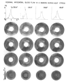
Stability of heterogeneity of myocardial blood flow in normal awake baboons
- PMID: 4017198
- PMCID: PMC3426898
- DOI: 10.1161/01.res.57.2.285
Stability of heterogeneity of myocardial blood flow in normal awake baboons
Abstract
Regional myocardial blood flow has been thought to be relatively uniform, in accord with the singular function of myocardial cells. However, considerable spatial heterogeneity has been observed in the hearts of anesthetized animals and in isolated hearts. Studies were undertaken in a total of 13 baboons. Eleven were awake, healthy animals sitting in chairs at rest or feeding, some performed mild leg exercise (wheel turning), and others were subjected to whole body heating; two were anesthetized, methodological controls. Microspheres (15 +/- 3 micron diameter, 0.5 X 10(6)/kg body weight) were injected via a catheter into the apex of the left ventricle while arterial blood was sampled at a constant rate for calculating cardiac output. Microspheres with different labels were injected at six intervals of 20 minutes to several hours. On sacrifice, the hearts were sectioned into 204 locatable pieces (left ventricle, 168; right ventricle, 27; and atria, 9). Average resting myocardial flow was 2.1 +/- 0.2 ml/g per min (mean +/- SD, n = 11). Left and right ventricles and atria comprised 70 +/- 2% (n = 13), 20 +/- 2%, and 10 +/- 2% respectively of the total heart mass while receiving 80 +/- 3%, 16 +/- 2%, and 4 +/- 2% of the total myocardial flow. Thus, mean left ventricular flow was 114 +/- 5% of the average for the whole heart, right ventricular flow was 81 +/- 13%, and atrial flow was 41 +/- 13%. Myocardial flow heterogeneity was marked; in left ventricle, regional flows ranged from one-third to two times the mean, the relative dispersion (= standard deviation/mean) of regional flows, corrected for methodological scatter and temporal variation, was 0.33 +/- 0.06 (n = 67) in the whole heart, 0.26 +/- 0.07 in left ventricle, 0.32 +/- 0.11 in right ventricle, and 0.22 +/- 0.19 in the atria. The pattern of regional flows in each heart tended to remain stable with time. In each piece averaged over time, the relative dispersion due to temporal heterogeneity was 0.11 +/- 0.03 (n = 2040) in the whole heart, 0.09 +/- 0.03 in the left ventricle, 0.15 +/- 0.05 in the right ventricle, and 0.23 +/- 0.06 in the atria. The conclusion is that the degree of spatial heterogeneity of local myocardial flows in conscious primates is similar to that of anesthetized animals and isolated hearts, and is much greater than that due to temporal fluctuations.
Figures


References
-
- Barlow CH, Chance B. Ischemic areas in perfused rat hearts: Measurement by NADH fluorescence photography. Science. 1976;193:909–910. - PubMed
-
- Bassingthwaighte JB. Calculation of transorgan transport functions for a multipath capillary-tissue system. 1982. Report UW/ BIOENG 82/1 (available from NTIS, Springfield, VA 22161)
-
- Bassingthwaighte JB, Goresky CA. Modeling in the analysis of solute and water exchange in the microvascularure. In: Renkin EM, Michel CC, editors. Hand-book of Physiology. vol 4. American Physiological Society; Washington, D.C.: 1984. pp. 549–626. sec II. The Cardiovascular System. part 1, The Microcirculation.
Publication types
MeSH terms
Grants and funding
LinkOut - more resources
Full Text Sources
Miscellaneous

