. 2025 Apr 15;64(8):1770-1787.
doi: 10.1021/acs.biochem.4c00812. Epub 2025 Apr 2.

Characterization of the Two-Domain Peptide Binding Mechanism of the Human CGRP Receptor for CGRP and the Ultrahigh Affinity ssCGRP Variant

Affiliations

Characterization of the Two-Domain Peptide Binding Mechanism of the Human CGRP Receptor for CGRP and the Ultrahigh Affinity ssCGRP Variant

Katie M Babin et al. Biochemistry. .

Abstract

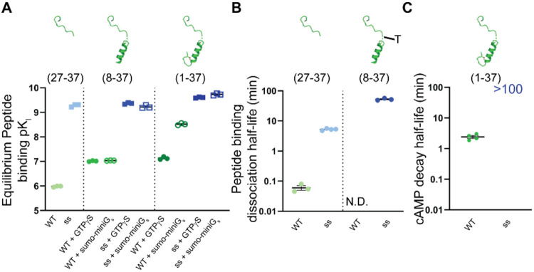
Calcitonin gene-related peptide (CGRP) is a 37-amino acid neuropeptide that functions in pain signaling and neuroimmune communication. The CGRP receptor, CGRPR, is a class B GPCR that is a drug target for migraine headache and other disorders. Here, we used nanoBRET receptor binding and cAMP biosensor signaling assays and theoretical modeling to characterize the CGRPR "two-domain" peptide binding mechanism. Single-site extracellular domain (ECD)-binding and two-site ECD/transmembrane domain (TMD)-binding peptides were examined for CGRP and a high-affinity variant "ssCGRP" with modifications in the C-terminal region. Wildtype and ssCGRP(27-37) bound the ECD with affinities of 1 μM and 0.5 nM, and residence times of 5 s and 8 min, respectively. The (8-37) antagonist fragments had affinities of 100 nM for wildtype and 0.5 nM for ss and exhibited behavior consistent with two-site ECD/TMD binding. ssCGRP(8-37) had a residence time of 76 min. CGRP(1-37) agonist had 25-fold higher affinity for the G protein-coupled state of the CGRPR (Ki = 3 nM) than the uncoupled state (Ki = 74 nM), and elicited short-duration cAMP signaling. In contrast, ssCGRP(1-37) had similar strong affinities for both receptor states (Ki = 0.2 to 0.25 nM), and induced long-duration signaling. An equilibrium reaction network mathematical model of CGRPR activation that includes peptide and G protein binding was developed. This captured wildtype CGRP binding experiments well, but the ssCGRP binding properties were not fully reproduced, suggesting that it may exhibit a distinct binding mechanism. Together, these results advance our quantitative understanding of the CGRPR two-domain mechanism and support the ss variants as potential long-acting therapeutics.


Conflict of interest statement

The authors declare the following competing financial interest(s): AP is inventor on a patent covering the ssCGRP variant. The other authors report no conflicts.

Figures

Figure 1
Figure 1
Structural Depiction of the different lengths of peptides and mutations in ssCGRP. (A) Structural depiction of CGRPR bound to αCGRP and G protein. (B) Structural depiction of the different lengths of peptides used throughout these studies. (C) The C-terminus of CGRP bound to CGRPR ECD (left). Model of the ssCGRP mutations (tan) in the C-terminus of the peptide bound to CGRPR ECD (right). All structural depictions were modeled and modified from PDB 6E3Y.
Figure 2
Figure 2
Equilibrium and kinetic binding of CGRP and ssCGRP (27-37) peptides. (A) nanoBRET competition equilibrium in COS-7 membranes competing against 10 nM CGRP(27-37)*-TAMRA probe. (B and C) nanoBRET competition simultaneous addition kinetics in COS-7 membranes of CGRP(27-37) (B) or ssCGRP(27-37) (C) competing against 20 nM CGRP(27-37)*-TAMRA probe. Data were analyzed using a Motulsky-Mahan equation that incorporates kinetic drift. The kinetic curve fits are shown in red.
Figure 3
Figure 3
Equilibrium and kinetic binding of CGRP and ssCGRP (8-37) peptides. (A) nanoBRET competition equilibrium in COS-7 membranes competing against 30 nM AM2/IMD(8-47)-TAMRA probe in the G protein uncoupled state (50 μM GTPγS) or 3 nM probe in the G protein coupled state (30 μM mGs). (B) Equilibrium binding of ssCGRP(8-37)-TAMRA probe in COS-7 membranes. (C) Association kinetics of ssCGRP(8-37)-TAMRA probe in COS-7 membranes. (D) Linear plot of association rates from C plotted against probe concentration. Plot combines all n = 3 independent replicates with mean ± SEM. (E) Dissociation kinetics of 1 nM ssCGRP(8-37)-TAMRA probe in COS-7 membranes. Dissociation was initiated by addition of 10 μM unlabeled ssCGRP(8-37). Kinetic curve fits are shown in red.
Figure 4
Figure 4
Two-domain binding of CGRP and ssCGRP peptides. (A) Structural depictions of N-terminal agonist cAMP CAMYEL competition assay. (B) N-terminal agonist cAMP CAMYEL competition assay in COS-7 cells. Graph showing combined n = 3 independent replicates with mean ± SEM.
Figure 5
Figure 5
CGRP and ssCGRP effects on heterodimer thermostability. (A) Heterodimer thermostability native PAGE assay with membranes expressing MBP-CLR-eGFP and MBP-RAMP1 from HEK293S GnT1 cells. The detergent-solubilized membranes were incubated at the indicated temperatures in the absence or presence of the indicated peptides followed by native PAGE analysis. Gels were imaged using eGFP in-gel fluorescence. Gels are representative of three independent experiments and show the CGRPR heterodimer band. (B and C) Densitometry analysis showing the melting curves with CGRP (B) or ssCGRP (C). The no peptide control was plotted on both graphs for reference. Plots combined n = 3 independent replicates with mean ± SEM.
Figure 6
Figure 6
Equilibrium binding and signaling kinetics of CGRP and ssCGRP (1-37) peptides. (A) nanoBRET competition equilibrium in COS-7 membranes competing against 30 nM AM2/IMD(8-47)-TAMRA probe in the G protein uncoupled state (50 μM GTPγS) or 3 nM probe in the G protein coupled state (30 μM mGs). (B, C) cAMP signaling kinetics in COS-7 cells expressing CGRPR. Cells were stimulated with 100 nM CGRP(1-37) (B) or ssCGRP(1-37) (C) followed by 10 μM addition of ssCGRP(8-37) antagonist or buffer addition (gray).
Figure 7
Figure 7
Summary scatter plots of peptide affinity and half-life values. (A) Scatter plot summarizing the pKi values from nanoBRET competition equilibrium assays for each length of CGRP and ssCGRP peptide. (B) Scatter plot summarizing the peptide binding half-life values from nanoBRET competition Motulsky-Mahan kinetics for CGRP and ssCGRP for (27-37) lengths and the slow half-life value of ssCGRP(8-37)-TAMRA. The “T” is for the TAMRA label in the peptide depiction for the (8--37) peptide. (C) Scatter plot summarizing the cAMP signaling decay half-life values for CGRP and ssCGRP (1-37). ssCGRP(1-37) was not able to be determined experimentally. All plots show the mean ± SEM of at least three individual replicates.
Figure 8
Figure 8
The reaction network model and mechanistic analysis of CLR activation and peptide binding in the presence and absence of G proteins. (A) The reaction network illustrates peptide and G protein binding steps for CLR in both active and inactive states and the corresponding equilibrium constant (K) for each step is shown (see “the reaction network” section in Methods for a detailed explanation). (B) Top: CGRP primary sequence. The sequences of two binding segments are color coded. Bottom: Depiction of two-stage binding mechanism of CGRP peptide to CLRact. (C) Depiction of conformational change and G protein binding mechanism of active (CLRact) and inactive (CLRinact) CLR. (D) Fraction of CLR occupied by both CGRP (green) and ssCGRP (blue) in the presence (solid line/filled circles) and absence (dashed line/open circles) of G proteins. This accounts for all four fully CGRP-bound species (CLRact:CGRPfull, CLRinact:CGRPfull,, G:CLRact:CGRPfull, and G:CLRinact:CGRPfull). (E) G protein sensitivity is plotted against the binding affinity of the TMD-binding segment of the peptide to CLRact (Ktmd-bind1). This is shown for CGRP (green), ssCGRP (blue) and intermediate points with gradual change between CGRP and ssCGRP. All G protein sensitivity plots are obtained from a ratio of half-maximum values in the absence and presence of G proteins, which is calculated by fitting the binding curves (e.g., panel D) to a sigmoid function. The Ktmd-bind1 value used to obtain plot D is shown with a gray dashed line. (F) G protein sensitivity vs binding affinity of the ECD-binding segment of peptide (Kecd-bind) is shown. Specific Kecd-bind values for CGRP and ssCGRP are marked by vertical green and blue dashed lines, respectively. Intermediate values are shown with solid vertical lines, transitioning from blue to green to indicate the gradual change. Images created with Biorender.com.

Similar articles

Cited by

References

    1. Russo A. F.; Hay D. L. CGRP physiology, pharmacology, and therapeutic targets: migraine and beyond. Physiol. Rev. 2023, 103, 1565–1644. 10.1152/physrev.00059.2021. - DOI - PMC - PubMed
    1. Russell F. A.; King R.; Smillie S. J.; Kodji X.; Brain S. D. Calcitonin gene-related peptide: physiology and pathophysiology. Physiol. Rev. 2014, 94, 1099–1142. 10.1152/physrev.00034.2013. - DOI - PMC - PubMed
    1. Iyengar S.; Ossipov M. H.; Johnson K. W. The role of calcitonin gene-related peptide in peripheral and central pain mechanisms including migraine. Pain 2017, 158, 543–559. 10.1097/j.pain.0000000000000831. - DOI - PMC - PubMed
    1. Russo A. F. Calcitonin gene-related peptide (CGRP): a new target for migraine. Annu. Rev. Pharmacol. Toxicol. 2015, 55, 533–552. 10.1146/annurev-pharmtox-010814-124701. - DOI - PMC - PubMed
    1. Edvinsson L. CGRP and migraine: from bench to bedside. Rev. Neurol. 2021, 177, 785–790. 10.1016/j.neurol.2021.06.003. - DOI - PubMed

Publication types

MeSH terms

Substances

LinkOut - more resources