. 2025 Apr 2;109(1):80.
doi: 10.1007/s00253-025-13466-6.

Monkeypox virus H3L protein as the target antigen for developing neutralizing antibody and serological assay

Affiliations

Monkeypox virus H3L protein as the target antigen for developing neutralizing antibody and serological assay

I-Hsiang Huang et al. Appl Microbiol Biotechnol. .

Abstract

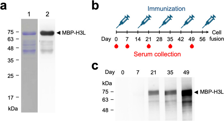
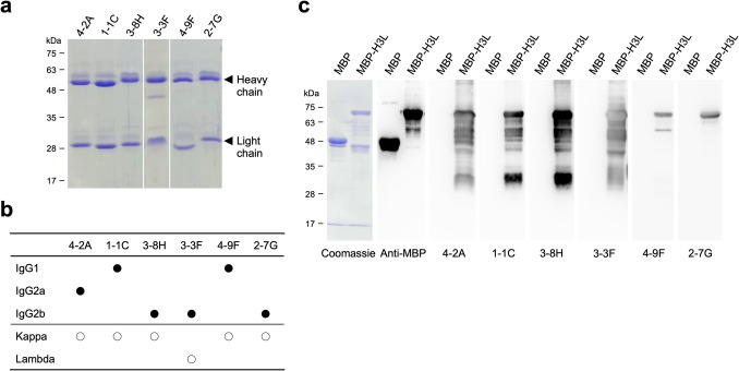
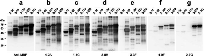
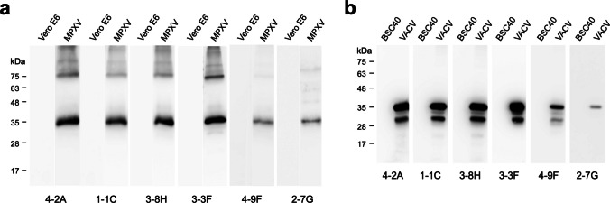
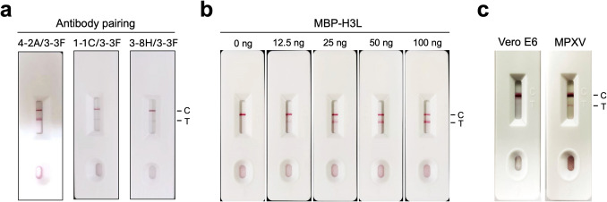
The large number of atypical monkeypox (Mpox) cases caused by emerging monkeypox virus (MPXV) strains was recently found in countries and regions where the Mpox was not reported before. Diagnostic tools and therapeutic agents are important countermeasures for preventing Mpox outbreak. H3L protein is the important surface antigen of MPXV for binding to host cell receptors and mediating viral infection. A broad range of murine anti-MPXV H3L monoclonal antibodies (mAbs) recognizing various binding epitopes have been generated in the study. The rapid test composed of the mAbs 4-2A and 3-3F can specifically detect H3L protein and MPXV virion. The mAb 3-3F exhibited strong MPXV neutralizing activity in a complement-dependent manner. Notably, 3-3F binds to a unique epitope within residues 35-89 of H3L protein. The serum samples collected from Mpox patients barely bound to the N-terminal portion of H3L protein ranging from 2 to 89 residues, indicating that the content of the 3-3F-like antibody is very low in Mpox patient sera. In contrast, the seropositivity was mostly observed using the C-terminal portion of H3L protein ranging from 185 to 282 residues as the target antigen in the immunoblot analysis. Taken together, the anti-MPXV H3L mAb can be developed as the Mpox diagnostic and therapeutic agents. Furthermore, H3L protein is the promising biomarker for serological analysis. KEY POINTS: •Anti-H3L mAbs can cross-react with H3L proteins in MPXV and VACV virions. •The LFIA rapid test using the mAbs 4-2A and 3-3F can specifically detect MPXV. •MPXV was neutralized by mAb 3-3F in a complement-dependent manner.

Keywords: Complement; H3L protein; Lateral flow immunochromatographic assay; Monkeypox virus (MPXV); Neutralizing antibody; Serological assay.


Conflict of interest statement

Declarations. Ethical statement: The animals’ care and protocol have been reviewed and approved by the Institutional Animal Care and Use Committee (IACUC) of National Taiwan University (approval number: NTU-112-EL-00080). A written informed consent was obtained from all individual Mpox patients in the study, which was approved by the Institutional Review Board of National Taiwan University Hospital. Conflict of interest: The authors declare no competing interest.

Figures

Fig. 1
Fig. 1
Immunization of mice with the recombinant MPXV MBP-H3L protein. a The purified MBP-H3L was analyzed on SDS-PAGE, coomassie staining (left panel), and WB with anti-His tag antibody (right panel). b BALB/c mice were immunized with five doses of the purified MBP-H3L in a 2-week interval. The mouse antisera were collected five times at days 0 (before immunization), 7, 21, 35, and 49 as indicated with red blood symbols in the diagram. c The collected mouse antisera were applied in the WB for monitoring the induction of the MBP-H3L-specific antibodies throughout the prime-boost immunization process
Fig. 2
Fig. 2
Characterization of the anti-H3L mAbs. a The purified 4-2A, 1-1C, 3-8H, 3-3F, 4-9F, and 2-7G were analyzed on SDS-PAGE under the reduction condition and further revealed by coomassie staining. b The isotypes of the anti-H3L mAbs were determined by using the Rapid ELISA Mouse mAb Isotyping Kit according to the manufacturer’s instruction. The isotypes of the heavy chains and light chains for each mAbs were indicated with solid and open circles, respectively. c The specificity of the anti-H3L mAbs was identified by WB analysis using MBP and MPB-H3L as the antigens. Anti-MBP was used as the control antibody to detect both of MBP and MBP-H3L
Fig. 3
Fig. 3
Characterization of the binding epitopes of anti-H3L mAbs. The truncated H3L fragments (2–34, 2–89, 2–184, 2–239, and 2–282) fused with an N-terminal MBP-tag were analyzed on SDS-PAGE and subjected to WB analysis with anti-MBP (a), 4-2A (b), 1-1C (c), 3-8H (d), 3-3F (e), 4-9F (f), and 2-7G (g), respectively
Fig. 4
Fig. 4
Recognition of the authentic H3L proteins in MPXV and VACV virions by anti-H3L mAbs. a The culture media collected from the control Vero E6 cells or the MPXV-infected Vero E6 cells, which produced MPXV virions, were analyzed on SDS-PAGE and subjected to WB analysis with 4-2A, 1-1C, 3-8H, 3-3F, 4-9F, or 2-7G, respectively. b The culture media collected from the control BSC40 cells or the VACV-infected BSC40 cells, which produced VACV virions, were analyzed on SDS-PAGE and subjected to WB analysis with 4-2A, 1-1C, 3-8H, 3-3F, 4-9F, or 2-7G, respectively
Fig. 5
Fig. 5
The anti-H3L mAb-based LFIA rapid test. a The mAb pairing 4-2A/3-3F, 1-1C/3-3F, or 3-8H/3-3F-based LFIA rapid test was assembled for evaluation of the binding efficiency for MBP-H3L. C, control line. T, test line. b The various amounts of MBP-H3L protein (0, 12.5, 25, 50, and 100 ng) were added to the 4-2A/3-3F-based LFIA rapid tests for determining the values of the limit of detection (LOD). c Vero E6 cell lysates and MPXV virions are also used to examine the detection specificity of the 4-2A/3-3F-based LFIA rapid test
Fig. 6
Fig. 6
Neutralization of MPXV by 3-3F in the presence of complements. a The MPXV virions were pre-incubated with PBS, 4-2A, 1-1C, 3-8H, 3-3F, 4-9F, or 2-7G (50 μg/mL) and then added to Vero E6 cell monolayers for performing the PRNT assays, respectively. Percentage inhibition = [(control − antibody)/control] × 100. Control stands for the counting numbers of plaques formed in the PRNT assays with PBS. Antibody stands for the counting numbers of plaques formed in the PRNT assays with the indicated mAb. Data are presented as means ± SD of three biological replicates (n = 3). b The MPXV virions were pre-incubated with PBS or 3-3F (50 μg/mL) in the absence or presence of 10% baby rabbit complements (-/ + complement) and then added to Vero E6 cell monolayers for performing the PRNT assays. After incubation for 7 days, cells were fixed with formaldehyde and stained with crystal violet. Three biological replicates were conducted for each experimental group. c The counting numbers of plaques derived from b were calculated by using the formula described previously and presented as percentage inhibition. Control, without 10% baby rabbit complements. + C, with 10% baby rabbit complements
Fig. 7
Fig. 7
The antibody profiles of the Mpox patient sera against the truncated H3L fragments. The different lengths of the truncated H3L fragments (2–34, 2–89, 2–184, 2–239, and 2–282) fused with an N-terminal MBP-tag were analyzed on SDS-PAGE and subjected to WB analysis with anti-MBP (a) or the human sera (1:1000) collected from Mpox patients P1-10 (cl) 2 weeks after admission to the hospital, respectively

Similar articles

Cited by

References

    1. Benhnia MR, McCausland MM, Laudenslager J, Granger SW, Rickert S, Koriazova L, Tahara T, Kubo RT, Kato S, Crotty S (2009) Heavily isotype-dependent protective activities of human antibodies against vaccinia virus extracellular virion antigen B5. J Virol 83(23):12355–12367 - PMC - PubMed
    1. Benhnia MR, McCausland MM, Moyron J, Laudenslager J, Granger S, Rickert S, Koriazova L, Kubo R, Kato S, Crotty S (2009) Vaccinia virus extracellular enveloped virion neutralization in vitro and protection in vivo depend on complement. J Virol 83(3):1201–1215 - PMC - PubMed
    1. Benhnia MR, Maybeno M, Blum D, Aguilar-Sino R, Matho M, Meng X, Head S, Felgner PL, Zajonc DM, Koriazova L, Kato S, Burton DR, Xiang Y, Crowe JE Jr, Peters B, Crotty S (2013) Unusual features of vaccinia virus extracellular virion form neutralization resistance revealed in human antibody responses to the smallpox vaccine. J Virol 87(3):1569–1585 - PMC - PubMed
    1. Cheng YC, Chang SC (2021) Development and biochemical characterization of the monoclonal antibodies for specific detection of the emerging H5N8 and H5Nx avian influenza virus hemagglutinins. Appl Microbiol Biotechnol 105(1):235–245 - PubMed
    1. Cho W, Park S, Kim HJ, Lee M, Choi YS, Yeo SG, Lee J, Koyanagi A, Jacob L, Smith L, Rahmati M, Ahmad S, Fond G, Boyer L, Rhee SY, Lee SW, Shin JI, Woo HG, Yon DK (2024) Clinical characteristics and outcomes of patients with mpox during the 2022 mpox outbreak compared with those before the outbreak: a systematic review and meta-analysis. Rev Med Virol 34(1):e2508 - PubMed