. 2025 Apr 2;20(4):e0313816.
doi: 10.1371/journal.pone.0313816. eCollection 2025.

Targeting NLRP3 and AIM2 signaling pathways by Viscosol alleviates metabolic dysregulations induced inflammatory responses in diabetic neuro- and nephropathy: An in silico and in vivo study

Affiliations

Targeting NLRP3 and AIM2 signaling pathways by Viscosol alleviates metabolic dysregulations induced inflammatory responses in diabetic neuro- and nephropathy: An in silico and in vivo study

Summan Thahiem et al. PLoS One. .

Abstract

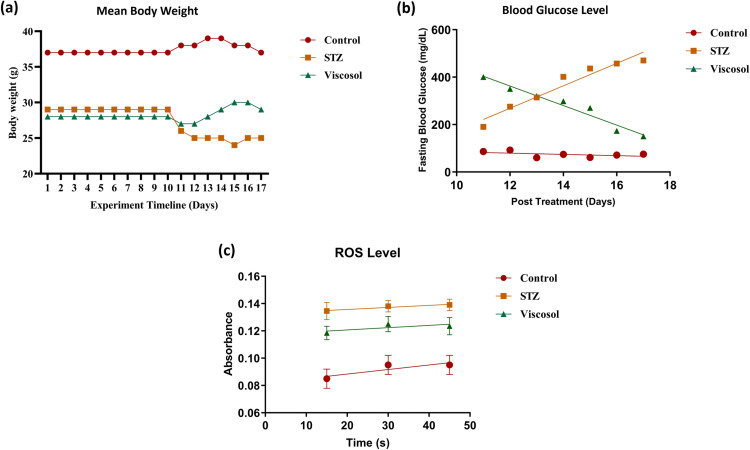
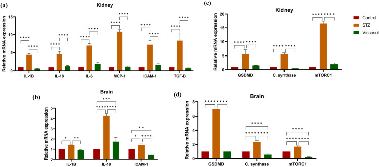
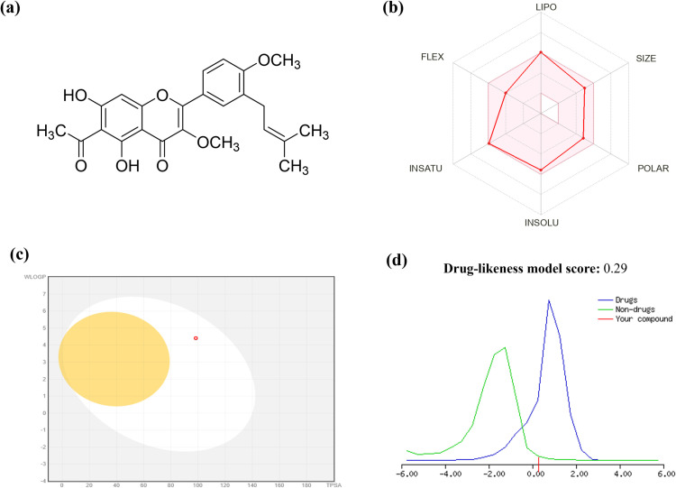
Type 2 Diabetes (T2D) is a chronic metabolic disorder, considered the fastest growing pandemic of the 21stcentury. Meta-inflammation is a pivotal characteristic of T2D. Hyperactivated PTP1B, NLRP3, and AIM2 inflammasomes are considered the major regulators of metabolic inflammation. The concept of diabetes as an inflammatory disease has changed the pathogenic vision of T2D and hence, the compounds that mitigateinflammation in the setting of T2D are under the limelight of research. Current study aimed to evaluatethe anti-inflammatory potency of Viscosol, a novel PTP1B inhibitor, isolated from Dodonaea viscosa, in the STZ-HFD-induced T2D mouse model. Herein, male mice(C57BL/6), were administrated with Streptozotocin (STZ) (40mg/kg) and Viscosol (33mg/kg), intraperitoneally. Computational profiling revealed good absorption, distribution, metabolism and excretion (ADME) properties, least toxicity, and high docking score of Viscosol with PTP1B(-6.4 kcal/mol), NLRP3(-7.2 kcal/mol), and AIM2(-7.4 kcal/mol). Viscosol treatment significantly restored normal body weight (p < 0.0001), decreased the blood glucose level (p < 0.001), serum ROS level(p < 0.05) and diminished the severity of histopathological lesions, inflammatory lobules and increased the cell count of both brain and kidney tissues. The RT-qPCR analysis showed that Viscosol significantly reduced the mRNA expression of PTP1B, NF-κB, NLRP3, and AIM2up to 2.7-folds, 2.6-folds, 5.7-folds and 14.2-folds in the kidney tissues and 1.6-folds, 1.2-folds, 10.2-folds and 1.5-folds in brain tissues. Conclusively, inhibition of PTP1B via Viscosol could attenuate meta-inflammation by suppressing the aberrant NLRP3 and AIM2 inflammasome signaling in diabetes-linked pathophysiology.


Conflict of interest statement

The authors have declared that no competing interests exist.

Figures

Fig 1
Fig 1. Baseline characteristics and oxidative profiling.
(a) Mean body weight (g) of all mice against experimental days. A significant regain in body weight was observed in the Viscosol treated group(****p < 0.0001). (b) Blood glucose profiling. The graphshowsadecrease in blood glucose level in the presence of Viscosol (***p < 0.001). (c) ROS profiling.Viscosol treatment significantly reduced the ROS level (*p < 0.05).
Fig 2
Fig 2. Photomicrographs of H & E-stained kidney and brain section at 10X.
(a) Normal morphology of the kidney tissue; STZ group, presenting histological changes; tubular dilation (*), epithelial cell necrosis (α), degeneration of renal tubules (β), tubular interstitial fibrosis (box), glomeruli shrinkage and thickening of bowman capsule (ε) mesangial matrix deposition (γ); partial retrieval towards normal morphology in Viscosol treated group. The second panel shows brain normal tissues; HFD-STZ group, presenting altered tissue morphology having increased axonal swelling (arrowhead), loss of axons (neon arrows), lost myelin sheath (ε), loss of Schwann cells (arrow), and neuronal disorganization (*); Viscosol treated group, showing partial retrieval towards normal morphology. (b) Cell count score wassignificantly higher in Viscosol treated group as compared to STZ group [Kidney: STZ =  0.76 ±  0.01, Viscosol =  0.84 ±  0.015, ****p < 0.0001); (Brain: STZ =  0.64 ±  0.01, Viscosol =  0.924 ±  0.01, ****p < 0.0001]. (c) Inflammatory lobules score showed significant reduction in inflammatory lobules in the Viscosol treated group as compared to STZ group [Kidney: STZ =  1.15 ±  0.006, Viscosol =  1.02 ±  0.005, ****p < 0.0001; Brain: STZ =  1.08 ±  0.01, Viscosol =  1.02 ±  0.01, ****p < 0.0001].
Fig 3
Fig 3. The expression profile of lipogenic genes, oxidative stress mediators as well as UPRER and mitochondrial stress markers.
(a) Relative abundance of CD36, chREBP, and SREBP1c mRNA levels in diabetic mice group whose expression successfully reduced in the Viscosol treated group. (b)Relative expressionofHMGB1 and COX2 level, in all three groups, respectively. (c, d)Relative mRNA level of UPRER markers, i.e., ATF-6a, PERK, IRE-1, and mitochondrial stress markers, i.e., DRP1, ATF5, and TXNIP as the fold change activity in the Viscosol treated group, compared to the STZ group. Values are mean ±  SD, with their standard errors denoted by vertical bars. Ordinary two-way ANOVA followed by Tukey’s test was applied, and results were found to be significant. (****p < 0.0001, ***p < 0.0001, **p <  0.01 and * p < 0.05).
Fig 4
Fig 4. Effect of Viscosol on upstream mediators of inflammasomes activation, PTP1B and inflammasome complexes.
(a, b)Relative mRNA level of P2X4, CaSR, NEK7, and CSTB in the kidney and brain samples. Increased expression was observedin the STZ group whereas reduced expression in the Viscosol treated group. (c, d)Relative mRNA expression of PTP1B, NF-κB, NLRP3, and AIM2.Viscosol treatment significantly reduced their expression as compared to STZ group. Ordinary two-way ANOVA and Tukey’s test was performed, and results were found to be significant. (Data represented as Mean ±  SD, ****p < 0.0001, ***p < 0.0001, **p < 0.01 and * p <  0.05).
Fig 5
Fig 5. Effect of Viscosol on the expression of inflammatory cytokines, chemokines as well as cell death pathways.
(a, b) Relative mRNA expression of inflammatory cytokines; IL-1β, 1L-18, and IL-6 and chemokines; MCP-1, ICAM-1, and TGF-β. An increased trendline in the expression of respective markers was observed in STZ group, whereas decreased expression was observed in Viscosol treated group. (c, d) Relative mRNA expression of mediators of cell death pathways; pyroptosis, apoptosis, and autophagy.An increased expression of GSDMD and cardiolipinsynthasewas observed in STZ group and reduced expression was observed in Viscosol treated group, respectively. Expression of mTORC1 was elevated in the STZ group whereas reduced in the Viscosol treated group. The results shown are represented as mean ±  SD. Significant differences compared to control (****p < 0.0001, ***p < 0.0001, **p < 0.01 and * p <  0.05; two-wayANOVA and Tukey’s test).
Fig 6
Fig 6. Viscosol structure and ADME/T profiling.
(a) Chemical structure of Viscosol (b) Oral bioavailability radar, depicting Lipinski’s and Veber’s analysis. (c) BOILED-Egg model for Viscosol using SwissADME. The points in BOILED-yolk Egg’s region indicate the probability of molecules passively permeating the blood–brain barrier, whereas BOILED-white Egg’s region indicates the passive absorption by Gastrointestinal tract. (d) Drug-likeness score of Viscosol using MolSoft tool. Viscosol has a value of 0.29. The Non-drug-like behavior is denoted by (a green color curve), while drug-like behavior denoted by (a blue color curve). Compounds with a negative or zero value should not be considered drugs.
Fig 7
Fig 7. Schematic illustration of Viscosol mediated amelioration of meta-inflammation in T2D mice model.
Fig 8
Fig 8. Representative 2D and 3D images ofmolecular docking of Viscosol.
(a) 3D view of Viscosol in the binding pocket of PTP1B. (b) 2D configuration of interacting active amino acid residues of Viscosol with PTP1B (highlighting the H-bonding interaction with active amino acids GLU A:127; LYS A:128, and one pi-sigma bond through MET A: 133).(c)3D view of Viscosol in the binding pocket of NLRP3. (d)2D configuration of interacting active amino acid residues of Viscosol with NLRP3 (highlighting the H-bonding interaction withactive amino acids GLU A:644, HIS A:674). (e) 3D view of Viscosol in the binding pocket of AIM2. (f) 2D configuration of interacting active amino acid residues of Viscosol with AIM2 (highlighting the H-bonding interaction withactive amino acids GLU A:168, VAL A:330, LYS A:162).

References

    1. Aggarwal N, to PK-T 2 D-FP, 2020 undefined. Diabetes Microvascular Complications: An Overview of Epigenetic Modifications. books.google.com N Aggarwal, PK KareType 2 Diabetes-From Pathophysiology to Cyber Systems, 2020•books.google.com. [cited 19 Jun 2024]. Available: https://books.google.com/books?hl=en&lr=&id=87dFEAAAQBAJ&oi=fnd&pg=PA171...
    1. IDF Diabetes Atlas 2021 | IDF Diabetes Atlas. [cited 25 Mar 2023]. Available: https://diabetesatlas.org/atlas/tenth-edition/
    1. Zhang SX, Wang JJ, Kowluru RA. Cross talks between oxidative stress, inflammation and epigenetics in diabetic retinopathy. Cells. 2023;12(2):300. doi: 10.3390/cells12020300 - DOI - PMC - PubMed
    1. Jin Q, Liu T, Qiao Y, Liu D, Yang L, Mao H, et al.. Oxidative stress and inflammation in diabetic nephropathy: role of polyphenols. Front Immunol. 2023;14:1185317. doi: 10.3389/fimmu.2023.1185317 - DOI - PMC - PubMed
    1. Rohm T, Meier D, Olefsky J, Immunity MD-, 2022 undefined. Inflammation in obesity, diabetes, and related disorders. Elsevier. [cited 25 Mar 2023]. Available: https://www.sciencedirect.com/science/article/pii/S1074761321005495 - PMC - PubMed

MeSH terms

Substances