. 2025 Dec;30(1):2485666.
doi: 10.1080/13510002.2025.2485666. Epub 2025 Apr 2.

Spermidine prevents iron overload-induced impaired bone mass by activating SIRT1/SOD2 signaling in senile rat model

Affiliations

Spermidine prevents iron overload-induced impaired bone mass by activating SIRT1/SOD2 signaling in senile rat model

Zhi-Qing Du et al. Redox Rep. 2025 Dec.

Abstract

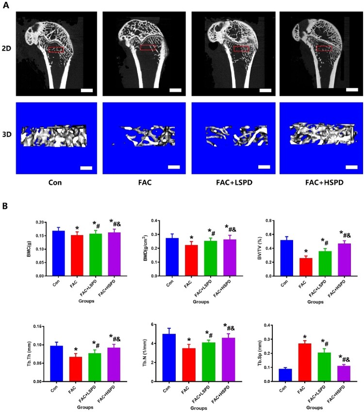
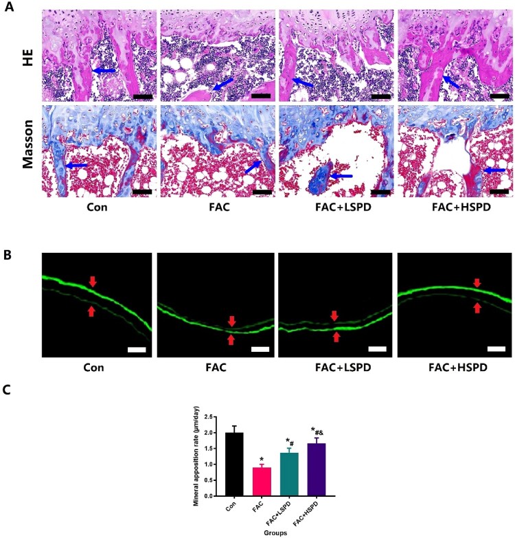
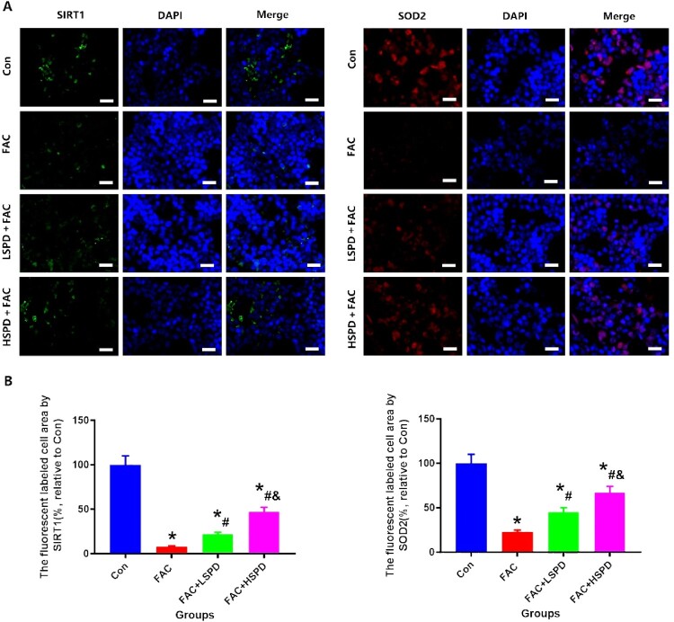
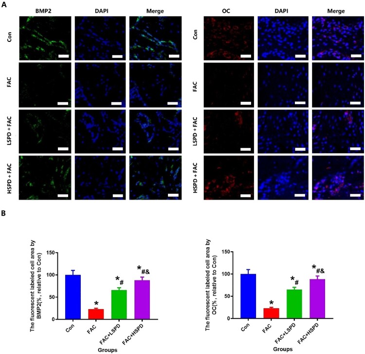
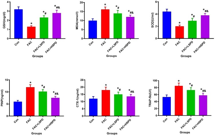
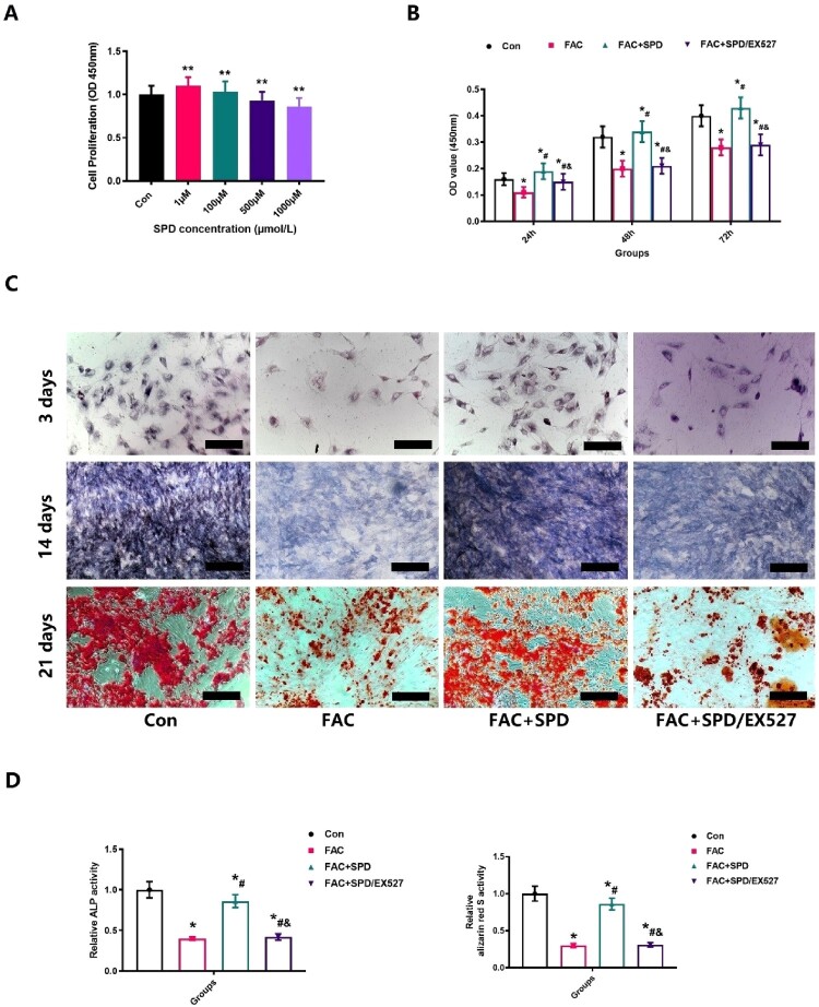
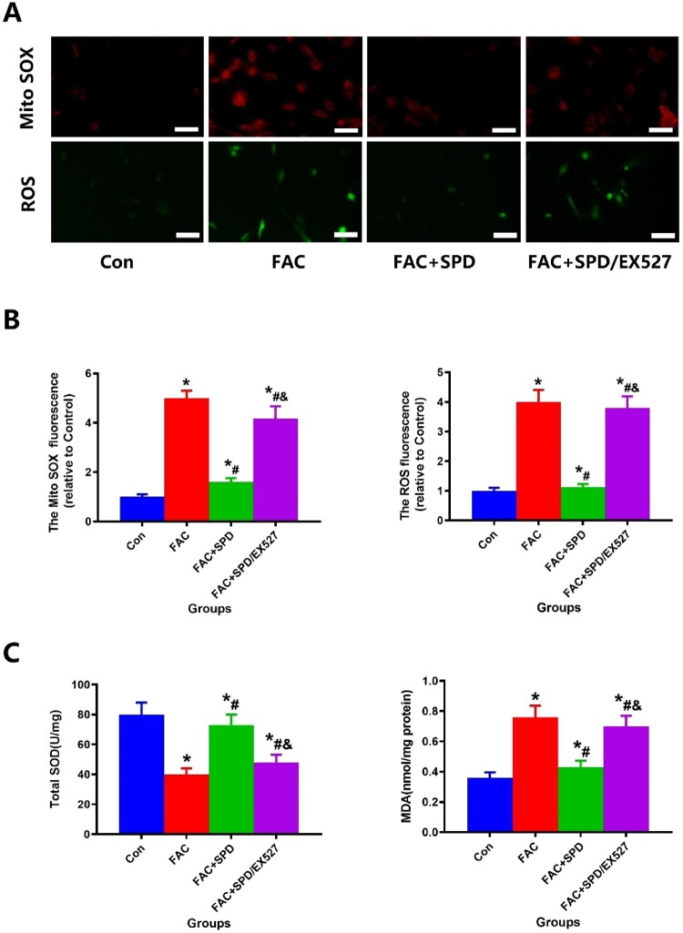
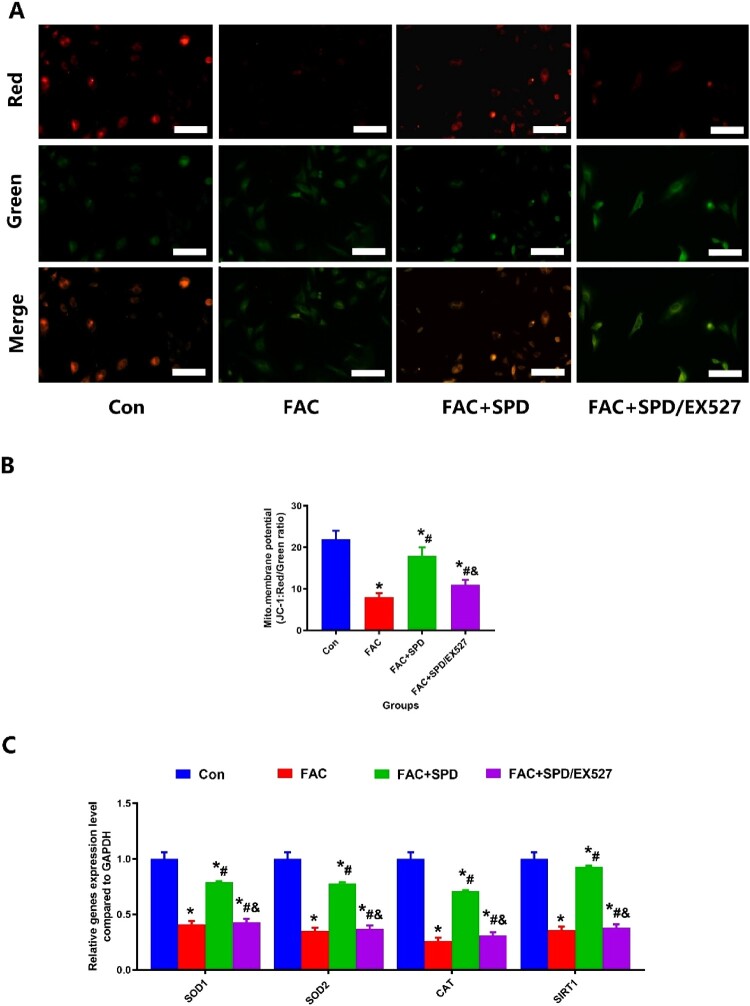
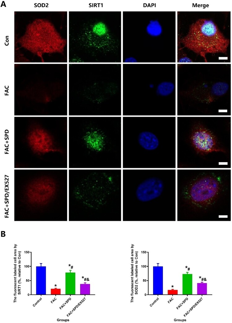
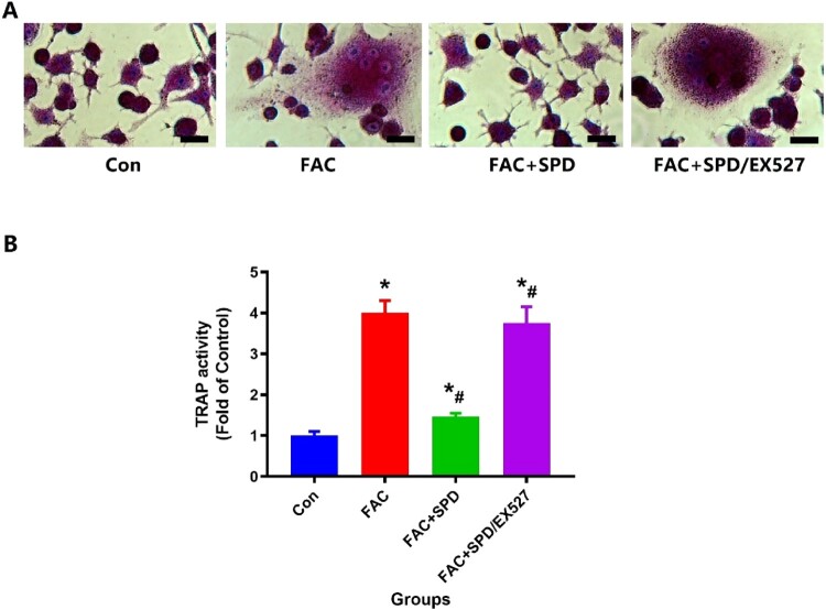
Spermidine (SPD) is an organic compound known for its powerful antioxidant stress and anti-aging properties, and whether SPD has the ability to reduce bone mass in elderly iron overload rats is unknown. The study aimed to assess SPD's impact on iron overload-induced bone loss in elderly rats. In our aged rat model, we found that iron overload negatively influences bone metabolism and remodeling, resulting in decreased bone mineral density and increased bone loss. However, SPD treatment effectively alleviated these harmful effects, as shown by reduced serum levels of MDA and increased SOD and GSH levels. Additionally, SPD-treated rats exhibited enhanced bone mass and higher expression of OC, BMP2, SIRT1, and SOD2 in their bones. Moreover, SPD restored the imbalance in bone metabolism by counteracting the inhibition of osteogenic differentiation and promoting osteoclast differentiation induced by iron overload in MC3T3-E1 and RAW264.7 cells affected by EX527. In summary, our findings suggest that SPD's antioxidant properties may exert anti-osteoporosis effects through activation of the SIRT1/SOD2 signaling pathway.

Keywords: Iron overload; MC3T3-E1; RAW264.7; SIRT1/SOD2 signaling pathway; bone mass; bone metabolism; oxidative stress; senile osteoporosis; spermidine.


Conflict of interest statement

No potential conflict of interest was reported by the author(s).

Figures

Figure 1.
Figure 1.
SPD has the potential to mitigate the negative impact of FAC on bone mass in elderly rats. A. Depicting alterations in the microstructure of distal femoral trabecular bone, this study presents representative 2D and 3D micro-CT images from the Con, FAC, FAC+LSPD, and FAC+HSPD groups (n = 5). The scale bar represents 2 mm. B. Quantitative outcomes of trabecular bone in the metaphysis of the femur, encompassing parameters such as bone mineral content (BMC), connectivity density (Conn.D), ratio of bone volume to total volume (BV/TV), trabecular number (Tb. N), trabecular thickness (Tb. Th), and trabecular spacing (Tb.Sp) (n = 5).
Figure 2.
Figure 2.
The potential of SPD to increase bone trabecular mass bone mineralization rate(BMR) in FAC treated rats from Con, FAC, FAC+LSPD, and FAC+HSPD groups. A. Representative HE and Masson staining observed changes in trabecular bone structure and medullary cavity cells in the distal femur (X40), blue arrow represents the trabecular bone. B. Representative fluorescence labeled images of changes in cortical BMR of femoral bone in each group of rats (X200) (n = 5), the red arrows represent the distance of bone mineralization; C. Quantitative detection of cortical BMR of fluorescently labeled femoral bone (n = 5).
Figure 3.
Figure 3.
Immunohistochemical analysis revealed that the administration of SPD systemically had the potential to modify SIRT1 and SOD2 expression in bone tissue located in the femoral metaphysis of aged rats with iron overload. A. Representative SIRT1 and SOD2 staining images in bone tissue from Con, FAC, FAC+LSPD, and FAC+HSPD groups. Scale bar: 25 µm (n = 5). B. The fluorescence-labeled bone tissue in the femoral metaphysis served as a representation of the quantitative outcomes obtained from SIRT1 and SOD2 analysis (n = 5).
Figure 4.
Figure 4.
Immunohistochemical analysis revealed that the administration of SPD systemically had the potential to modify BMP2 and OC expression in bone tissue located in the femoral metaphysis of aged rats with iron overload. A. Representative BMP2 and OC staining images in bone tissue from Con, FAC, FAC+LSPD, and FAC+HSPD groups. Scale bar: 25 µm (n = 5). B. The fluorescence-labeled bone tissue in the femoral metaphysis served as a representation of the quantitative outcomes obtained from BMP2 and OC analysis (n = 5).
Figure 5.
Figure 5.
Systemic administration of SPD exhibits notable efficacy in reducing oxidative stress markers and effectively restoring the imbalanced bone metabolism equilibrium in aged rats with iron overload (n = 5).
Figure 6.
Figure 6.
Effect of SPD on the proliferation rate and osteogenic differentiation of MC3T3-E1 cells with FAC treatment. A. The effect of SPD on cell proliferation rate at different concentrations (1, 100, 500, 1000 µM) was detected by CCK-8 for 24 h (n = 5); B. The effect of SPD and FAC on the rate of cell proliferation was detected by CCK-8 at a concentration of 1 µM (n = 5). C. After adding SPD, the expression of alkaline phosphatase and formation of calcified nodules increased significantly (n = 5). D. For the quantitative analysis of alkaline phosphatase staining and alizarin red staining positive areas (n = 5).
Figure 7.
Figure 7.
Assessment of the impact of SPD intervention on ROS and Mito SOX production in MC3T3-E1 cells treated with FAC. A. After the implementation of SPD, there was a noticeable reduction in the quantities of ROS and Mito SOX generated in MC3T3-E1 cells subjected to FAC treatment (n = 5). B. Quantitative analysis of ROS and Mito SOX via immunofluorescence staining (n = 5). C. Changes in the levels of SOD and MDA were noted after the application of SPD and FAC interventions (n = 5).
Figure 8.
Figure 8.
SPD therapy promotes recovery of mitochondrial membrane potential activity in the presence of iron overload by JC-1 staining. A. JC-1 staining was observed at high magnification to assess the ability of SPD to significantly improve the recovery of mitochondrial membrane potential in iron overload MC3TE-E1 (Scale bar = 25 µm) (n = 5); B. Quantitative analysis of changes in mitochondrial membrane potential in MC3TE-E1 with iron overload after SPD intervention (n = 5). C. RT-PCR were assess the ability of SPD to significantly improve the related genes expression in iron overload MC3TE-E1 (n = 5).
Figure 9.
Figure 9.
SPD therapy promotes SIRT1 and SOD2 expression in the presence of iron overload. A. immunofluorescence stainings were observed at high magnification to assess the levels of SIRT1 and SOD2 in MC3T3-E1 cells under conditions of iron overload (Scale bar = 25 µm) (n = 5); B. Quantitative analysis of changes in SIRT1 and SOD2 expression in MC3T3-E1 with iron overload after SPD intervention (n = 5).
Figure 10.
Figure 10.
The osteoclast capacity in RAW264.7 cells, which was enhanced due to iron overload, experienced disruption upon treatment with SPD. A. SPD treatment significantly decreased the higher expression of TRAP caused by FAC (Scale bar = 50 µm) (n = 5). B. For the assessment of TRAP staining positive regions using quantitative analysis (n = 5).

Similar articles

References

    1. Lee EN, Choe SY, Choi EH, et al. . Effects of parity and breast feeding duration on the risk of osteoporosis in postmenopausal Korean women: a systematic review and meta-analysis. J Menopausal Med. 2019 Aug;25(2):100–107. doi:10.6118/jmm.19197 - DOI - PMC - PubMed
    1. Park E, Kim J, Kim MC, et al. . Anti-osteoporotic effects of kukoamine B isolated from lycii radicis cortex extract on osteoblast and osteoclast cells and ovariectomized osteoporosis model mice. Int J Mol Sci. 2019 Jun 6;20(11):1–14. - PMC - PubMed
    1. Wasilewski GB, Vervloet MG, Schurgers LJ.. The bone-vasculature axis: calcium supplementation and the role of vitamin K. Front Cardiovasc Med. 2019;6:6. doi:10.3389/fcvm.2019.00006 - DOI - PMC - PubMed
    1. Fan L, Li J, Yu Z, et al. . The hypoxia-inducible factor pathway, prolyl hydroxylase domain protein inhibitors, and their roles in bone repair and regeneration. Biomed Res Int. 2014;2014:239356. - PMC - PubMed
    1. Tao ZS, Qiang Z, Tu KK, et al. . Treatment study of distal femur for parathyroid hormone (1–34) and beta-tricalcium phosphate on bone formation in critical size defects in rats. J Biomater Appl. 2015 Oct;30(4):484–491. doi:10.1177/0885328215592854 - DOI - PubMed

LinkOut - more resources