. 2025 Apr 4;11(14):eads3398.
doi: 10.1126/sciadv.ads3398. Epub 2025 Apr 2.

An engineering-reinforced extracellular vesicle-integrated hydrogel with an ROS-responsive release pattern mitigates spinal cord injury

Affiliations

An engineering-reinforced extracellular vesicle-integrated hydrogel with an ROS-responsive release pattern mitigates spinal cord injury

Jian Cao et al. Sci Adv. .

Abstract

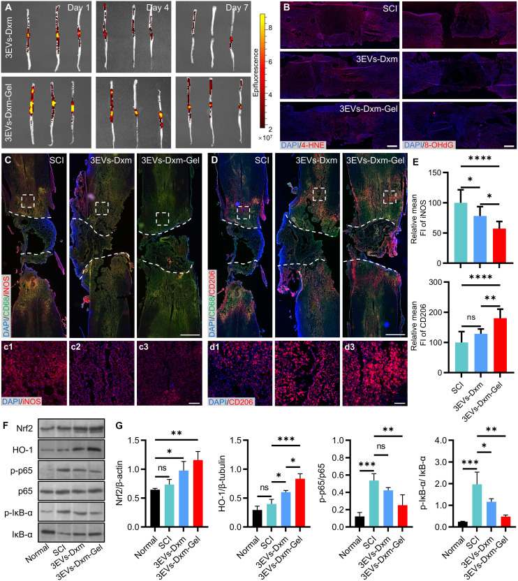
The local delivery of mesenchymal stem cell-derived extracellular vesicles (EVs) via hydrogel has emerged as an effective approach for spinal cord injury (SCI) treatment. However, achieving on-demand release of EVs from hydrogel to address dynamically changing pathology remains challenging. Here, we used a series of engineering methods to further enhance EVs' efficacy and optimize their release pattern from hydrogel. Specifically, the pro-angiogenic, neurotrophic, and anti-inflammatory effects of EVs were reinforced through three-dimensional culture and dexamethasone (Dxm) encapsulation. Then, the prepared Dxm-loaded 3EVs (3EVs-Dxm) were membrane modified with ortho-dihydroxy groups (-2OH) and formed an EV-integrated hydrogel (3EVs-Dxm-Gel) via the cross-link with phenylboronic acid-modified hyaluronic acid and tannic acid. The phenylboronic acid ester in 3EVs-Dxm-Gel enabled effective immobilization and reactive oxygen species-responsive release of EVs. Topical injection of 3EVs-Dxm-Gel in SCI rats notably mitigated injury severity and promoted functional recovery, which may offer opportunities for EV-based therapeutics in central nervous system injury.


Figures

Fig. 1.
Fig. 1.. Illustration of the stepwise preparation of 3EVs-Dxm-Gel and its proposed therapeutic mechanism in SCI treatment.
(A) 3EVs extracted from MS were loaded with Dxm using the probe sonication method to obtain 3EVs-Dxm. Subsequently, 3EVs-Dxm were modified with -2OH groups and cross-linked with HA-PBA and TA to form an injectable hydrogel (3EVs-Dxm-Gel). (B) 3EVs-Dxm-Gel with an antioxidative effect was injected into the injured spinal cord and released anti-inflammatory 3EVs-Dxm in an ROS-responsive pattern. 3EVs-Dxm-Gel treatment attenuated oxidative damage and neuroinflammation during the acute phase, while it improved neural repair and angiogenesis in the chronic phase, thereby aiding in the function restoration following SCI.
Fig. 2.
Fig. 2.. Preparation and characterization of 3EVs-Dxm-Gel.
(A) SEM images of MS (scale bar, 100 μm) and (B) its surface MSCs (scale bar, 10 μm). The white arrows indicate MSCs. (C) Expression of biomarkers of 2EVs and 3EVs including CD9, CD63, and TSG101. (D) TEM images of 2EVs and 3EVs. Scale bar, 200 nm. (E) Drug contents, (F) encapsulation ratios, (G) particle sizes, (H) zeta potentials, and (I) TEM images (scale bar, 200 nm) of 3EVs-Dxm fabricated with different sonication energies [(E) to (H) n = 3]. (J) Morphology, (K) inner structure, and (L) 3EVs-Dxm distribution of 3EVs-Dxm-Gel [scale bars, 50 μm (K) and 200 nm (L)]. (M) Injectable property of 3EVs-Dxm-Gel. (N) Cumulative release curve of EVs from 3EVs-Dxm-Gel and 3EVs-Dxm + Gel in PBS or H2O2 solutions (n = 3). **P < 0.01 and ***P < 0.001.
Fig. 3.
Fig. 3.. Assessment of the therapeutic effects of 3EVs-Dxm.
(A) HUVEC migration after 10 hours (scale bar, 100 μm) and (B) tube formation after 4 hours (scale bar, 100 μm) in the Ctrl, 2EVs, and 3EVs groups. (C) Semiquantitative analysis of cell migration within 10 hours (n = 3) and (D) total branching length of the tube (n = 3). (E) PC12 cell differentiation in Ctrl, NGF, NGF + 2EVs, and NGF + 3EVs groups (scale bar, 50 μm). The red oval indicates representative neurite (scale bar, 100 μm). (F to H) Semiquantitative analysis of differentiation rate (F) (n = 3; two regions per well were selected for statistical analysis), mean neurite counts per cell (G) (n = 3; two regions per well were selected for statistical analysis), and mean neurite length for PC12 cells with different treatments (H) (n = 3; 12 differentiated cells per well were selected for statistical analysis). The significance test of difference was conducted in NGF, NGF + 2EVs, and NGF + 3EVs groups. (I) iNOS and CD206 expressions of LPS-stimulated BV2 cells treated with 3EVs, Dxm, and 3EVs-Dxm and (J) their relative mean fluorescence intensity (FI) analysis (scale bars, 50 μm; n = 3). (K) TNF-α, IL-6, IL-4, and IL-10 secretions of LPS-stimulated BV2 cells with the treatments of 3EVs, Dxm, and 3EVs-Dxm (n = 3). nsP > 0.05, *P < 0.05, **P < 0.01, ***P < 0.001, and ****P < 0.0001.
Fig. 4.
Fig. 4.. In vivo fate of 3EVs-Dxm-Gel and its antioxidative and anti-inflammatory effects in SCI rats.
(A) EV distribution after injecting 3EVs-Dxm and 3EVs-Dxm-Gel in the spinal cord for 1, 4, and 7 days. (B) 4-HNE and 8-OHdG levels in the spinal cord from SCI, 3EVs-Dxm, and 3EVs-Dxm-Gel groups (scale bar, 500 μm). (C) iNOS and (D) CD206 expressions of spinal cord tissues in SCI, 3EVs-Dxm, and 3EVs-Dxm-Gel groups (scale bar, 1 mm) and (E) their relative mean fluorescence intensity analysis (n = 3; three regions per section were selected for statistical analysis). [(C), 1 to (C), 3 and (D), 1 to (D), 3] are the zoomed-in images from white squares in the upper images. Scale bars, 100 μm. The white dashed lines indicate incision of spinal cord. (F) Nrf2, HO-1, p-p65, p65, p-IκB-α, and IκB-α expressions of the spinal cord from Normal, SCI, 3EVs-Dxm, and 3EVs-Dxm-Gel groups and (G) their semiquantitative analysis (n = 3). nsP > 0.05, *P < 0.05, **P < 0.01, ***P < 0.001, and ****P < 0.0001.
Fig. 5.
Fig. 5.. Locomotor and sensory promotion effects of 3EVs-Dxm-Gel in SCI rats.
(A) Body weight and (B) BBB score changes of rats in Normal, SCI, 3EVs-Dxm, and 3EVs-Dxm-Gel groups (n = 6). The blue or red asterisk (*) represents the statistical difference between the SCI group and the 3EVs-Dxm or 3EVs-Dxm-Gel group. w, weeks. (C) Pie graphs showing the distribution of BBB scores of rats on week 6. (D) Schematic diagram of labeled joints of hindlimb and (E) their trajectories during the movement within each group. The red lines refer to the swing phase, and the black lines signify the stance phase. (F) Stride length, (G) fluctuation range of body weight support height, (H) knee height, (I) ankle height, and fluctuation range of (J) knee angle and (K) ankle angle of rat hindlimbs with different treatments (n = 3; 12 strides of each rat were selected for statistical analysis). (L) 50% PWT of rats under the mechanical stimulation in Normal, SCI, 3EVs-Dxm, and 3EVs-Dxm-Gel groups (n = 6). (M) Reaction time of rats under the heat stimulation in each group (n = 6). nsP > 0.05, *P < 0.05, **P < 0.01, ***P < 0.001, and ****P < 0.0001.
Fig. 6.
Fig. 6.. Tissue recovery promoted by 3EVs-Dxm-Gel in SCI rats.
(A) Glial fibrillary acidic protein (GFAP) and NF expressions in the spinal cord in SCI, 3EVs-Dxm, and 3EVs-Dxm-Gel groups (scale bar, 1 mm). The white dashed lines indicate the boundary of lesion. (B) Zoomed-in images of white squares in (A) (scale bar, 100 μm). (C) Quantitative analysis of NF+ area fraction (n = 3; three regions per section were selected for statistical analysis) and NF+ fiber length (n = 3; nine fibers per section were selected for statistical analysis) in the lesion. (D) CD31 expression in the spinal cord in SCI, 3EVs-Dxm, and 3EVs-Dxm-Gel groups (scale bar, 200 μm). (E) Zoomed-in images of white squares in (D) (scale bar, 100 μm). The white arrows present the neovascularization. *P < 0.05, ***P < 0.001, and ****P < 0.0001.

References

    1. Ahuja C. S., Wilson J. R., Nori S., Kotter M. R. N., Druschel C., Curt A., Fehlings M. G., Traumatic spinal cord injury. Nat. Rev. Dis. Primers. 3, 17018 (2017). - PubMed
    1. Rubiano A. M., Carney N., Chesnut R., Puyana J. C., Global neurotrauma research challenges and opportunities. Nature 527, S193–S197 (2015). - PubMed
    1. Guan B., Anderson D. B., Chen L., Feng S., Zhou H., Global, regional and national burden of traumatic brain injury and spinal cord injury, 1990–2019: A systematic analysis for the Global Burden of Disease Study 2019. BMJ Open 13, e075049 (2023). - PMC - PubMed
    1. GBD Spinal Cord Injuries Collaborators , Global, regional, and national burden of spinal cord injury, 1990–2019: A systematic analysis for the Global Burden of Disease Study 2019. Lancet Neurol. 22, 1026–1047 (2023). - PMC - PubMed
    1. Zheng B., Tuszynski M. H., Regulation of axonal regeneration after mammalian spinal cord injury. Nat. Rev. Mol. Cell Biol. 24, 396–413 (2023). - PubMed

MeSH terms

LinkOut - more resources