. 2025 Apr 2;16(1):237.
doi: 10.1038/s41419-025-07542-y.

SGK1 upregulation in GFAP+ neurons in the frontal association cortex protects against neuronal apoptosis after spinal cord injury

Affiliations

SGK1 upregulation in GFAP+ neurons in the frontal association cortex protects against neuronal apoptosis after spinal cord injury

Anbiao Wu et al. Cell Death Dis. .

Abstract

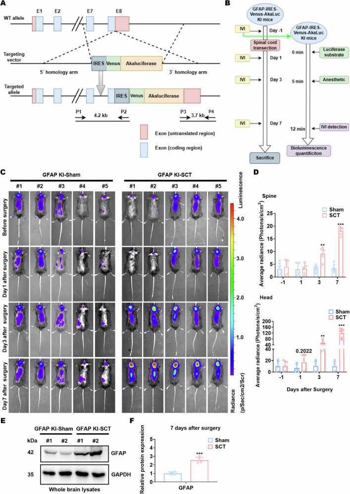
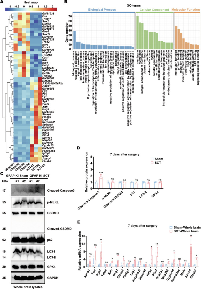
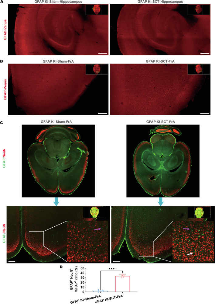
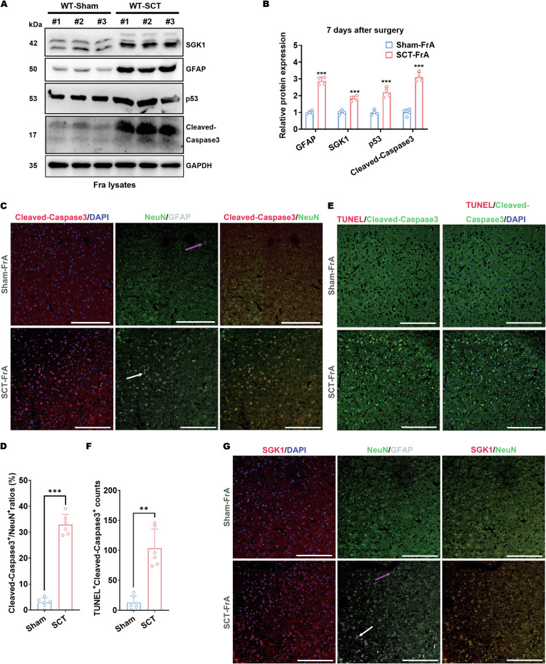
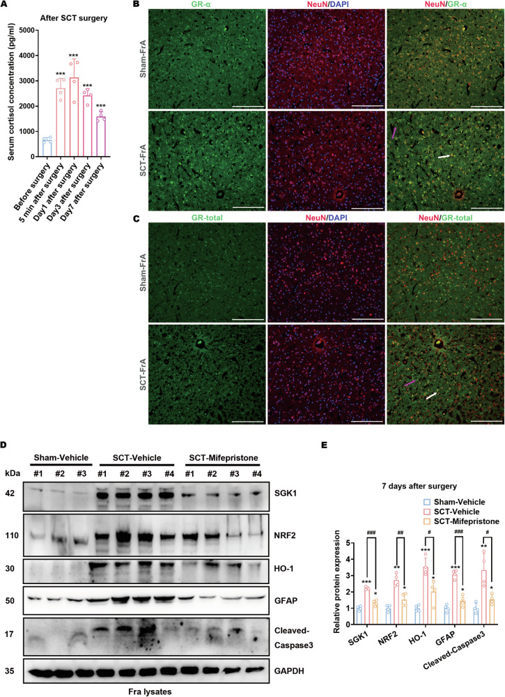
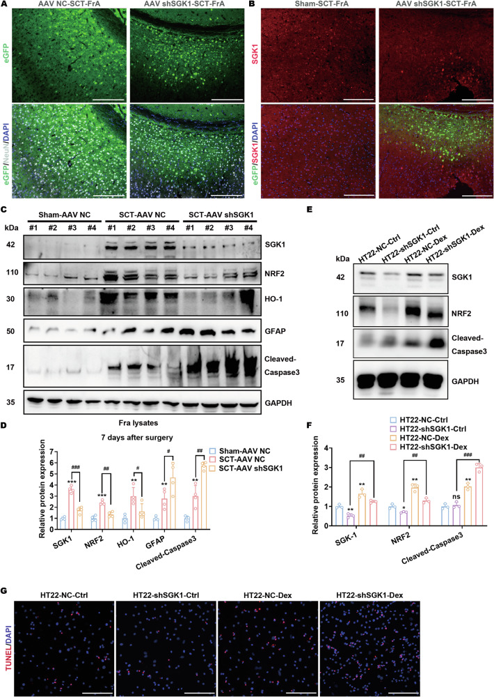
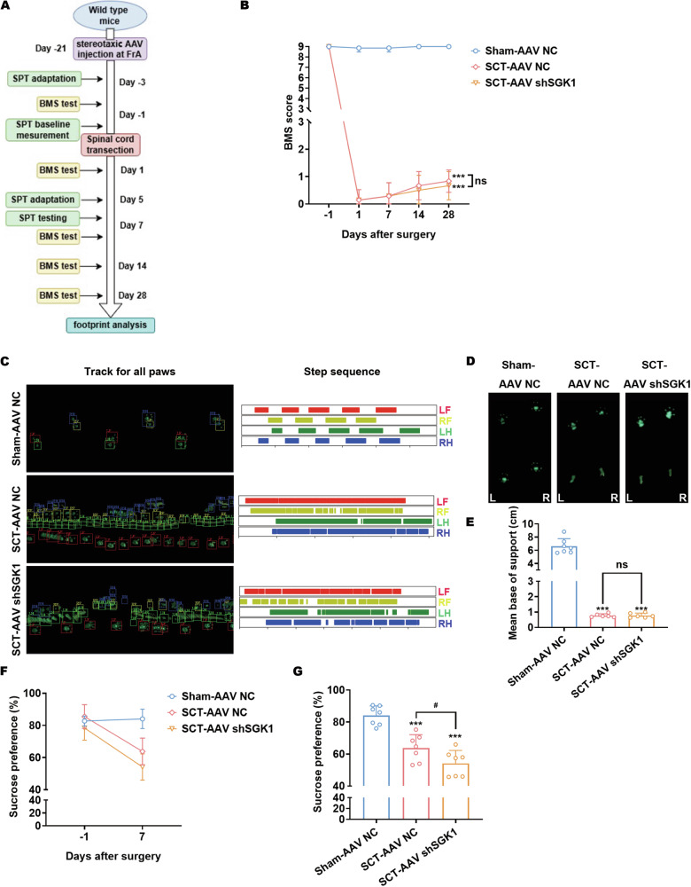
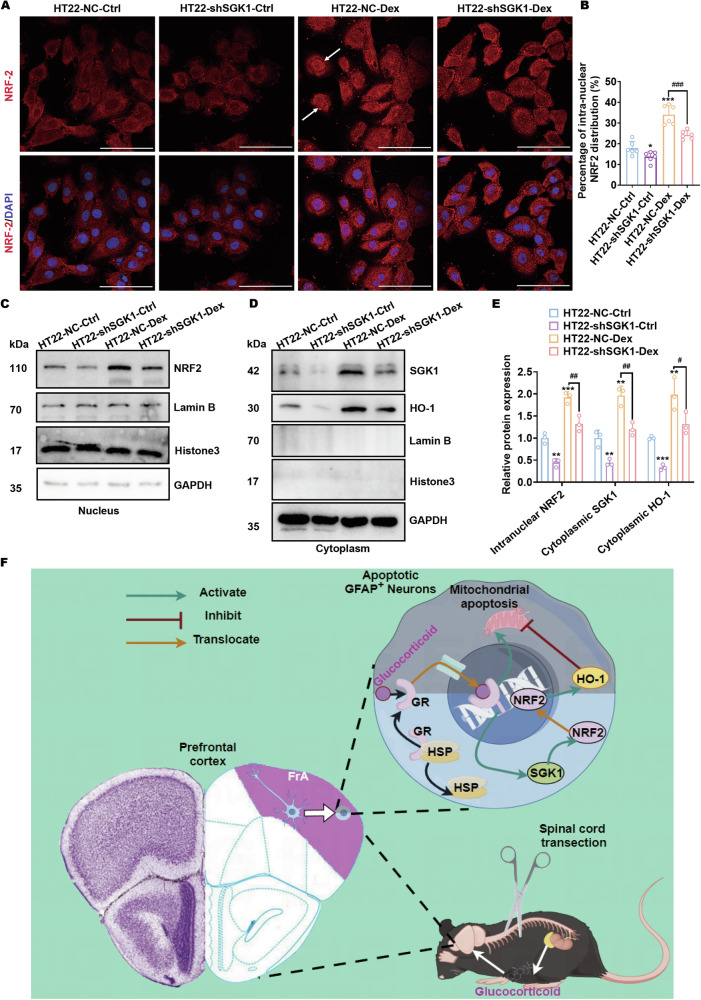
Spinal cord injury (SCI) casts devastating and long-lasting impacts on the well-being of patients. Cognitive deficits and emotional disorders are common in individuals with SCI, yet the underlying mechanisms are not completely understood. Astrogliosis and glial scar formation occur during the subacute phase post-injury, playing complicated roles in remyelination and neurite regrowth. Therefore, we constructed a GFAP-IRES-Venus-AkaLuc knock-in mouse model for the corresponding studies. Surprisingly, complete spinal cord transection (SCT) surgery led to earlier and more prominent augmentation of bioluminescence in the brain than in the spinal cord. Bulk RNA sequencing revealed the activation of apoptotic signaling and the upregulation of serum and glucocorticoid-regulated kinase 1 (SGK1). The pattern of GFAP signals changed throughout the brain after SCT, as indicated by tissue clearing and immunostaining. Specifically, GFAP signals were intensified in the frontal association cortex (FrA), an encephalic region involved in associative learning and recognition memory processes. Further exploration unraveled that intensified GFAP signals in the FrA were attributed to apoptotic neurons with SGK1 upregulation, which was induced by sustained high glucocorticoid levels after SCT. The introduction of SGK1 silencing vectors confirmed that SGK upregulation in these FrA neurons exerted anti-apoptotic effects through NRF2/HO-1 signaling. In addition, SGK1 knockdown in FrA neurons aggravated the post-SCI depressive-like behaviors. Thus, ectopic SGK1 expression designated for limbic neurons could serve as a promising therapeutic target for the future development of treatments for spinal cord injuries.


Conflict of interest statement

Competing interests: The authors declare no competing interests.

Figures

Fig. 1
Fig. 1. GFAP signals are intensified in mouse brains after spinal cord transection.
A Schematic representation of the mouse GFAP genomic locus and the designated targeting strategy used to produce and identify GFAP-IRES-Venus-AkaLuc KI mice. B Schematic description of spinal cord transection (SCT) and in vivo imaging procedures for KI mice. IVI, in vivo imaging. C Representative images of in vivo imaging detecting the bioluminescence of KI mice at various time points of sham surgery or SCT. D Statistical analysis of bioluminescence signals in the spine (top) and head (bottom) of the KI mice shown in (C). **p < 0.01, ***p < 0.001 vs. Sham; n = 5 biologically independent samples. E, F Protein expression of GFAP in the whole-brain lysates of KI mice 7 days after sham surgery or SCT. Representative images (E) and statistical analyses (F) are shown. ***p < 0.001 vs. Sham-whole brain; n = 4 biologically independent samples.
Fig. 2
Fig. 2. SGK1 upregulation and apoptotic signaling are activated in the mouse brain after spinal cord transection.
A Heatmap of DEGs generated from bulk RNA-sequencing of whole brains of KI mice 7 days after sham surgery or SCT. B Bar plots of the GO enrichment of DEGs shown in (A). C, D Protein expression of cleaved-Caspase-3, p-MLKL (Ser345), GSDMD, p62, LC3, and GPX4 in the whole-brain lysates of KI mice 7 days after surgery. Representative images (C) and statistical analyses (D) are shown. ***p < 0.001 vs. Sham-whole brain; n = 4 biologically independent samples. E qPCR analysis of the upregulated genes shown in (A). *p < 0.05 and **p < 0.01 vs. Sham-whole brain; n = 4 biologically independent samples.
Fig. 3
Fig. 3. GFAP-positive neurons emerge in the mouse frontal association cortex after spinal cord transection.
A, B Representative horizontal section images of the hippocampus (A) and frontal association cortex (FrA, B) of an SCT-treated KI mouse and its sham surgery control after tissue clearing without immunostaining (PEGASOS). Scale bar, 500 μm. C Representative horizontal section images of the whole brain of an SCT-treated KI mouse and its sham surgery control after immunostaining-compatible tissue clearing (iDISCO) with antibodies against GFAP (green) and NeuN (red). GFAP+ NeuN- cells were indicated with magenta arrows whereas GFAP+ NeuN+ cells were indicated with white arrows. Scale bar, 500 μm. D Statistical analysis of GFAP+ NeuN+ cell ratios in all GFAP+ cells within the FrA sections of sham/SCT-treated KI mice. ***p < 0.001 vs. GFAP KI-Sham-FrA; n = 5 distinct transverse sections.
Fig. 4
Fig. 4. Cellular apoptosis correlates with SGK1 upregulation in GFAP-positive neurons in the frontal association cortex of mice with spinal cord transection.
A, B Protein expression of SGK1, GFAP, p53, and cleaved-caspase-3 in the frontal association cortex tissue from SCT-treated, C57 wild-type mice 7 days after the surgery. Representative images (A) and statistical analyses (B) are shown. ***p < 0.001 vs. Sham-FrA; n = 4 biologically independent samples. Coronal sections of the FrA tissue from sham- or SCT-treated mice 7 days after surgery were subjected to the following co-staining: C, D Co-staining with antibodies against NeuN (green), GFAP (white) and cleaved-Caspase-3 (red). Representative images (C, scale bar, 250 μm) and statistical analyses (D) are shown. ***p < 0.001 vs. Sham-FrA; n = 6 biologically independent samples. E, F Co-staining with an antibody against cleaved-Caspase-3 (green) and TUNEL probes (red). Representative images (E, scale bar, 250 μm) and statistical analyses (F) are shown. **p < 0.01 vs. Sham-FrA; n = 6 biologically independent samples. G Co-staining with antibodies against NeuN (green), GFAP (white), and SGK1 (red). Representative images (scale bar, 250 μm) are shown.
Fig. 5
Fig. 5. Hypercortisolism after spinal cord transection leads to neuronal apoptosis and the upregulation of GFAP and SGK1.
A Mouse serum cortisol levels at designated time points before and after SCT surgery were measured by ELISA ***p < 0.001 vs. Sham; n = 4 biologically independent samples. B, C Coronal sections of the FrA tissue from sham- or SCT-treated mice 7 days after surgery were co-stained with antibodies against GR-α (green) and NeuN (red) (B) or with antibodies against GR-total (green) and NeuN (red) (C). Scale bar, 250 μm. Typical GR-α+NeuN-/GR-total+NeuN- cells were indicated with magenta arrows, whereas GR-α+NeuN+/GR-total+NeuN+ cells were indicated with white arrows. D, E Protein expression of SGK1, NRF2, HO-1, GFAP, and cleaved-Caspase-3 in the FrA of mifepristone-pretreated mice after surgery. Representative images (D) and statistical analyses (E) are shown. *p < 0.05, **p < 0.01 and ***p < 0.001 vs. Sham-Vehicle; ##p < 0.01 and ###p < 0.001 vs. SCT-Vehicle; n = 4 biologically independent samples.
Fig. 6
Fig. 6. SGK1 upregulation exerts neuroprotective effects against glucocorticoid-mediated neuronal apoptosis after spinal cord transection.
A Coronal sections of the FrA from SCT-treated mice preinjected with AAV-NC or AAV-shSGK1 carrying the eGFP encoding sequence were stained with anti-NeuN (white). Scale bar, 250 μm. B Coronal sections of the FrA from SCT-treated mice preinjected with AAV-shSGK1 were stained with anti-SGK1 (red). Scale bar, 250 μm. C, D Protein expression of SGK1, NRF2, HO-1, GFAP, and cleaved-Caspase-3 in the FrA of SCT-treated mice preinjected with AAV-NC or AAV-shSGK1. Representative images (C) and statistical analyses (D) are shown. **p < 0.01 and ***p < 0.001 vs. Sham-AAV NC; ##p < 0.01, ###p < 0.001 vs. SCT-AAV NC; n = 4 biologically independent samples. (E and F) Protein expression of SGK1, NRF2, and cleaved-Caspase-3 in AAV-shSGK1-infected HT22 cells treated with dexamethasone. Representative images (E) and statistical analyses (F) are shown. *p < 0.05, **p < 0.01 and ***p < 0.001 vs. HT22-NC-Ctrl; ##p < 0.01, ###p < 0.001 vs. HT22-NC-Dex; n = 3 biologically independent samples. G TUNEL staining of AAV-shSGK1-infected HT22 cells treated with dexamethasone. Scale bar, 500 μm.
Fig. 7
Fig. 7. SGK1 knockdown in mouse FrA neurons aggravated the post-SCI depressive-like behaviors.
A Schematic representation of functional and behavioral tests evaluating SCT-treated mice preinjected with AAV-NC or AAV-shSGK1 in the FrA. B BMS scores from open-field tests evaluating hindlimb locomotion of sham/SCT-treated mice preinjected with AAV-NC or AAV-shSGK1. The tests were performed weekly after surgery. ***p < 0.001 vs. Sham-AAV NC; n = 6–7 biologically independent samples (One mouse in the SCT-AAV NC group and one mouse in the SCT-AAV-shSGK1 group died during day 14–day 28 after surgery). Wilcoxon rank-sum test was used for statistics. C Representative images of gait analysis evaluating hindlimb locomotion of sham/SCT-treated mice preinjected with AAV-NC or AAV-shSGK1, performed 4 weeks after surgery (LF, left front; LH, left hind; RF, right front; RH, right hind). D, E Footprints and the corresponding average stride length of sham/SCT-treated mice preinjected with AAV-NC or AAV-shSGK1, obtained from the gait analysis. Representative images (D) and statistical analyses (E) are shown. ***p < 0.001 vs. Sham-AAV NC; n = 6–7 biologically independent samples as stated above. Brown-Forsythe and Welch ANOVA was used for statistics. F, G Sucrose preference tests of sham/SCT-treated mice preinjected with AAV-NC or AAV-shSGK1. The sucrose preference before and after surgery was demonstrated (F) and the effects of SGK1 knockdown were analyzed. ***p < 0.001 vs. Sham-AAV NC; #p < 0.05 vs. SCT-AAV NC; n = 7 biologically independent samples.
Fig. 8
Fig. 8. SGK1 signaling causes NRF2 intranuclear translocation and HO-1 upregulation in apoptotic neurons.
A, B AAV-NC- or AAV-shSGK1-infected HT22 cells treated with or without dexamethasone were stained with an antibody against NRF2 (red). Representative confocal microscopy images (A, scale bar, 100 μm) and the corresponding analysis of intranuclear NRF2 distribution (B) are shown. *p < 0.05 and ***p < 0.001 vs. HT22-NC-Ctrl; ###p < 0.001 vs. HT22-NC-Dex; n = 6 biologically independent samples. Typical nuclear localization of NRF2 is indicated with white arrows. CE Protein levels of the intranuclear NRF2 and cytoplasmic SGK1 and HO-1 in AAV-NC- or AAV-shSGK1-infected HT22 cells treated with or without dexamethasone. Representative images (C and D) and statistical analyses (E) are shown. *p < 0.05, **p < 0.01 and ***p < 0.001 vs. HT22-NC-Ctrl; #p < 0.05, ##p < 0.01 vs. HT22-NC-Dex; n = 3 biologically independent samples. F Schematic summary of hypercortisolism-induced neuronal apoptosis synchronized with SGK1-induced neuronal protection in GFAP+ neurons in the FrA of mice after SCT.

References

    1. McDonald JW, Sadowsky C. Spinal-cord injury. Lancet. 2002;359:417–25. 10.1016/S0140-6736(02)07603-1. - PubMed
    1. Ahuja CS, Wilson JR, Nori S, Kotter MRN, Druschel C, Curt A, et al. Traumatic spinal cord injury. Nat Rev Dis Prim. 2017;3:17018. 10.1038/nrdp.2017.18. - PubMed
    1. Hu X, Xu W, Ren YL, Wang ZJ, He XL, Huang RZ, et al. Spinal cord injury: molecular mechanisms and therapeutic interventions. Signal Transduct Target Ther. 2023;8:245. 10.1038/s41392-023-01477-6. - PMC - PubMed
    1. Hellenbrand DJ, Quinn CM, Piper ZJ, Morehouse CN, Fixel JA, Hanna AS. Inflammation after spinal cord injury: a review of the critical timeline of signaling cues and cellular infiltration. J Neuroinflammation. 2021. 10.1186/s12974-021-02337-2. - PMC - PubMed
    1. Ahuja CS, Fehlings M. Concise review: Bridging the gap: novel neuroregenerative and neuroprotective strategies in spinal cord injury. Stem Cells Transl Med. 2016;5:914–24. 10.5966/sctm.2015-0381. - PMC - PubMed

MeSH terms

Substances