. 2025 Apr 2;16(1):3144.
doi: 10.1038/s41467-025-58350-z.

Structural mimicry of UM171 and neomorphic cancer mutants co-opts E3 ligase KBTBD4 for HDAC1/2 recruitment

Affiliations

Structural mimicry of UM171 and neomorphic cancer mutants co-opts E3 ligase KBTBD4 for HDAC1/2 recruitment

Zhuoyao Chen et al. Nat Commun. .

Abstract

Neomorphic mutations and drugs can elicit unanticipated effects that require mechanistic understanding to inform clinical practice. Recurrent indel mutations in the Kelch domain of the KBTBD4 E3 ligase rewire epigenetic programs for stemness in medulloblastoma by recruiting LSD1-CoREST-HDAC1/2 complexes as neo-substrates for ubiquitination and degradation. UM171, an investigational drug for haematopoietic stem cell transplantation, was found to degrade LSD1-CoREST-HDAC1/2 complexes in a wild-type KBTBD4-dependent manner, suggesting a potential common mode of action. Here, we identify that these neomorphic interactions are mediated by the HDAC deacetylase domain. Cryo-EM studies of both wild-type and mutant KBTBD4 capture 2:1 and 2:2 KBTBD4-HDAC2 complexes, as well as a 2:1:1 KBTBD4-HDAC2-CoREST1 complex, at resolutions spanning 2.7 to 3.3 Å. The mutant and drug-induced complexes adopt similar structural assemblies requiring both Kelch domains in the KBTBD4 dimer for each HDAC2 interaction. UM171 is identified as a bona fide molecular glue binding across the ternary interface. Most strikingly, the indel mutation reshapes the same surface of KBTBD4 providing an example of a natural mimic of a molecular glue. Together, the structures provide mechanistic understanding of neomorphic KBTBD4, while structure-activity relationship (SAR) analysis of UM171 reveals analog S234984 as a more potent molecular glue for future studies.


Conflict of interest statement

Competing interests: The authors declare no competing interests.

Figures

Fig. 1
Fig. 1. CoREST complex recruitment requires the full-length KBTBD4R313PRR dimer.
a Representative immunoblots of lysates and anti-FLAG immunoprecipitates from HEK293 cells expressing FLAG-KBTBD4R313PRR full-length or Kelch domain protein. Immunoprecipitation was performed twice with similar results. Source data in this figure are provided in the Source Data file. b Size-exclusion chromatography showing oligomerization states of KBTBD4R313PRR and its monomeric variant (mono KBTBD4). c Representative immunoblots of lysates and anti-FLAG immunoprecipitates from HEK293 cells expressing FLAG-KBTBD4 variants as indicated. Immunoprecipitation was performed twice with similar results. Source data in this figure are provided in the Source Data file.
Fig. 2
Fig. 2. KBTBD4R313PRR is recruited to CoREST N-terminal domains.
a Domain organization of CoREST1 indicating the construct designs used for interaction site mapping. b Representative immunoblots of lysates and anti-HA immunoprecipitates from HEK293 cells expressing HA-CoREST1 fragments, as well as FLAG-KBTBD4R313PRR as indicated. Immunoprecipitation was performed at least twice with similar results and source data are provided in the Source Data file. c For microscale thermophoresis (MST) experiments, unlabelled CoREST1NT2-HDAC2 or LSD1 was titrated at indicated concentrations into 40 nM fluorescently-labelled KBTBD4R313PRR. CoREST1NT2-HDAC2 bound to KBTBD4R313PRR with a dissociation constant (KD) of 0.75 µM. All MST experiments were performed in triplicates. Data points and error bars represent the means and standard deviations. Source data are provided in the Source Data file.
Fig. 3
Fig. 3. CoREST1NT2-HDAC2 is the common binding entity for KBTBD4R313PRR and KBTBD4WT-UM171.
a For crosslinking experiments, 2.5 μM of indicated proteins were mixed and incubated with 4 mM BS3 crosslinker. The reactions were terminated by laemmli buffer and then analyzed in parallel with uncrosslinked controls on an SDS-PAGE gel, followed by Coomassie staining. Crosslinked or uncrosslinked species were labelled alongside the gel image. b Unlabelled CoREST1NT2-HDAC2 was titrated at indicated concentrations into 40 nM fluorescently-labelled KBTBD4 variants, with or without 25 μM UM171, for MST measurements. The apparent KD values of CoREST1NT2-HDAC2 binding to KBTBD4R313PRR and KBTBD4WT-UM171, respectively, were determined to be 0.75 µM and 15 µM, while negligible binding was observed to KBTBD4WT alone. c Unlabelled UM171 was titrated at indicated concentrations into 40 nM fluorescently-labelled KBTBD4WT with 5 μM CoREST1NT2-HDAC2 for MST measurements (apparent KD = 14 µM). All MST experiments were performed in triplicates. Data points and error bars represent the means and standard deviations. Source data are provided in the Source Data file. d Various E2 enzymes were mixed with CoREST1NT2-HDAC2, KBTBD4R313PRR, neddylated CUL3, E1 and ubiquitin, and incubated in the presence of ATP and MgCl2 to measure ubiquitylation. The reactions were analyzed on an SDS-PAGE gel and visualised by Coomassie stain. Ubiquitylated HDAC2 and KBTBD4R313PRR showed higher molecular weight ladders as labelled in the figure. e Immunoblots of in vitro ubiquitylation reaction mixtures using UBE2D1 E2 enzyme. KBTBD4 variants and UM171 were added to the reaction mixtures as indicated. The ubiquitylation experiments were performed at least twice with similar results. Source data are provided in the Source Data file.
Fig. 4
Fig. 4. Cryo-EM structures reveal HDAC2 as the direct interactor of UM171 and neomorphic mutants of KBTBD4.
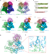
a Cryo-EM map of the 2:2 KBTBD4R313PRR-HDAC2 complex coloured by individual subunit (contour level = 0.15). BTB-BACK and Kelch regions in KBTBD4 are labelled. b Ribbon representation of the 2:2 KBTBD4R313PRR-HDAC2 structure with each subunit coloured as in a. The dimer interface α1 and β1 in the KBTBD4 BTB domain are labelled. c Domain organizations of KBTBD4WT, KBTBD4R313PRR and HDAC2. df Cryo-EM maps of UM171-induced KBTBD4WT-substrate complexes at 2:1 (KBTBD4:HDAC2, contour level = 0.08), 2:1:1 (KBTBD4:HDAC2:CoREST1, contour level = 0.08) and 2:2 (KBTBD4:HDAC2, contour level = 0.08). UM171 is coloured in yellow. Asterisk indicates the unoccupied central pocket of the KBTBD4 Kelch domain. g Ribbon representation of the KBTBD4WT homodimer binding to UM171 and HDAC2. Subunits are coloured as in df. Each HDAC2 subunit interacts with residues at sites 1 and 2 in the KBTBD4 dimer as boxed and labelled by text. The distance between the two Kelch domains is measured by the side chains of E381. The interaction interface between the N-terminus of KBTBD4 chain a and the Kelch domain of KBTBD4 chain b is indicated with a red box. h Close-up view of the interaction interface between the N-terminus of KBTBD4 chain a and the Kelch domain of KBTBD4 chain b. KBTBD4 chains are coloured as in g and shown in ribbon representation. Key interacting residues are shown in sticks.
Fig. 5
Fig. 5. Cryo-EM structure of the KBTBD4R313PRR-HDAC2 complex reveals the basis of neomorphic substrate recruitment in cancer.
a Ribbon representation of the KBTBD4R313PRR Kelch domain. The Kelch β-propeller consists of blades I to VI with each blade consisting of four antiparallel β strands (labelled A-D). Loops connecting βB and βB in blade II and IV are labelled as BC II and BC IV, respectively. The R313PRR cancer mutation in BC II is shown as red spheres. b Ribbon representation of HDAC2 binding to KBTBD4R313PRR_a at site 1 (BC II) and KBTBD4R313PRR_b at site 2 (BC IV). The second HDAC2 chain in the model is a symmetric duplication of the first and, therefore, not shown for simplicity. c Close-up view of site 1 interface. HDAC2 is shown as yellow surface and KBTBD4R313PRR_a as cyan ribbon. The key interacting arginine residues in KBTBD4R313PRR_a are shown as sticks in cyan. df Close-up views of site 1 key interfacial residues shown in sticks with the same colour scheme as c. Hydrogen bonding is indicated by dashed lines. g Close-up view of the site 2 interface. HDAC2 is shown as yellow surface and KBTBD4R313PRR_b as grey ribbon. The key interacting residues in KBTBD4R313PRR are shown as grey sticks. hj Close-up views of site 2. Key interfacial residues are shown in sticks with the same colour scheme as in g.
Fig. 6
Fig. 6. Cryo-EM structures show UM171 recruits HDAC2 to KBTBD4WT as a molecular glue.
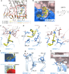
a Superposition of 2:1 and 2:2 complexes at HDAC2-interacting sites 1 and 2 in ribbon representation. UM171 is shown in stick representation. KBTBD4WT BC loops involved in both interaction sites are labelled. b Chemical structure of UM171 and a close-up view of UM171 binding at site 1 in the KBTBD4WT-HDAC2 interface. UM171 is shown in yellow sticks, KBTBD4WT and HDAC2 are shown in blue and pink as surface representations. ce Close-up views of UM171 binding showing key interfacial residues as sticks. Hydrogen bonds are indicated by dashed lines. The subunits are coloured as in b. f Surface representations show the induced-fit of UM171 bound to KBTBD4WT_a (top panel) relative to the equivalent surface in unbound KBTBD4WT_b (lower panel). Loops involved in the induced fit are labelled. g Superposition of UM171-bound KBTBD4WT_a (blue) and unbound KBTBD4WT_b (light blue) at site 1 in ribbon and stick representations. Distances are measured to show the enlarged cleft in the UM171-bound KBTBD4WT_a. h Superposition of KBTBD4WT site 2 in HDAC2-bound KBTBD4WT_b and unbound KBTBD4WT_a. KBTBD4WT chains are shown in ribbon representation. L406 in BC IV in both KBTBD4WT chains is shown in sticks and the distances between side chains is measured to demonstrate the loop movement.
Fig. 7
Fig. 7. Structural mimicry of UM171 and cancer mutant KBTBD4R313PRR.
Superposition of UM171 and KBTBD4R313PRR at site 1 (a) or site 2 (b). UM171 and key interacting residues are shown as sticks. Subunits are coloured as following: in the UM171 model, KBTBD4WT chains in blue and light blue, UM171 in yellow and HDAC2 in pink; in the KBTBD4R313PRR model, KBTBD4R313PRR in purple and light purple and HDAC2 in green.

References

    1. Jeffery, C. J. Proteins with neomorphic moonlighting functions in disease. IUBMB Life63, 489–494 (2011). - PubMed
    1. Takiar, V., Ip, C. K., Gao, M., Mills, G. B. & Cheung, L. W. Neomorphic mutations create therapeutic challenges in cancer. Oncogene36, 1607–1618 (2017). - PMC - PubMed
    1. Mo, X. et al. Systematic discovery of mutation-directed neo-protein-protein interactions in cancer. Cell185, 1974–1985.e1912 (2022). - PMC - PubMed
    1. Kliche J. et al. Proteome-scale characterisation of motif-based interactome rewiring by disease mutations. Mol Syst Biol20, 1025–1048 (2024). - PMC - PubMed
    1. Schreiber, S. L. Molecular glues and bifunctional compounds: Therapeutic modalities based on induced proximity. Cell Chem. Biol.31, 1050–1063 (2024). - PubMed

MeSH terms

LinkOut - more resources