. 2025 Apr 2;16(1):3164.
doi: 10.1038/s41467-025-58536-5.

Amyloid-reoriented enzyme catalysis

Affiliations

Amyloid-reoriented enzyme catalysis

Taka Sawazaki et al. Nat Commun. .

Abstract

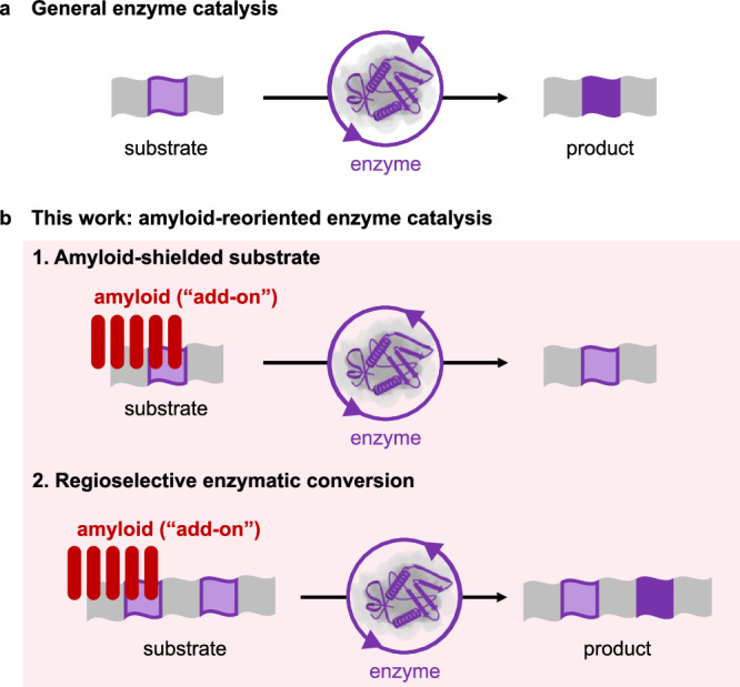
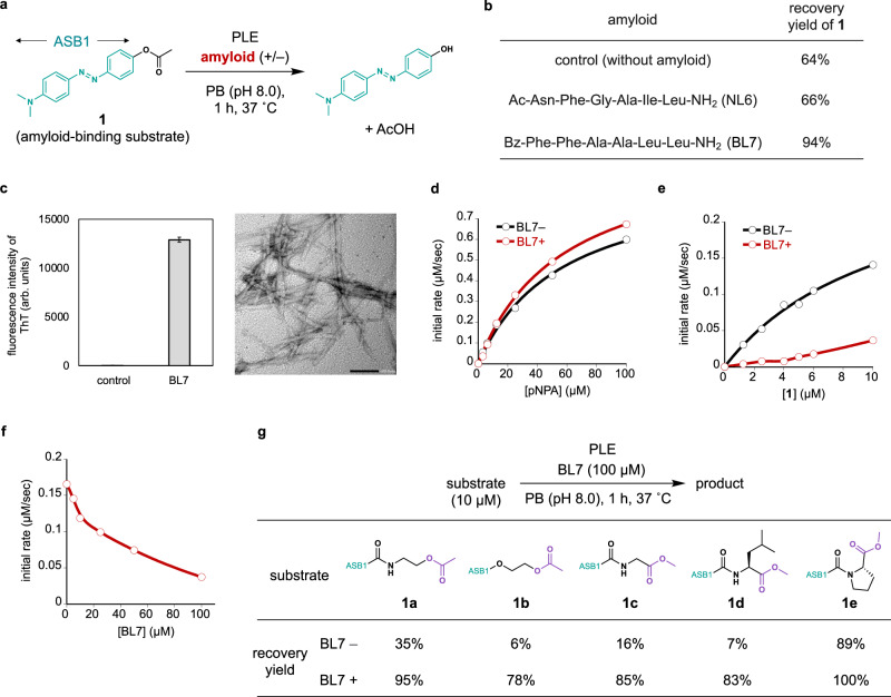
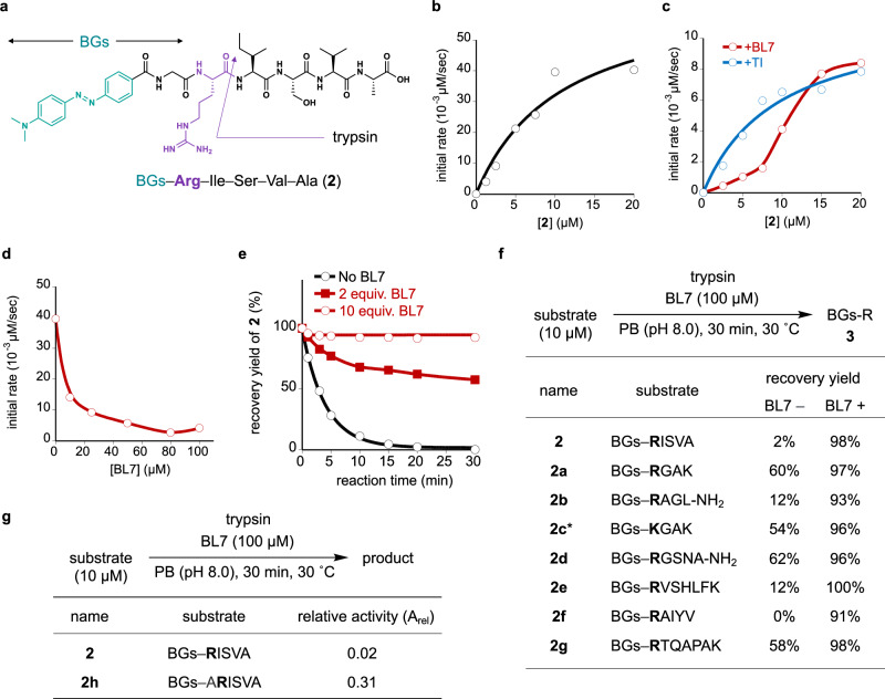
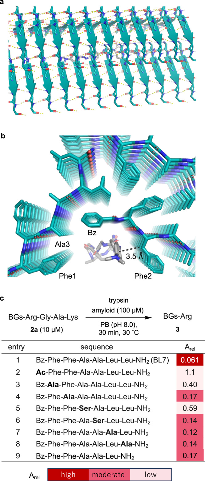
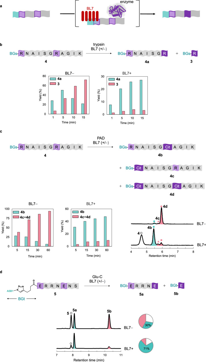
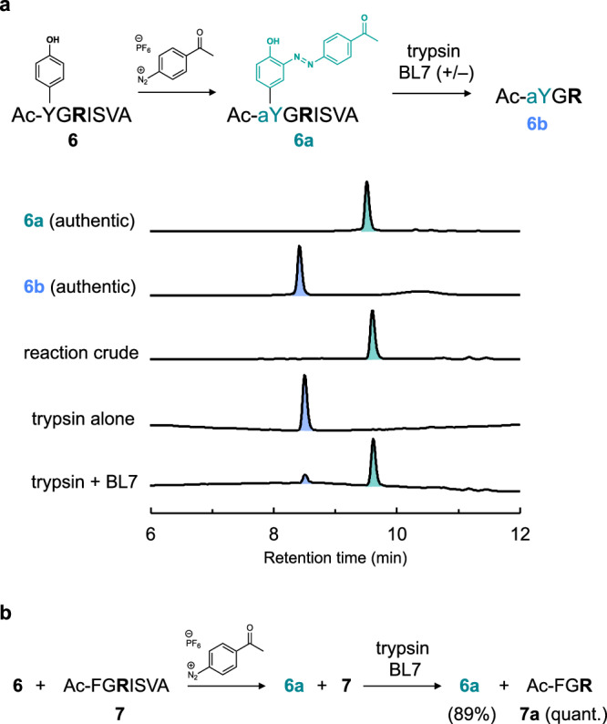
Enzyme catalysis is essential for molecular transformations. Here, we make use of amyloid, a fibrillar aggregate formed by stacking peptides with β-sheet, which offers unique selectivity in enzymatic reactions. Azo-stilbene derivative (ASB), the amyloid-recognition motif, is incorporated into the substrate, which allows the amyloid consisting of Bz-Phe-Phe-Ala-Ala-Leu-Leu-NH2 (BL7) to shield the substrates from the approaching enzyme. X-ray crystallographic analysis and structure-shielding effect relationship studies of BL7 reveal that the benzene rings present in the N-terminal benzoyl group and Phe1 side chain are particularly important for the shielding effect on the substrate. The finding results in a selective transformation system in which the reactive site close to ASB is protected by amyloid, while a site far from ASB is converted by the enzymes (trypsin, protein arginine deiminase [PAD], and Staphylococcus aureus V-8 Protease [Glu-C]). Further, the amyloid-shielded enzyme catalysis is compatible with an intact peptide, as the side chain of Tyr can be converted to the amyloid-recognizing motif. The enzymatic reactions combining amyloid provide unique selectivity for molecular transformation which may be used in diverse fields, including in synthetic chemistry.


Conflict of interest statement

Competing interests: The authors declare no competing interests.

Figures

Fig. 1
Fig. 1. Conceptual illustrations to explain amyloid-reoriented enzyme catalysis.
a General enzyme catalysis. The substrate undergoes an enzymatic transformation to give a product. b This work. (1) Amyloid-shielded substrate is no longer under the control of the enzyme, and the structure is unchanged. (2) A potential modification site is shielded by the amyloid and is therefore inaccessible to the enzyme, while the other site far from amyloid undergoes enzymatic conversion. This results in a regioselective conversion.
Fig. 2
Fig. 2. Studies on PLE-catalyzed reactions.
a Hydrolysis of ester bond in substrate 1 (10 µM) possessing amyloid-binding ASB group. PB = phosphate buffer. b Recovery yields of 1 in the reaction conditions depicted in (a) without or with 100 µM of amyloid (NL6 or BL7). c Characterization of BL7 amyloid using ThT fluorescence assay (left) and TEM image (right). For ThT fluorescence assays, the mean ± SEM of relative fluorescence intensity values from three independent experiments (i.e., n = 3) are shown. d Michaelis–Menten analysis of PLE for pNPA without or with BL7 (100 µM). e Michaelis–Menten analysis of PLE for 1 without or with BL7 (100 µM). f Initial reaction rates of PLE for 1 (10 µM) with varied concentrations of BL7 (0–100 µM). g Substrate scope of PLE-catalyzed hydrolysis without or with BL7. All recovery yields were determined using analytical high-performance liquid chromatography (HPLC).
Fig. 3
Fig. 3. Studies on trypsin-catalyzed reactions.
a Tryptic cleavage of amide bond at the C-terminal side of arginine residue in substrate 2 possessing the amyloid binding group (BGs) composed of ASB with a glycyl linker. The reaction was conducted in phosphate buffer (pH 8.0) at 30 °C. b Michaelis–Menten analysis of trypsin for 2. c Michaelis–Menten analysis of trypsin for 2 in the presence of TI (25 µM) or BL7 (100 µM). d Initial reaction rates of trypsin for 2 (10 µM) with varied concentrations of BL7. e Time-course of trypsin-catalyzed reactions of 2 (10 µM) in the absence (black line) or presence of BL7 (red line with filled squares: 20 µM, red line with white circles: 100 µM). f Substrate scope of trypsin-catalyzed reactions without or with BL7. All recovery yields were determined using analytical HPLC. *The product is BGs-K. g Comparison of the shielding effect of BL7 for 2 and 2h. Arel = (100 – recovery yield in the presence of BL7) / (100 – recovery yield in the absence of BL7).
Fig. 4
Fig. 4. Insights into the shielding effect of BL7.
a, b The three-dimensional structure of BL7 amyloid determined by X-ray crystallographic analysis and the docking study with substrate 2a. a Side view. b Cross-sectional view. These structures are shown by sticks with oxygen, nitrogen, and carbon colored in red, blue, and light green (except for carbon in 2a, which is colored in gray). c Chemical modification studies of BL7 and the shielding effect of BL7 derivatives for 2a. Arel = (100 – recovery yield in the presence of BL7 derivative)/(100 – recovery yield in the absence of BL7 derivative).
Fig. 5
Fig. 5. Regioselective transformations of peptides enabled by the shielding effect of BL7 amyloid.
a Conceptual illustration of regioselective, enzymatic transformation with amyloid. The light blue part represents the amyloid binding group (BG). The substrate portion close to the BG is shielded by amyloid and is therefore unaffected by the enzyme (remaining light purple color), whereas the substrate portion far from the BG is converted by the enzyme (changing from light to dark purple color). b Trypsin-catalyzed reaction of 4. Reaction conditions: 4 (10 µM), BL7 (none or 100 µM), and trypsin in phosphate buffer (pH 8.0) at 30 °C. c PAD-catalyzed reactions of 4. Reaction conditions: 4 (10 µM), BL7 (none or 100 µM), and PAD in HEPES buffer (pH 7.6 containing 10 mM CaCl2 and 5 mM dithiothreitol) at 37 °C. HPLC charts show the data at t = 60 min (detected at 475 nm). d Glu-C-catalyzed reactions of 5. Reaction conditions: 5 (10 µM), BL7 (none or 100 µM), and Glu-C in phosphate buffer (pH 8.0) at 37 °C. The HPLC (detected at 475 nm) and pie charts show the data at t = 24 h.
Fig. 6
Fig. 6. Binding of BL7 to BG constructed at the late stage using an intact peptide.
a Reaction scheme (upper) and associated HPLC charts (lower, detected at 350 nm). Authentic 6a and 6b were synthesized by a stepwise solid phase peptide synthesis using Ac-aY-OH. b Application to substrate-selective amide bond cleavage.

References

    1. Chen, K. & Arnold, F. H. Engineering new catalytic activities in enzymes. Nat. Catal.3, 203–213 (2020).
    1. Wang, Y. et al. Directed evolution: methodologies and applications. Chem. Rev.121, 12384–12444 (2021). - PubMed
    1. Emmanuel, M. A. et al. Photobiocatalytic strategies for organic synthesis. Chem. Rev.123, 5459–5520 (2023). - PMC - PubMed
    1. Zhang, X. et al. Enzymatic synthesis of organoselenium compounds via C‒Se bond formation mediated by sulfur carrier proteins. Nat. Synth.3, 477–487 (2024).
    1. Huffman, M. A. et al. Design of an in vitro biocatalytic cascade for the manufacture of islatravir. Science366, 1255–1259 (2019). - PubMed

LinkOut - more resources