. 2025 Apr 2;22(1):35.
doi: 10.1186/s12987-025-00639-8.

Rutin ameliorates stress-induced blood‒brain barrier dysfunction and cognitive decline via the endothelial HDAC1‒Claudin-5 axis

Affiliations

Rutin ameliorates stress-induced blood‒brain barrier dysfunction and cognitive decline via the endothelial HDAC1‒Claudin-5 axis

Zhao-Wei Sun et al. Fluids Barriers CNS. .

Abstract

Background: Emerging evidence suggests that chronic stress compromises blood‒brain barrier (BBB) integrity by disrupting brain microvascular endothelial cells (BMECs), contributing to the development of cognitive impairments. Thus, targeting the BBB is expected to be a promising treatment strategy. The biological function of rutin has been investigated in neurological disorders; however, its regulatory role in stress-induced BBB damage and cognitive decline and the underlying mechanisms remain elusive.

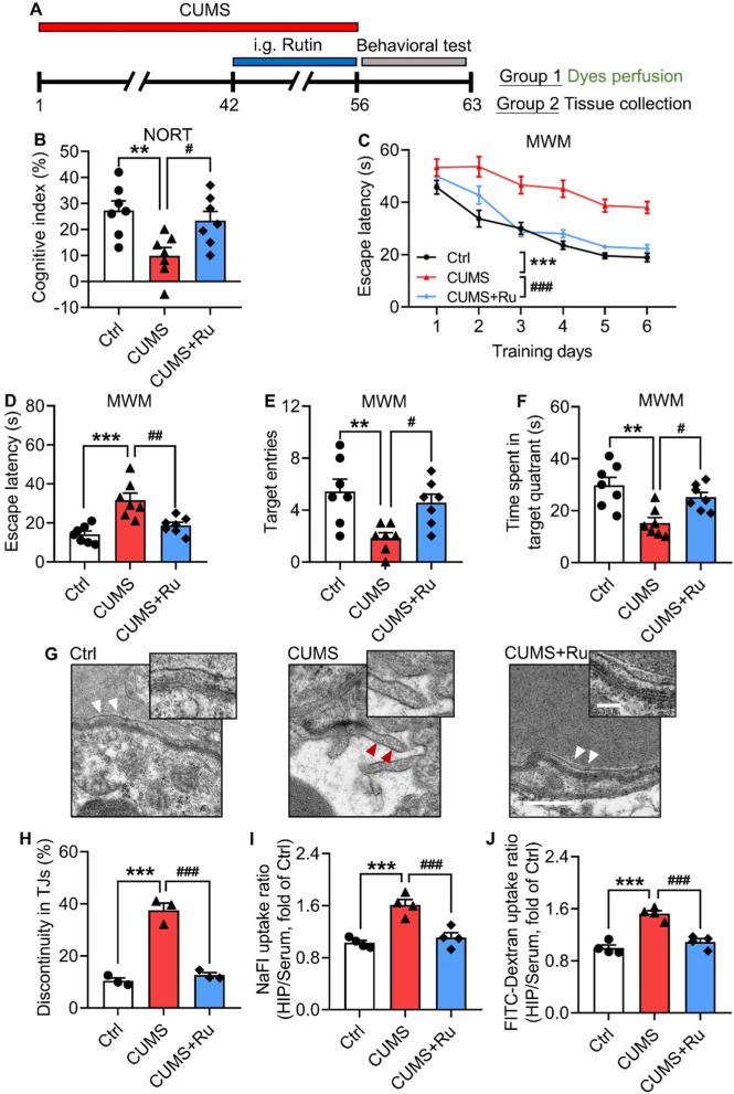
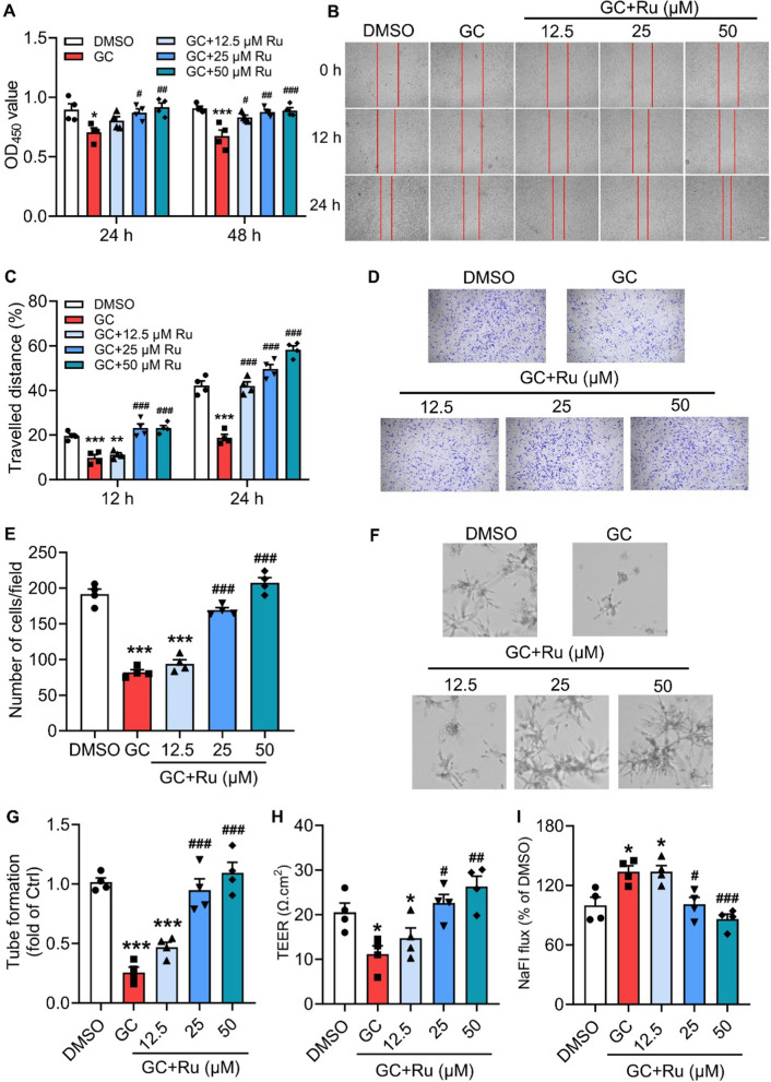
Methods: In a chronic unpredictable mild stress (CUMS) mouse model, a fluorescent dye assay and behavioral tests, including a novel object recognition test and Morris water maze, were performed to evaluate the protective effects of rutin on BBB integrity and cognition. The effects of rutin on BMEC function were also investigated in hCMEC/D3 cells (a human brain microvascular endothelial cell line) in vitro. Furthermore, the molecular mechanisms by which rutin restores BBB endothelium dysfunction were explored via RNA-seq, quantitative real-time PCR, western blotting, immunofluorescence and chromatin immunoprecipitation. Finally, biotinylated tumor necrosis factor-α (TNF-α) was employed to test the influence of rutin on the ability of circulating TNF-α to cross the BBB.

Results: We identified that rutin attenuated BBB hyperpermeability and cognitive impairment caused by the 8-week CUMS procedure. Moreover, rutin promoted the proliferation, migration and angiogenesis ability of BMECs, and the integrity of the cellular monolayer through positively regulating the expression of genes involved. Furthermore, rutin impeded histone deacetylase 1 (HDAC1) recruitment and stabilized H3K27ac to increase Claudin-5 protein levels. Ultimately, normalization of the hippocampal HDAC1‒Claudin-5 axis by rutin blocked the infiltration of circulating TNF-α into the brain parenchyma and alleviated neuroinflammation.

Conclusions: This work establishes a protective role of rutin in regulating BMEC function and BBB integrity, and reveals that rutin is a potential drug candidate for curing chronic stress-induced cognitive deficits.

Keywords: BBB; BMEC function; Claudin–5; H3K27ac modification; Rutin; Stress–induced cognitive decline.


Conflict of interest statement

Declarations. Ethics approval and consent to participate: All animal experiments and experimental procedures were authorized by the Animal Care and Use Committee at the AMS (IACUC-DWZX-2022–738). Consent for publication: Not applicable. Competing interests: The authors declare no competing interests.

Figures

Fig. 1
Fig. 1
Rutin ameliorates cognitive and BBB deficits in CUMS mice. A Experimental timeline of the CUMS procedure and rutin treatment in mice. B Cognitive index of Ctrl, CUMS and CUMS with rutin treatment (CUMS + Ru) mice in the NORT (One–way ANOVA with Tukey’s post hoc test, n = 7 mice for each group). C Escape latency to reach the platform of the mice during the training trials in the MWM (One–way ANOVA with Tukey’s post hoc test, n = 7 mice for each group). DF Escape latency (D), target entries (E) and time spent in the target quadrant (F) by the mice in the probe trial of the MWM (One–way ANOVA with Tukey’s post hoc test, n = 7 mice for each group). G Representative TEM images of TJ structure in Ctrl, CUMS and CUMS + Ru mice. Scale bar, 500 nm (white arrowheads: intact TJs; red arrowheads: discontinuous TJs). H Quantification of the percentages of discontinuous TJs in the (G) (One–way ANOVA with Tukey’s post hoc test, n = 3 mice for each group, 50–60 TJs per mouse). I, J Hippocampal permeability of the BBB to NaFI (I) and FITC–Dextran (J) in the mice (One–way ANOVA with Tukey’s post hoc test, n = 4 mice for each group). The data are presented as the mean ± SEM. **p < 0.01, ***p < 0.001, vs Ctrl; #p < 0.05, ##p < 0.01, ###p < 0.001, vs CUMS
Fig. 2
Fig. 2
Rutin promotes the integrity of cerebral microvascular endothelial cells. A Proliferation of rutin–treated hCMEC/D3 cells after 24 and 48 h determined by a CCK8 assay (One–way ANOVA with Tukey’s post hoc test, n = 4 biological replicates). B Representative images of wound healing in rutin–treated cells at 0, 12 and 24 h. Scale bar: 200 μm. C Quantification of the distances traveled during wound healing in the (B) (One–way ANOVA with Tukey’s post hoc test, n = 4 biological replicates). D Representative images of transwell assays in rutin–treated cells. Scale bar: 200 μm. E Quantification of the numbers of migrated cells in the (D) (One–way ANOVA with Tukey’s post hoc test, n = 4 biological replicates). F Representative images of tube formation in rutin–treated cells. Scale bar, 50 μm. G Quantification of the tubes in the (F) (One–way ANOVA with Tukey’s post hoc test, n = 4 biological replicates). H The TEER value of rutin-treated cells (One–way ANOVA with Tukey’s post hoc test, n = 4 biological replicates). I The flux of NaFI in rutin-treated cells (One–way ANOVA with Tukey’s post hoc test, n = 4 biological replicates). The data are presented as the mean ± SEM. *p < 0.05, **p < 0.01, ***p < 0.001, vs DMSO; #p < 0.05, ##p < 0.01, ###p < 0.001, vs GC
Fig. 3
Fig. 3
Genome–wide identification of rutin-targeting genes in hCMEC/D3 cells via whole–transcriptome RNA sequencing. A Volcano plot of the DEGs in hCMEC/D3 cells with rutin treatment. B Heatmap of the DEGs in hCMEC/D3 cells with rutin treatment. C The biological process categories of the DEGs by GO analysis. D Representative DEG mRNA levels in rutin-treated hCMEC/D3 cells determined by RT‑qPCR (One–way ANOVA with Tukey’s post hoc test, n = 3 biological replicates). The data are presented as the mean ± SEM. **p < 0.01, ***p < 0.001
Fig. 4
Fig. 4
Rutin maintains H3K27ac modification to rescue Claudin–5 expression in CUMS mice. A Representative images of Claudin–5 protein levels in the hippocampus of mice as determined by western blotting. B Quantification of the Claudin–5 protein levels in the (A) (One–way ANOVA with Tukey’s post hoc test, n = 4 biological replicates). C Representative images of H3K27ac protein levels in the hippocampus of Ctrl, CUMS and CUMS + Ru mice by western blotting. D Quantification of the H3K27ac protein levels in the (C) (One–way ANOVA with Tukey’s post hoc test, n = 4 biological replicates). E Hdac1, Hdac2 and Ep300 mRNA levels in the hippocampus of the mice (One–way ANOVA with Tukey’s post hoc test, n = 4 biological replicates). F Representative images of HDAC1 protein levels in the hippocampus of mice by western blotting. G Quantification of the HDAC1 protein levels in the (F) (One–way ANOVA with Tukey’s post hoc test, n = 4 biological replicates). H Diagram of the designed primer pairs for the indicated regions. I Enrichment of H3K27ac modifications at different sites of the Cldn5 promoter in the hippocampus of mice (One–way ANOVA with Tukey’s post hoc test, n = 3 biological replicates). J Enrichment of HDAC1 600 bp upstream from the Cldn5 TSS in the hippocampus of mice (One–way ANOVA with Tukey’s post hoc test, n = 3 biological replicates). The data are presented as the mean ± SEM. *p < 0.05, ***p < 0.001, vs Ctrl; #p < 0.05, ##p < 0.01, ###p < 0.001, vs CUMS
Fig. 5
Fig. 5
Rutin enhances the binding of C/EBPα to the Cldn5 promoter in CUMS mice. A Representative images of C/EBPα protein levels in the hippocampus of Ctrl, CUMS and CUMS + Ru mice by western blotting. B Quantification of the C/EBPα protein levels in the (A) (One–way ANOVA with Tukey’s post hoc test, n = 4 biological replicates). C Diagram of the predicted binding site of C/EBPα to the Cldn5 promoter. D Enrichment of C/EBPα 600 bp upstream from the Cldn5 TSS in the hippocampus of mice (One–way ANOVA with Tukey’s post hoc test, n = 4 biological replicates). E Enrichment of C/EBPα 600 bp upstream from the Cldn5 TSS in CUMS mice with pyroxamide (Pyro) treatment (One–way ANOVA with Tukey’s post hoc test, n = 4 biological replicates). F Enrichment of C/EBPα 600 bp upstream from the Cldn5 TSS in hCMEC/D3 cells treated with 0, 10 and 20 μM Pyro (One–way ANOVA with Tukey’s post hoc test, n = 4 biological replicates). The data are presented as the mean ± SEM. *p < 0.05, **p < 0.01, ***p < 0.001, vs Ctrl; ###p < 0.001, vs CUMS
Fig. 6
Fig. 6
Rutin inhibits the passage of circulating TNF–α into the hippocampus of CUMS mice. A, B TNF–α protein levels in the blood (A) and hippocampus (B) of Ctrl, CUMS and CUMS + Ru mice (One–way ANOVA with Tukey’s post hoc test, n = 6 mice for each group). C Experimental timeline of CUMS, rutin treatment and biotinylated TNF–α detection. D Representative images of DAPI (blue), biotinylated TNF–α (green) and Claudin–5 (red) immunostaining in the hippocampus of Ctrl, CUMS and CUMS + Ru mice. Scale bar, 2 μm. E Representative images of DAPI (blue), biotinylated TNF–α (green) and CD31 (red) immunostaining in the hippocampus of Ctrl, CUMS and CUMS + Ru mice. Scale bar, 2 μm. F Schematic showing that rutin treatment promoted endothelial cell proliferation, migration and angiogenesis. Moreover, rutin inhibited HDAC1–dependent H3K27 deacetylation to facilitate the transcriptional activity of C/EBPα at the Cldn5 promoter. As a result, rutin reversed chronic stress–induced Cldn5 loss and BBB breakdown. Furthermore, the restoration of rutin on the BBB blocked the infiltration of circulating TNF–α into the hippocampus and attenuated cognitive dysfunction. The data are presented as the mean ± SEM. ***p < 0.001, vs Ctrl; ###p < 0.001, vs CUMS

References

    1. Saeedi M, Rashidy-Pour A. Association between chronic stress and Alzheimer’s disease: therapeutic effects of Saffron. Biomed Pharmacother. 2021;133:110995. - PubMed
    1. Lupien SJ, Juster RP, Raymond C, Marin MF. The effects of chronic stress on the human brain: from neurotoxicity, to vulnerability, to opportunity. Front Neuroendocrinol. 2018;49:91–105. - PubMed
    1. Escher CM, Sannemann L, Jessen F. Stress and Alzheimer’s disease. J Neural Transm. 2019;126:1155–61. - PubMed
    1. Lyons CE, Bartolomucci A. Stress and Alzheimer’s disease: a senescence link? Neurosci Biobehav Rev. 2020;115:285–98. - PMC - PubMed
    1. Zhang YL, Wang J, Zhang ZN, Su Q, Guo JH. The relationship between amyloid-beta and brain capillary endothelial cells in Alzheimer’s disease. Neural Regen Res. 2022;17:2355–63. - PMC - PubMed

MeSH terms

LinkOut - more resources