. 2025 Apr;29(7):e70519.
doi: 10.1111/jcmm.70519.

NUPR1 Promotes Radioresistance in Colorectal Cancer Cells by Inhibiting Ferroptosis

Affiliations

NUPR1 Promotes Radioresistance in Colorectal Cancer Cells by Inhibiting Ferroptosis

Yimin Fang et al. J Cell Mol Med. 2025 Apr.

Abstract

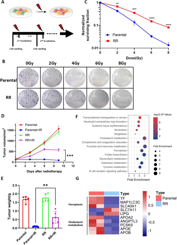
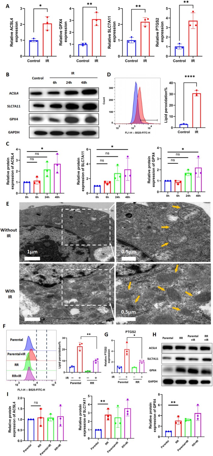
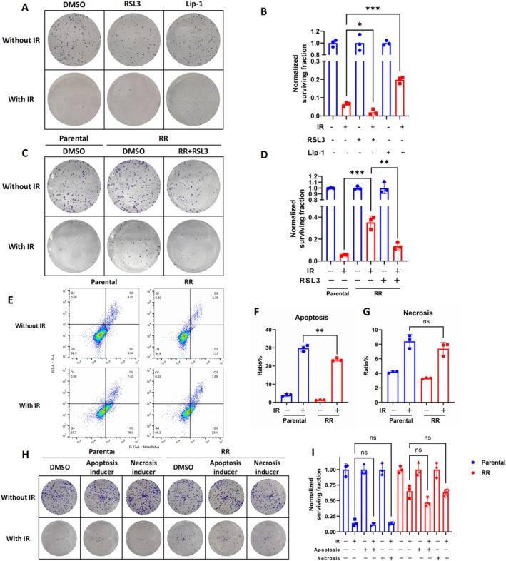
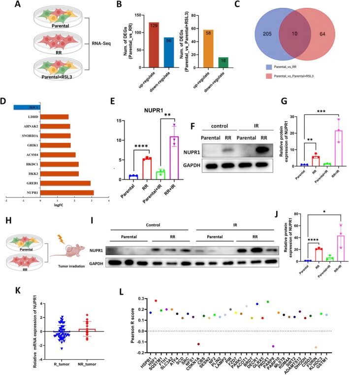
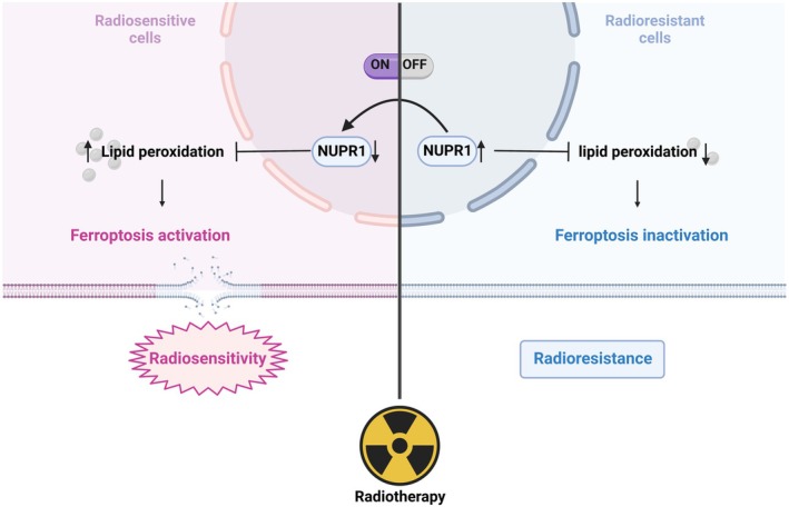
Radioresistance is a major clinical challenge and the underlying mechanism has not been thoroughly elucidated. In this study, a radioresistant (RR) cell line is established to explore the transcriptomic signatures of radioresistance in colorectal cancer (CRC). KEGG enriched pathway analysis demonstrated that ferroptosis is inactivated in RR cells. Further detection confirmed that radiotherapy can promote ferroptosis, and ferroptosis inactivation is one of the hallmarks of radioresistance in CRC. What's more, induction of ferroptosis can restore the radiosensitivity of CRC cells. Then, we performed RNA sequencing to compare gene expression between parental and RR cells, and cells pretreated with or without RSL3. Via high-throughput screening, NUPR1 was identified as a potential candidate for ferroptosis-mediated radioresistance in CRC. CRC cells can acquire radiation resistance by NUPR1-mediated ferroptosis suppression in the NUPR1-overexpressing cell line. More importantly, ZZW-115, an NUPR1 inhibitor, can sensitise RR cells to radiotherapy. Overall, our findings identify ferroptosis inactivation linked with resistance to radiotherapy. Besides, NUPR1 can promote radiation resistance by inhibiting ferroptosis, and targeting NUPR1 may be a potential strategy to relieve radioresistance associated with ferroptosis in CRC.

Keywords: NUPR1; colorectal cancer; ferroptosis; radiotherapy resistance.


Conflict of interest statement

The authors declare no conflicts of interest.

Figures

FIGURE 1
FIGURE 1
Transcriptomic signatures of radioresistant CRC cells. (A) Flow chart of induction of RKO‐RR cells. (B, C) Clonogenic survival curves for RKO cells and RKO‐IRresist cells receiving radiotherapy (from 0 to 8 Gy). The survival data were normalised to those of unirradiated cells. n = 3, **p < 0.01, ***p < 0.001, ****p < 0.0001 by two‐tailed unpaired Student's t‐test. (D, E) In subcutaneous tumorigenesis model, RKO and RR tumour growth (D) and tumour weight (E) after radiotherapy (8 Gy twice). n = 5, **p < 0.01, ***p < 0.001 by two‐tailed unpaired Student's t‐test. (F) Kyoto Encyclopedia of Genes (KEGG) pathway enrichment analysis. (G) Heatmap of RNA sequencing between Parental cells and RR cells.
FIGURE 2
FIGURE 2
Ferroptosis inactivation is one of the hallmarks of radioresistance in CRC. (A) qRT‐PCR analysis of ACSL4, GPX4, SLC7A11 and PTGS2 in RKO cells at 24 h after receiving radiotherapy (6 Gy). n = 3, *p < 0.05, **p < 0.01, ****p < 0.0001 by two‐tailed unpaired Student's t‐test. (B) WB detected the protein levels of ACSL4, SLC7A11 and GPX4 in RKO cells at 6, 24 and 48 h after receiving radiotherapy (6 Gy). (C) Bar chart shows relative protein expression of ACSL4, SLC7A11 and GPX4 in RKO cells at 6, 24 and 48 h after receiving radiotherapy (6 Gy). n = 3, *p < 0.05 by ANOVA. (D) Lipid peroxidation level of RKO cells receiving radiotherapy (6 Gy). Bar charts show relative levels of lipid peroxidation in RKO cells. n = 3, ****p < 0.0001 by two‐tailed unpaired Student's t‐test. (E) Transmission electron microscopy images of RKO cells at 24 h received radiotherapy (6 Gy). Yellow arrows: mitochondria. (F) Lipid peroxidation levels were measured in RKO cells and RR cells at 24 h after radiotherapy (6 Gy). Bar chart shows relative levels of lipid peroxidation in RKO cells and RR cells. n = 3, **p < 0.01 by two‐tailed unpaired Student's t‐test. (G) qRT‐PCR analysis of PTGS2 in RKO cells and RR cells at 24 h after radiotherapy (6Gy). n = 3, *p < 0.05 by two‐tailed unpaired Student's t‐test. (H) WB detected the protein levels of ACSL4, SLC7A11 and GPX4 in RKO cells and RR cells at 24 h after receiving radiotherapy (6Gy). (I) Bar chart shows relative protein expression of ACSL4, SLC7A11 and GPX4 in RKO cells and RR cells at 24 h after receiving radiotherapy (6Gy). n = 3, **p < 0.01 by ANOVA.
FIGURE 3
FIGURE 3
Induction of ferroptosis restores the radiation sensitivity of CRC cells. (A, B) Clonogenic survival of RKO cells pretreated with 100 nM RSL3, 5 μM lip‐1 or DMSO for 24 h followed by radiotherapy (4 Gy). The survival data were normalised to those of unirradiated cells. n = 3, ***p < 0.001,*p < 0.05 by two‐tailed unpaired Student's t‐test. (C, D) Clonogenic survival of RR cells that were pretreated with 100 nM RSL3 or DMSO for 24 h followed by radiotherapy (4 Gy). The survival data were normalised to those of unirradiated cells. n = 3, ***p < 0.001, **p < 0.01 by two‐tailed unpaired Student's t‐test. (E, F, G) Apoptosis and necrosis levels were measured in RKO and RR cells at 24 h after radiotherapy (6Gy). n = 3, **p < 0.01 by two‐tailed unpaired Student's t‐test. (H, I) Clonogenic survival of RKO and RR cells pretreated with apoptosis inducer (TNF‐α and SM‐164) and necrosis inducer (TNF‐α, SM‐164 and Z‐VAD‐FMK) for 24 h followed by radiotherapy (4 Gy). The survival data were normalised to those of unirradiated cells, n = 3.
FIGURE 4
FIGURE 4
Identification of NUPR1 potential for ferroptosis‐mediated radioresistance in CRC. (A) Flow Chart of RNA Sequencing. (B) The DEGs between Parental cells and RR cells and DEGs between RKO cells and RKO cells treated with 1 μM RSL3. (C) Venn diagram to identify DEGs between parental cells and RR cells that were correlated with ferroptosis. (D) The differential expression of those 10 genes in RR cells and RKO cells. (E) qRT‐PCR analysis of NUPR1 in RKO cells and RR cells at 24 h after radiotherapy (6 Gy). n = 3, **p < 0.01, ****p < 0.0001 by two‐tailed unpaired Student's t‐test. (F) WB detected the protein levels of NUPR1 in RKO cells and RR cells at 24 h after radiotherapy (6 Gy). (G) Bar chart shows relative protein expression of NUPR1 in RKO cells and RR cells at 24 h after radiotherapy (6 Gy). n = 3, **p < 0.01, ***p < 0.001 by ANOVA. (H–I) WB detected the protein levels of NUPR1 in RKO cells and RR cells with or without radiotherapy in subcutaneous tumorigenesis model. (J) Bar chart shows relative protein expression of NUPR1 in RKO cells and RR cells with or without radiotherapy in subcutaneous tumorigenesis model. n = 3, *p < 0.05, ****p < 0.0001 by ANOVA. (K) The expression of NUPR1 in rectal cancer patients receiving neoadjuvant chemoradiation in the TCGA datasets. (L) The relationship between NUPR1 and ferroptosis suppressors.
FIGURE 5
FIGURE 5
NUPR1 promotes radioresistance in CRC cells by inhibiting ferroptosis. (A) WB was used to verify the expression of NUPR1 in RKO cells and RKO‐NUPR1 cells. B Bar chart shows relative protein expression of NUPR1 in RKO cells and RKO‐NUPR1 cells. n = 3, **p < 0.01 by two‐tailed unpaired Student's t‐test. (C) Expression of PTGS2 in RKO cells, RR cells and RKO‐NUPR1 cells 24 h after radiotherapy (6 Gy). n = 3, **p < 0.01 by two‐tailed unpaired Student's t‐test. (D) Lipid peroxidation levels were measured in RKO cells, RR cells, and RKO‐NUPR1 cells at 24 h after radiotherapy (6 Gy). Bar chart shows relative levels of lipid peroxidation in RKO cells, RR cells and RKO‐NUPR1 cells. n = 3, ***p < 0.001 by two‐tailed unpaired Student's t‐test. (E, F) Clonogenic survival of RKO cells, RR cells and RKO‐NUPR1 cells receiving radiotherapy (4 Gy). The survival data were normalised to those of unirradiated cells. n = 3, *p < 0.05 by two‐tailed unpaired Student's t‐test. (G) Lipid peroxidation levels were measured in RKO cells and RR cells pretreated with 1 μM ZZW‐115 at 24 h after radiotherapy (6 Gy). Bar chart shows relative levels of lipid peroxidation in RKO cells, RR cells, and RKO‐NUPR1 cells. n = 3, **p < 0.01 by two‐tailed unpaired Student's t‐test. (H–I) Clonogenic survival of RKO cells RR cells and RR cells pretreated with 1 μM ZZW‐115 for 24 h followed by radiotherapy (4 Gy). The survival data were normalised to those of unirradiated cells. n = 3, **p < 0.01, *p < 0.05 by two‐tailed unpaired Student's t‐test. (J, K) Kaplan–Meier OS (I) and DFS (J) curves for patients in the high‐NUPR1 group (N = 81) and low‐NUPR1 group (N = 61).
FIGURE 6
FIGURE 6
A working model of the mechanism by which NUPR1 can promote radioresistance in colorectal cancer cells by inhibiting ferroptosis.

Similar articles

References

    1. “Erratum: Global Cancer Statistics 2018: GLOBOCAN Estimates of Incidence and Mortality Worldwide for 36 Cancers in 185 Countries,” CA: A Cancer Journal for Clinicians 70, no. 4 (2020): 313. - PubMed
    1. Sauer R., Liersch T., Merkel S., et al., “Preoperative Versus Postoperative Chemoradiotherapy for Locally Advanced Rectal Cancer: Results of the German CAO/ARO/AIO‐94 Randomized Phase III Trial After a Median Follow‐Up of 11 Years,” Journal of Clinical Oncology 30, no. 16 (2012): 1926–1933. - PubMed
    1. Bahadoer R. R., Dijkstra E. A., van Etten B., et al., “Short‐Course Radiotherapy Followed by Chemotherapy Before Total Mesorectal Excision (TME) Versus Preoperative Chemoradiotherapy, TME, and Optional Adjuvant Chemotherapy in Locally Advanced Rectal Cancer (RAPIDO): A Randomised, Open‐Label, Phase 3 Trial,” Lancet Oncology 22, no. 1 (2021): 29–42, 10.1016/S1470-2045(20)30555-6. - DOI - PubMed
    1. Ngan S. Y., Burmeister B., Fisher R. J., et al., “Randomized Trial of Short‐Course Radiotherapy Versus Long‐Course Chemoradiation Comparing Rates of Local Recurrence in Patients With T3 Rectal Cancer: Trans‐Tasman Radiation Oncology Group Trial 01.04,” Journal of Clinical Oncology 30, no. 31 (2012): 3827–3833. - PubMed
    1. Zhu J., Liu A., Sun X., et al., “Multicenter, Randomized, Phase III Trial of Neoadjuvant Chemoradiation With Capecitabine and Irinotecan Guided by UGT1A1 Status in Patients With Locally Advanced Rectal Cancer,” Journal of Clinical Oncology 38, no. 36 (2020): 4231–4239. - PMC - PubMed

MeSH terms

Substances