Case Reports
. 2025 Jan 16:39:e02162.
doi: 10.1016/j.idcr.2025.e02162. eCollection 2025.

Necrotizing soft tissue infection due to Nocardia brasiliensis in an immunocompetent host

Affiliations
Case Reports

Necrotizing soft tissue infection due to Nocardia brasiliensis in an immunocompetent host

Malleswari Ravi et al. IDCases. .

Abstract

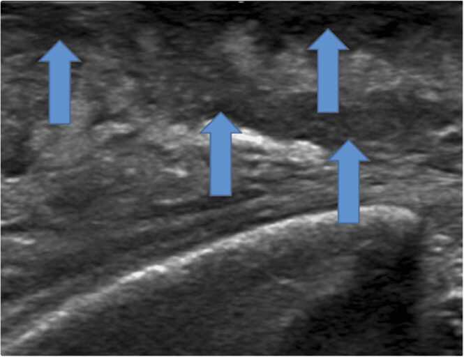
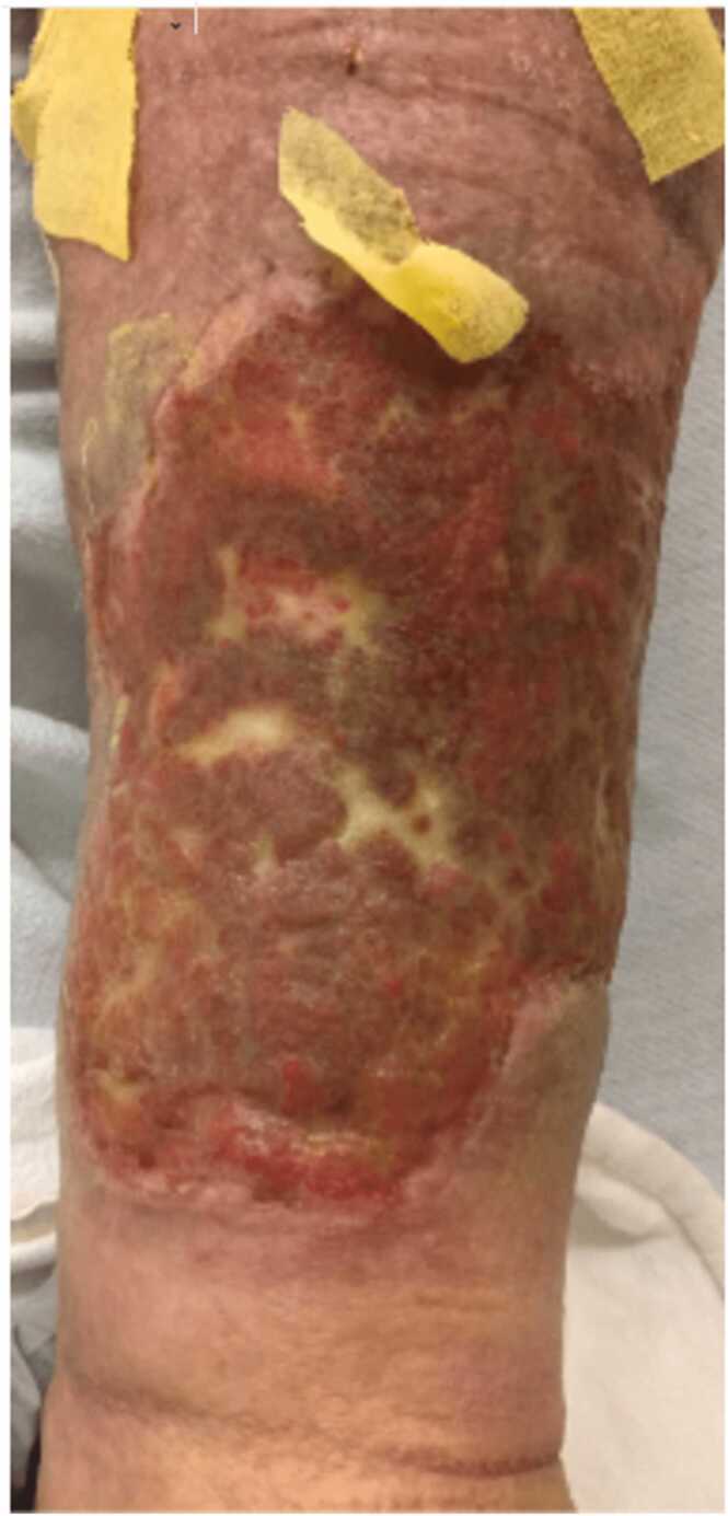
A male in early 70's with no known immunocompromising conditions developed progressive swelling, pain, and erythema of his left lower extremity (LLE) over 2 weeks following multiple abrasions to his LLE by a thorny bush while working in a brush field. He was found to have multiple purple bullae on the anterior surface of the distal half of the LLE, swelling, induration, and exquisite tenderness along with leukocytosis of 17,000 cells per milliliter, and elevated creatinine 1.75 mg/dl. and non loculated fluid collections under the cutaneous lesions on ultrasound. During excisional debridement, liquefied skin and subcutaneous tissue and necrotic subcutaneous fat were seen. Tissue cultures grew Nocardia brasiliensis (N.brasiliensis). Minocycline was given for 3 months due to renal dysfunction with Trimethoprim-sulfamethoxazole (TMP-SMX). He had skin grafting of the leg wound and had complete healing of his wound. N. brasiliensis is an infrequent cause of primary necrotizing skin infections in immunocompetent people.

Keywords: Immunocompetent; Necrotizing; Nocardia brasiliensis; Soft tissue infection.


Conflict of interest statement

The authors declare that they have no known competing financial interests or personal relationships that could have appeared to influence the work reported in this paper.

Figures

Fig. 1
Fig. 1
Arrows pointing to nodular skin lesions that coalesced to form bullae. The figure shows the patient's left leg with nodular purple skin lesions that coalesced over about 48 hours into bullae; the leg was red and with pitting edema extending from below the knee to involve the ankle and foot.
Fig. 2
Fig. 2
Ultrasound of the left leg with arrows pointing to ill-defined irregular non-loculated hypoechoic areas in the subcutaneous tissues of the leg.
Fig. 3
Fig. 3
Shows left leg post-surgical wound taken within the operating room immediately after surgical debridement of the left leg.
Fig. 4
Fig. 4
Shows the patient's leg wound at 2 months after skin grafting.

References

    1. Ricci J.A., Weil A.A., Eberlin K.R. Necrotizing cutaneous nocardiosis of the hand: a case report and review of literature. J Hand Microsurg. 2015;7(1):224–227. doi: 10.1007/s12593-015-0173-7. - DOI - PMC - PubMed
    1. Conville P.S., Brown-Elliott B.A., Smith T., Zelazny A.M. The complexities of nocardia taxonomy and identification. J Clin Microbiol. 2018;56(1) doi: 10.1128/JCM.01419-17. - DOI - PMC - PubMed
    1. Acevedo C.T., Imkamp F., Marques Maggio E., Brugger S.D. Primary cutaneous nocardiosis of the head and neck in an immunocompetent patient. BMJ Case Rep. 2021;14(5) doi: 10.1136/bcr-2020-241217. - DOI - PMC - PubMed
    1. Dodiuk-Gad R., Cohen E., Ziv M., et al. Cutaneous nocardiosis: report of two cases and review of the literature. Int J Dermatol. 2010;49(12):1380–1385. doi: 10.1111/j.1365-4632.2010.04554.x. - DOI - PubMed
    1. Wilson J.W. Nocardiosis: updates and clinical overview. Mayo Clin Proc. 2012;87(4):403–407. doi: 10.1016/j.mayocp.2011.11.016. - DOI - PMC - PubMed

Publication types

LinkOut - more resources