. 2025 Mar 19:49:437-455.
doi: 10.1016/j.bioactmat.2025.02.008. eCollection 2025 Jul.

Protein to biomaterials: Unraveling the antiviral and proangiogenic activities of Ac-Tβ1-17 peptide, a thymosin β4 metabolite, and its implications in peptide-scaffold preparation

Affiliations

Protein to biomaterials: Unraveling the antiviral and proangiogenic activities of Ac-Tβ1-17 peptide, a thymosin β4 metabolite, and its implications in peptide-scaffold preparation

Khandoker Asiqur Rahaman et al. Bioact Mater. .

Abstract

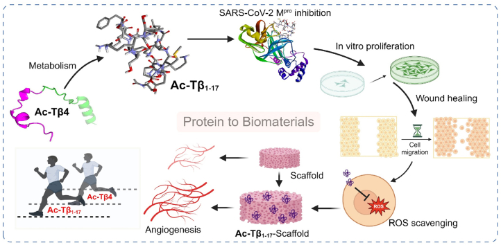
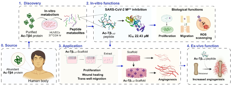
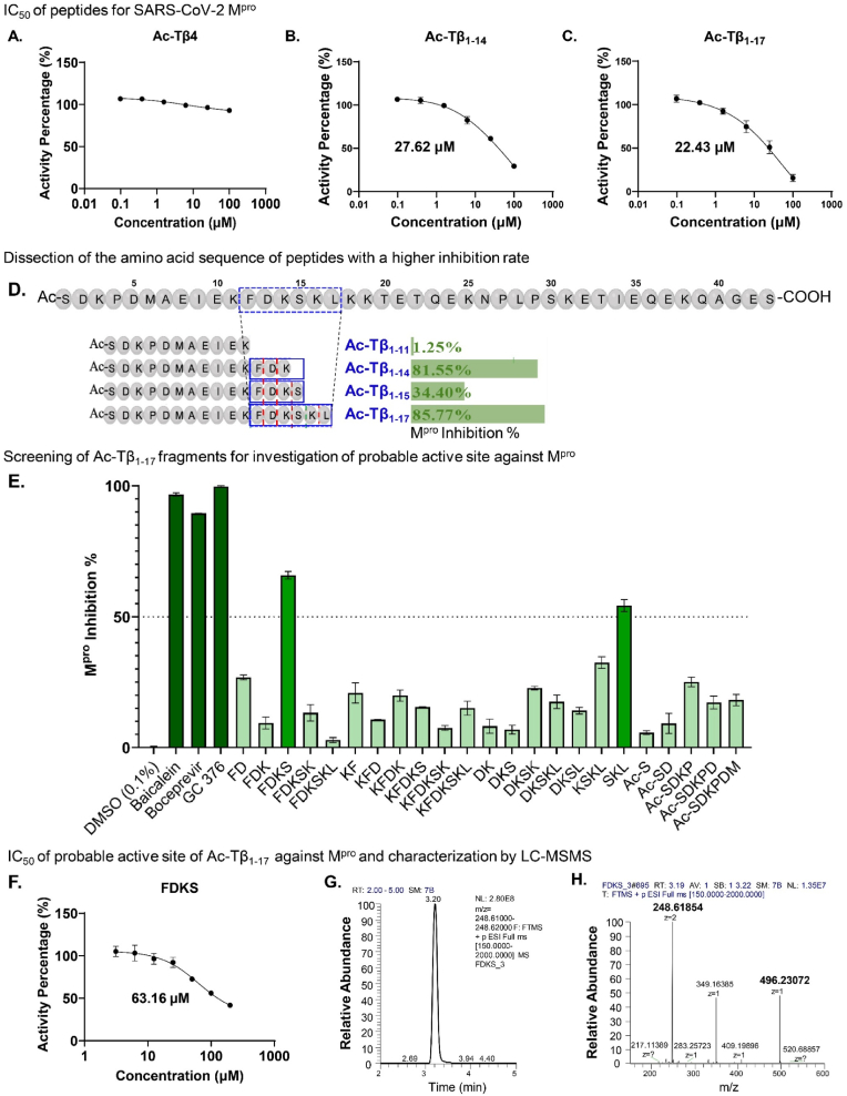
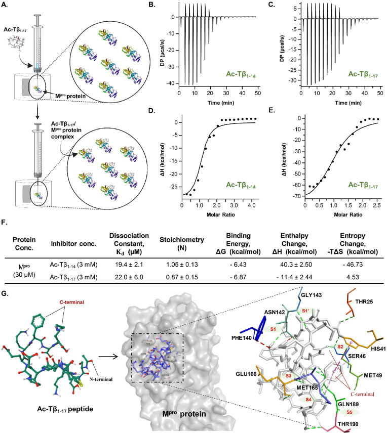
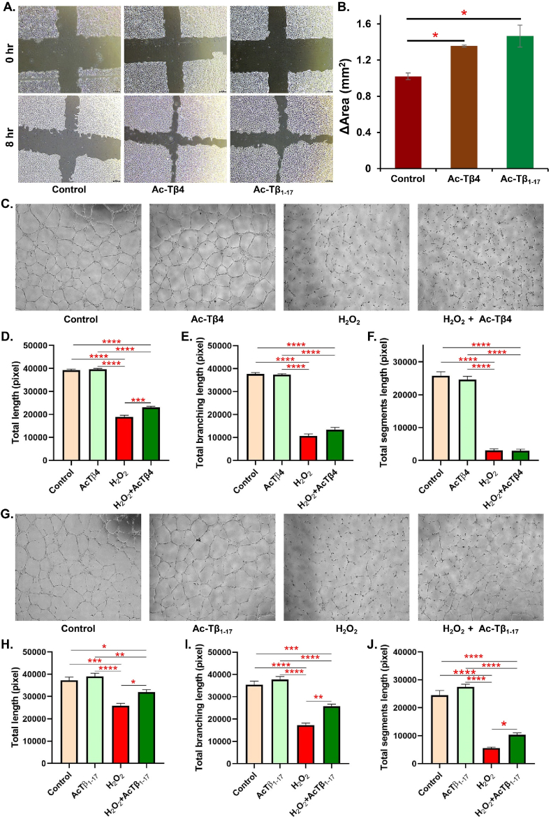
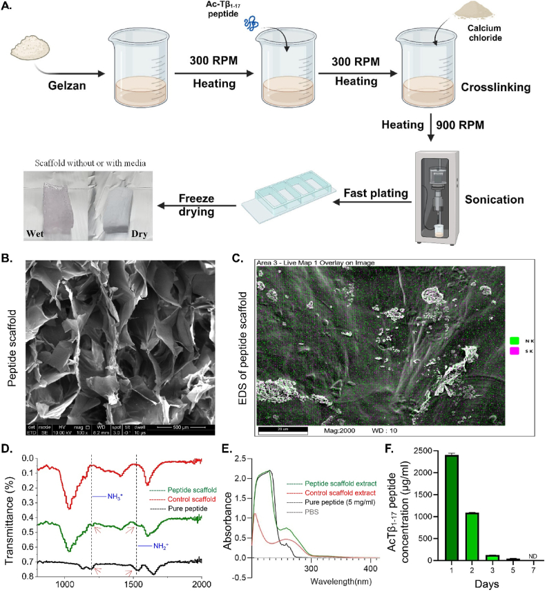
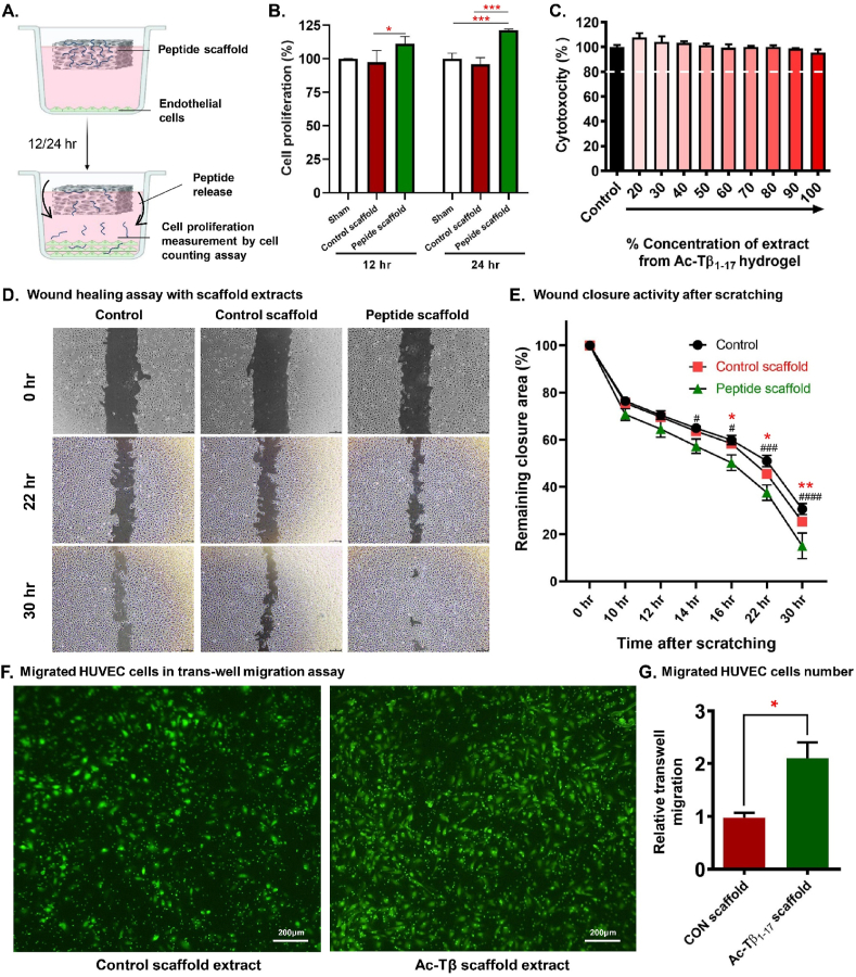
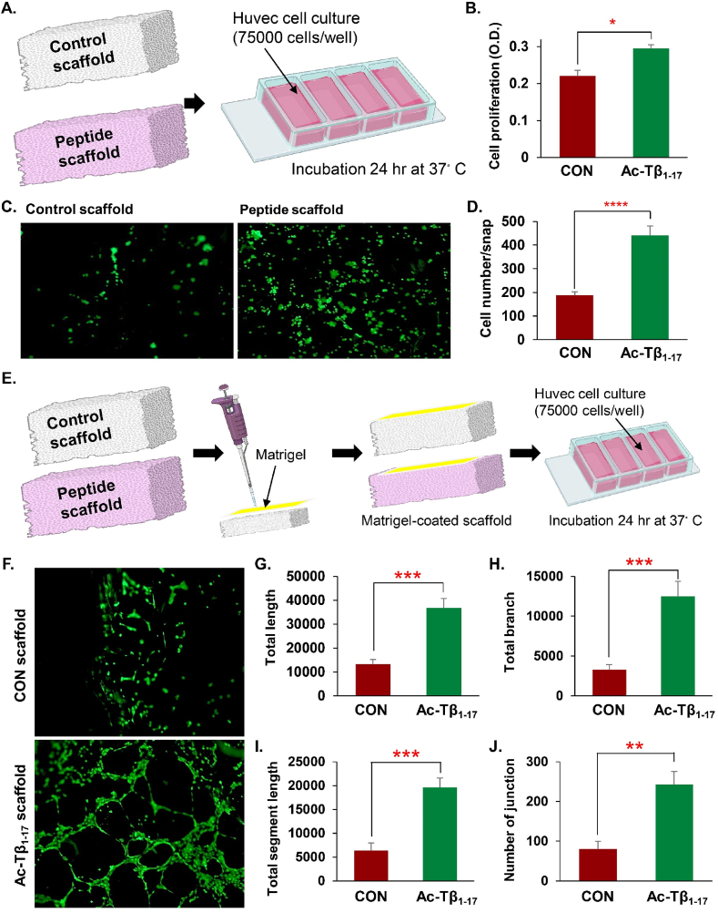
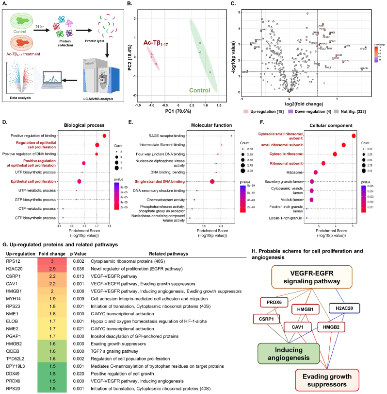
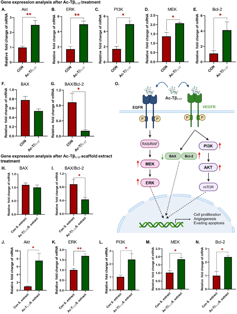
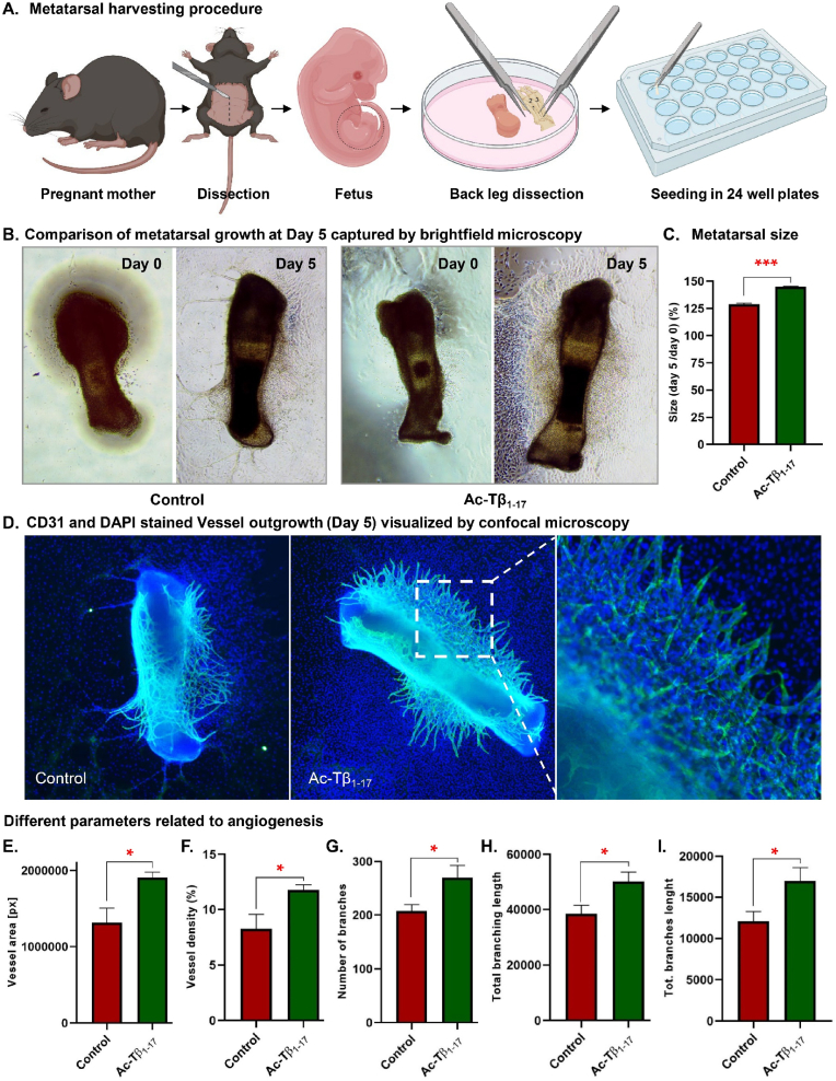
Peptide metabolites are emerging biomolecules with numerous possibilities in biomaterial-based regenerative medicine due to their inherent bioactivities. These small, naturally occurring compounds are intermediates or byproducts of larger proteins and peptides, and they can have profound effects, such as antiviral therapeutics, proangiogenic agents, and regenerative medicinal applications. This study is among the first to focus on using thymosin β4 protein-derived metabolites to pioneer novel applications for peptide metabolites in biomaterials. This study found that the novel peptide metabolite acetyl-thymosin β4 (amino acid 1-17) (Ac-Tβ1-17) exhibited significant protease inhibition activity against SARS-CoV-2, surpassing its precursor protein. Additionally, Ac-Tβ1-17 demonstrated beneficial effects, such as cell proliferation, wound healing, and scavenging of reactive oxygen species (ROS) in human umbilical vein endothelial cells (HUVEC). Integrating Ac-Tβ1-17 into a peptide-based scaffold facilitated cell growth and angiogenesis inside the scaffold and through gradual release into the surrounding environment. The Ac-Tβ1-17 peptide treatment induced significant biochemical responses in HUVEC, increasing Akt, ERK, PI3K, MEK, and Bcl-2 gene expression and proangiogenic proteins. Ac-Tβ1-17 peptide treatment showed similar results in ex vivo by enhancing mouse fetal metatarsal growth and angiogenesis. These findings highlight the potential of natural protein metabolites to generate biologically active peptides, offering a novel strategy for enhancing biomaterial compatibility. This approach holds promise for developing therapeutic biomaterials using peptide metabolites, presenting exciting prospects for future research and applications.

Keywords: Ac-Tβ1-17 peptide; Antiviral peptide; Peptide metabolites; Peptide-based biomaterial; Regenerative medicine; Thymosin β4.


Conflict of interest statement

Hyung-Seop Han is an editorial board member for Bioactive Materials and was not involved in the editorial review or the decision to publish this article. The authors declare the following personal relationships which may be considered as potential competing interests: Hyung-Seop Han is currently employed by Elecell Corporation.

Figures

Image 1
Graphical abstract
Fig. 1
Fig. 1
Illustration of the use of protein-derived peptide metabolites for application in biomaterials. The human body produces the Ac-Tβ4 protein, which, when metabolized in vitro, produces the Ac-Tβ1-17 peptide. This research demonstrated that Ac-Tβ1-17 peptide, a metabolite of Ac-Tβ4 protein, exhibits amplified antiviral and proangiogenic activities compared to the parent Ac-Tβ4 protein and has implications in peptide-scaffold preparation.
Fig. 2
Fig. 2
Exploring the Ac-Tβ1-17 sequence for identifying potential active sites targeting Mpro. The half-maximal inhibitory concentration (IC50) of the parent protein and two metabolite peptides against SARS-CoV-2 Mpro are determined (A–C). Dissection of the amino acid sequence of peptides in silico compared to the higher inhibition rate in vitro indicated the significance of the FDKSKL part (D). In vitro screening of Ac-Tβ1-17 fragments to assess probable active sites against Mpro. FDKS and SKL exhibited >50 % inhibition activity (E). The IC50 of FDKS peptide against SARS-CoV-2 Mpro is assessed (F). The structure of the FDKS peptide is analyzed using liquid chromatography-tandem mass spectrometry (LC-MS/MS) (G & H).
Fig. 3
Fig. 3
Isothermal titration calorimetry (ITC) analysis and molecular docking of Ac-Tβ1-14 and Ac-Tβ1-17. Diagrammatic representation of the ITC process (A). Representative thermodynamic profiles of Ac-Tβ1-14 and Ac-Tβ1-17 binding with SARS-CoV-2 Mpro in solution (B, C). The one-site theoretical binding model is fitted to the resulting data (D, E). Thermodynamic parameters of the interaction between peptides and Mpro (F). Molecular docking results of Ac-Tβ1-17 and Mpro. The dashed line indicates polar interactions between Ac-Tβ1-17 and Mpro (G).
Fig. 4
Fig. 4
Cytotoxicity screening of Ac-Tβ4 and its metabolites in fibroblasts (A), human umbilical vein endothelial cells (HUVECs) (B), and red blood cells (RBCs) (C). Treatment with Ac-Tβ4, Ac-Tβ1-14, and Ac-Tβ1-17 at various doses did not exhibit any toxicity with cell viability above 90 % dash line (A–B). Incubation of Ac-Tβ4 protein, Ac-Tβ1-14, and Ac-Tβ1-17 peptides with rat RBCs demonstrated no hemolytic activity (OD values < 1.5 % compared to 0.1 % sodium dodecyl sulfate [SDS]). Ac-Tβ1-17 peptides with rat RBCs are illustrated in the inset compared to 0.1 % SDS (C). The data are presented as the mean ± SEM, with three samples per group.
Fig. 5
Fig. 5
Wound healing and reactive oxygen species (ROS) scavenging activity of Ac-Tβ1-17 peptide. Microscopic images of wounds at 0 and 8 h post-treatment with 25 μg/mL of Ac-Tβ4 protein and Ac-Tβ1-17 peptide (A). The wound healing activity is measured using ImageJ, and comparing the area covered by regular, 25 μg/mL dose of Ac-Tβ4 protein and Ac-Tβ1-17 peptide. Asterisk (∗) indicates significant differences compared with the control group (B). ROS inhibition by Ac-Tβ4 protein under simultaneous H2O2 treatment (C–F). ROS inhibition by Ac-Tβ1-17 peptide under simultaneous H2O2 treatment (G–J). The data are presented as the mean ± SEM, with three samples per group. A p-value is considered statistically significant for all tests (ANOVA; ∗p < 0.05, ∗∗p < 0.01, ∗∗∗p < 0.001, and ∗∗∗∗p < 0.0001).
Fig. 6
Fig. 6
Characterization of Ac-Tβ1-17 scaffolds. Preparation of Ac-Tβ1-17-integrated scaffold (A). Surface assessment of Ac-Tβ1-17 scaffold using scanning electron microscopy (B). EDS characterization of Ac-Tβ1-17 scaffold using SEM for nitrogen and sulfur integration on the scaffold surface (C). Transmittance (%) analysis of pure peptide, control, and Ac-Tβ1-17 scaffolds using Fourier transform infrared (FTIR) spectroscopy. NH2+ and NH3+ generated peaks (red) from peptide (D). Extracts (100 %) from the control and Ac-Tβ1-17 scaffolds are measured using an ultraviolet (UV) spectrophotometer at 230 nm and compared with the 5 mg/mL pure Ac-Tβ1-17 peptide. The Ac-Tβ1-17 scaffolds exhibited a peak similar to that of the pure peptide peak at a wavelength of 230 nm (E). The release of the Ac-Tβ1-17 peptide from the scaffold was tracked over time at 1, 2, 3, 5, and 7 days (F).
Fig. 7
Fig. 7
Peptide releasing scheme of Ac-Tβ1-17-integrated scaffold (A). Level of cell proliferation after the incubation of human umbilical vein endothelial cells (HUVECs) with control and Ac-Tβ1-17 scaffolds floating on the media (B). Cell cytotoxicity is measured using various concentrations of the scaffold extracts collected after 24 h (C). A wound healing assay is performed using regular media (control), 100 % control scaffold extract, and 100 % Ac-Tβ1-17 scaffold (D). Wound closure activity is measured by calculating the remaining closure area compared to the individual's 0 h area. A two-way ANOVA was conducted to determine the differences between groups (n = 4) (“#” indicates significance between control and peptide scaffold, and “∗” indicates significance between control scaffold and peptide scaffold) (E). Migrated HUVECs in Transwell migration assay are assessed using calcein acetoxymethyl ester (AM) staining and fluorescence microscope (F). The relative number of migrated cells is compared with that of the control scaffold (G). The data are presented as the mean ± SEM, with three samples per group. A p-value is considered statistically significant for all tests (Student's t-test; ∗p < 0.05, ∗∗p < 0.01, ∗∗∗p < 0.001).
Fig. 8
Fig. 8
Effects of the acetyl-thymosin β4 (117) (Ac-Tβ1-17) scaffold on cell proliferation and angiogenesis. Scheme of human umbilical vein endothelial cells (HUVECs) cultured on the scaffold (A). Cell proliferation is measured using calcein acetoxymethyl ester (AM) staining of cells cultured on the scaffold with or without the Ac-Tβ1-17 peptide (B). Live cells on the scaffold are imaged using a fluorescence microscope (C). Cell numbers were quantified using ImageJ software (D). Scheme of human umbilical vein endothelial cells (HUVECs) tube formation assay on the scaffold (E). Tube formation is assessed using calcein AM staining of cells cultured on the scaffold with or without the Ac-Tβ1-17 peptide (F). The total length, branch, segment length, and number of junctions on the scaffold with or without the Ac-Tβ1-17 peptide were analyzed (G–J). The data are presented as the mean ± SEM, with three samples per group. A p-value is considered statistically significant for all tests (Student's t-test; ∗p < 0.05, ∗∗p < 0.01, ∗∗∗p < 0.001).
Fig. 9
Fig. 9
Protein expression assessment of acetyl-thymosin β4 (117) (Ac-Tβ1-17)-treated human umbilical vein endothelial cells (HUVECs). Proteomic scheme of Ac-Tβ1-17 peptide-treated HUVECs (A). The principal component analysis (PCA) score plot comparing the control and. Ac-Tβ1-17 treatment groups (B). Volcano plot of upregulated and downregulated protein genes in control vs. Ac-Tβ1-17-treated HUVECs (C). Gene Ontology (GO) enrichment pathway assessment of upregulated proteins identified from Ac-Tβ1-17 treatment (D–F). List of pregulated proteins and their associated proteins in control vs. Ac-Tβ1-17-treated HUVECs (G). Scheme of upregulated proteins associated with angiogenesis (H).
Fig. 10
Fig. 10
Ability of Ac-Tβ1-17peptide to regulate various gene expressions. Treatment with 25 μg/mL Ac-Tβ1-17 peptide for 24 h upregulates protein kinase B (Akt), extracellular signal-regulated kinase (ERK), phosphoinositide 3-kinase (PI3K), MEK and B-cell lymphoma 2 (Bcl-2) gene expression (A–E). Bcl-2-associated X protein (BAX) expression is reduced after Ac-Tβ1-17 peptide treatment but is insignificant (F). BAX/Bcl-2 ratio was significantly down regulated (G). Human umbilical vein endothelial cells (HUVECs) are also cultured with 100 % extract from the control scaffold and Ac-Tβ1-17 peptide scaffold. Akt, ERK, PI3K, MEK and Bcl-2 gene expression increased significantly (J–N), whereas BAX expression is unaffected (H). But BAX/Bcl-2 ratio was significantly down regulated (I). The molecular mechanism assessed from the study was illustrated in Fig. 10 (O). The data are presented as the mean ± SEM, with three samples per group. A p-value is considered statistically significant for all tests (Student's t-test; ∗p < 0.05, ∗∗p < 0.01).
Fig. 11
Fig. 11
Effects of Ac-Tβ1-17peptide in ex vivo vascularization of fetus metatarsal. Schematic illustration of fetal mouse metatarsal harvesting procedure (A). Treatment with 25 μg/mL Ac-Tβ1-17 peptide for 5 days significantly increased the metatarsal size (B–C). Representative fluorescent images of CD31 positive vessel outgrowth from metatarsal growth under control and 25 μg/mL Ac-Tβ1-17 peptide treatment conditions (D). Vessel-covered area (%) and vessel density (%) are measured using WimScatch (web‐based software from Wimasis) (E–F). ImageJ software measured the number of junctions, total branching length, and total branch length (G–I). The data are presented as the mean ± SEM, with four samples per group. A p-value is considered statistically significant for all tests (Student's t-test; ∗p < 0.05, ∗∗p < 0.01, ∗∗∗p < 0.001).

Similar articles

References

    1. Su M., Ling Y., Yu J., Wu J., Xiao J. Small proteins: untapped area of potential biological importance. Front. Genet. 2013;4:286. - PMC - PubMed
    1. Hamley I.W. Small bioactive peptides for biomaterials Design and therapeutics. Chem Rev. 2017;117(24):14015–14041. - PubMed
    1. Lachowicz J.I., Pichiri G., Piludu M., Fais S., Orru G., Congiu T., Piras M., Faa G., Fanni D., Dalla Torre G., Lopez X., Chandra K., Szczepski K., Jaremko L., Ghosh M., Emwas A.H., Castagnola M., Jaremko M., Hannappel E., Coni P. Thymosin beta4 is an endogenous Iron Chelator and molecular Switcher of Ferroptosis. Int. J. Mol. Sci. 2022;23(1) - PMC - PubMed
    1. Sosne G., Qiu P., Goldstein A.L., Wheater M. Biological activities of thymosin beta4 defined by active sites in short peptide sequences. FASEB J. 2010;24(7):2144–2151. - PubMed
    1. Xing Y., Ye Y., Zuo H., Li Y. Progress on the function and application of thymosin beta4. Front. Endocrinol. 2021;12 - PMC - PubMed

LinkOut - more resources