. 2025 Mar 17;7(2):fcaf113.
doi: 10.1093/braincomms/fcaf113. eCollection 2025.

Analysis of DNA from brain tissue on stereo-EEG electrodes reveals mosaic epilepsy-related variants

Affiliations

Analysis of DNA from brain tissue on stereo-EEG electrodes reveals mosaic epilepsy-related variants

Alissa M D'Gama et al. Brain Commun. .

Erratum in

Abstract

Somatic mosaic variants contribute to focal epilepsy, with variants often present only in brain tissue and not in blood or other samples typically assayed for genetic testing. Thus, genetic analysis for mosaic variants in focal epilepsy has been limited to patients with drug-resistant epilepsy who undergo surgical resection and have resected brain tissue samples available. Stereo-EEG (sEEG) has become part of the evaluation for many patients with focal drug-resistant epilepsy, and sEEG electrodes provide a potential source of small amounts of brain-derived DNA. We aimed to identify, validate, and assess the distribution of deleterious mosaic variants in epilepsy-associated genes in DNA extracted from trace brain tissue on individual sEEG electrodes. We enrolled a prospective cohort of 10 paediatric patients with drug-resistant epilepsy who had sEEG electrodes implanted for invasive monitoring. We extracted unamplified DNA and in parallel performed whole-genome amplification from trace brain tissue on each sEEG electrode. We also extracted DNA from resected brain tissue and blood/saliva samples where available. We performed deep sequencing (panel and exome) and analysis for candidate germline and mosaic variants. We validated candidate mosaic variants and assessed the variant allele fraction in amplified and unamplified electrode-derived DNA and across electrodes. We extracted unamplified DNA and performed whole-genome amplification from >150 individual electrodes from 10 individuals. Immunohistochemistry confirmed the presence of neurons in the brain tissue on electrodes. Deep sequencing and analysis demonstrated similar depth of coverage between amplified and unamplified DNA samples but significantly more potential mosaic variants in amplified samples. We validated four deleterious mosaic variants in epilepsy-associated genes in electrode-derived DNA in three patients who underwent laser ablation and did not have resected brain tissue samples available. Three of the four variants were detected in both amplified and unamplified electrode-derived DNA, with higher variant allele fraction observed in DNA from electrodes in closest proximity to the electrical seizure focus in one case. We demonstrate that mosaic variants can be identified and validated from DNA extracted from trace brain tissue on individual sEEG electrodes in patients with drug-resistant focal epilepsy, from both unamplified and amplified electrode-derived DNA. Our findings support a relationship between the extent of regional genetic abnormality and electrophysiology and suggest that with further optimization, this minimally invasive diagnostic approach holds promise for advancing precision medicine for patients with drug-resistant epilepsy as part of the surgical evaluation.

Keywords: drug-resistant epilepsy; epilepsy genetics; epilepsy surgery; somatic mosaicism; stereo-EEG.


Conflict of interest statement

The authors report no competing interests.

Figures
Graphical Abstract
Graphical Abstract
Figure 1
Figure 1
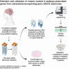
Study workflow and confirmation of neuronal cells adhered to sEEG electrodes. (A) Study workflow including sEEG procedure, individual electrode collection, confirmation of neuronal cells on electrodes, parallel extraction of genomic DNA and whole-genome amplification (WGA) from individual electrodes, deep sequencing, variant analysis, and amplicon sequencing validation of variants in DNA derived from individual electrodes. Created in BioRender (https://BioRender.com/s54s494). (B) Epifluorescence image of sEEG electrode-derived tissue cultured cells 2 days post-surgical removal with antibody labelling of microtubule-associated protein 2 (MAP2, green), a neuron-specific cytoskeletal protein isoform for identifying neuronal cells and visualization of dendritic processes. Nuclear staining was performed with Hoechst 33342 (blue).
Figure 2
Figure 2
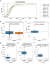
Deep sequencing coverage and variant calls. (A) The cumulative distribution of sequencing depth of all ES samples. The dashed line indicates the percentage of targeted regions that have been sequenced with 500× or less in each sample. The depth range is restricted to up to 5000× for visualization purposes. The average sequencing depth is not significantly different across amplified and unamplified samples in both panel sequencing (P = 0.76, B) and ES (P = 0.70, C). The number of mosaic variants is significantly higher in amplified samples than in unamplified samples for SNVs in panel sequencing (P = 4.5 × 10⁻¹⁶, D), indels in panel sequencing (P = 1.1 × 10⁻¹⁶, E) and SNVs in ES (P = 0.049, F). Avg, average; M.W.W., two-sided Mann–Whitney–Wilcoxon test with Bonferroni correction; A, whole-genome amplified; UA, unamplified.
Figure 3
Figure 3
MRI, sEEG electrode placement, and CNTNAP2 variant mosaic gradient for Patient 5. Blurring of the grey and white matter interface in the left posterior insula and posterior superior temporal gyrus (arrows) on (A) sagittal magnetization prepared rapid acquisition gradient echo with two inversion times (MP2RAGE) and (B) conventional sagittal magnetization prepared rapid acquisition gradient echo (MPRAGE) images. (C) Lateral view of the sEEG electrode implantation plan for Patient 5 using the standardized electrode nomenclature for stereo-EEG applications (SENSA) naming system. (D) Axial fused MP2RAGE and CT image with one of the active sEEG electrodes in the suspected areas overlaid on the MP2RAGE image. (E) Axial post-LITT ablation post-contrast MPRAGE shows the treated area, which matches the abnormality seen on prior MRIs. (F) sEEG plan overlaid with the mosaic gradient of the detected CNTNAP2 variant. Pink ovals represent electrodes corresponding to seizure onset or spread, and blue ovals represent non-involved electrodes. The number in each oval is the VAF detected in DNA extracted from that electrode. L, left; ND, not detected; R, right.
Figure 4
Figure 4
MRI and sEEG electrode placement for Patients 6 and 10. (A) Coronal T2-weighted MRI from an outside hospital for Patient 6 shows subependymal grey matter heterotopia adjacent to the right temporal horn (thin arrow) and subtle blurring of the grey and white matter interface in the right posterior insula and posterosuperior temporal gyrus (thick arrow). (B) Subsequent axial T2-weighted MRI obtained at our institution after the heterotopia had been ablated at the referring institution and before sEEG placement, again showing the subtle blurring at the grey and white matter interface in the right posterior insula (thick arrow) and right frontoparietal-predominant cerebral atrophy. (C) Lateral view of the sEEG electrode implantation plan for Patient 6 using the SENSA naming system. Coronal (D) and axial (E) T2-weighted MRI for Patient 10 show encephalomalacia in the left frontal lobe. (F) Lateral view of the sEEG electrode implantation plan for Patient 10 using the SENSA naming system. The shaded grey area represents the area of encephalomalacia. L, left; R, right.

Update of

References

    1. D'Gama AM, Poduri A. Precision therapy for epilepsy related to brain malformations. Neurotherapeutics. 2021;18(3):1548–1563. - PMC - PubMed
    1. Blumcke I, Spreafico R, Haaker G, et al. Histopathological findings in brain tissue obtained during epilepsy surgery. N Engl J Med. 2017;377(17):1648–1656. - PubMed
    1. Lopez-Rivera JA, Leu C, Macnee M, et al. The genomic landscape across 474 surgically accessible epileptogenic human brain lesions. Brain. 2023;146(4):1342–1356. - PMC - PubMed
    1. Winawer MR, Griffin NG, Samanamud J, et al. Somatic SLC35A2 variants in the brain are associated with intractable neocortical epilepsy. Ann Neurol. 2018;83(6):1133–1146. - PMC - PubMed
    1. D’Gama AM, Walsh CA. Somatic mosaicism and neurodevelopmental disease. Nat Neurosci. 2018;21(11):1504–1514. - PubMed

LinkOut - more resources