. 2025 Jun;266(2):177-191.
doi: 10.1002/path.6413. Epub 2025 Apr 3.

Histologic and molecular features shared between antibody-mediated rejection of kidney allografts and chronic histiocytic intervillositis support common pathogenesis

Affiliations

Histologic and molecular features shared between antibody-mediated rejection of kidney allografts and chronic histiocytic intervillositis support common pathogenesis

Léonie Albersammer et al. J Pathol. 2025 Jun.

Abstract

Chronic histiocytic intervillositis (CHI) is an inflammatory condition of the placenta, characterised by an abnormal, mainly macrophagic infiltrate within the intervillous space. Recent research suggests that CHI results from a 'maternal-foetal rejection' mechanism, because at least some CHI cases fulfil the criteria for antibody-mediated rejection (AMR) of kidney allografts according to the Banff classification [i.e. presence of anti-human leukocyte antigen (HLA) paternal antibodies activating the complement or foetal-specific antibodies (FSA), a macrophage-rich infiltrate, and positive C4d immunostaining]. To gain further insights into CHI pathogenesis, we aimed to refine the phenotype of the inflammatory infiltrate using a multiplex immunofluorescence technique and to compare the mRNA signatures between CHI and AMR of kidney allografts. Twelve patients with C4d+ FSA+ CHI were included in the study and compared to a control group of 5 patients without inflammatory lesions on placental examination. We developed a multiplex immunofluorescence panel to identify CD4+ and CD8+ T lymphocytes, CD68+/CD206- and CD68+/CD206+ macrophages, and NK cells in the villi and intervillous space. Molecular signatures were studied using NanoString® technology and the B-HOT panel recommended by the Banff classification for kidney allografts. Multiplex immunofluorescence revealed that the infiltrate in the intervillous space was mainly composed of CD68+/CD206- macrophages as well as a higher proportion of CD8+ lymphocytes in patients with CHI compared to controls. Densities of NK cells and CD4 T cells were very low. Molecular signatures showed an overexpression of HLA class II genes, an IFN-γ signature, and cytokine gene sets in C4d+ FSA+ CHI patients, also involved in kidney AMR. These results reinforce the paradigm of maternal-foetal rejection. © 2025 The Author(s). The Journal of Pathology published by John Wiley & Sons Ltd on behalf of The Pathological Society of Great Britain and Ireland.

Keywords: M1 type macrophages; chronic histiocytic intervillositis; maternal‐foetal rejection; molecular signatures; multiplex immunofluorescence.


Figures

Figure 1
Figure 1
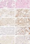
Comparison of stains of interest between a CHI placenta and a control placenta. (A) Haematoxylin–eosin–safran colouration on control placenta. Stars (⋆) are placed on villi. (B) HES staining of CHI placenta. The intervillous space is filled with macrophages (→) and fibrin (⋆). (C) CD68 staining of control placenta, showing a positivity on Hofbauer cells within the villi (⋆) and no macrophages in the intervillous space. (D) CD68 staining of CHI placenta showing numerous CD68‐positive cells in the intervillous space. (E) C4d staining of control placenta showing no positivity at syncytiotrophoblastic surface (inset at magnification ×55). (F) C4d staining of CHI placenta displaying a diffuse and intense positive staining at syncytiotrophoblastic surface (→). (G) HLA‐DR staining of control placenta showing no positivity at syncytiotrophoblastic surface. (H) HLA‐DR staining of CHI placenta showing a strong positivity on intervillous macrophages (→) and focal positivity on syncytiotrophoblastic surface (⊲) (inset at magnification ×55). (I) Beta2‐microglobulin negative staining of control placenta. (J) Beta2‐microglobulin staining of CHI placenta showing very focal positivity (⊲) (inset at magnification ×80). Scale bars, 100 μm.
Figure 2
Figure 2
Intervillous space infiltrate analysed using multiplex immunofluorescence. (A) Placenta with CHI. We observe the presence of numerous CD68+/CD206− macrophages (purple) and few CD8+ cells (red) in intervillous space, whereas Hofbauer cells express CD68 and CD206 (white) within villi, delineated by AE1/AE3 staining (green). Neither Nkp46+ nor CD4+ cells are present in this image. (B) Control placenta showing mainly CD68 + CD206+ macrophages and few CD8+ cells within the villi. Formalin‐fixed and paraffin‐embedded tissue sections were labelled for AE1/AE3 (pan‐cytokeratin staining) (green), CD8 (red), CD206 (white), CD68 (purple), NKp46 (cyan), and CD4 (yellow) using Opal reagents (Akoya Biosciences) then scanned using a Codex Fusion Akoya scanner. Original magnification ×20.
Figure 3
Figure 3
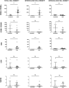
Placental cell densities. (A) Density (cells/mm2) of each cell population in intervillous space for control and CHI groups. (B) Density (cells/mm2) of each cell population in intravillous space for control and CHI groups. (C) Proportions of cell populations in intervillous space for control and CHI groups. (D) Proportions of cell populations in intravillous space for control and CHI groups.
Figure 4
Figure 4
Comparison of median cell densities of each population of interest depending on compartment. NS: not significant, *p < 0.05, **p < 0.01, ***p < 0.001.
Figure 5
Figure 5
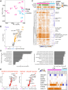
Transcriptomic changes associated with CHI assessed using a NanoString microarray. Microarray analyses using the B‐HOT panel from NanoString Technologies were performed on placental tissue from CHI cases (n = 12) and control pregnancies (n = 5). (A) Principal component analysis (PCA) plot illustrating transcriptome variation between CHI (pink) and control (blue) placentas, with each group colour‐coded. The percentage of variance explained by the first (x‐axis) and second (y‐axis) components is indicated in brackets. (B) Volcano plot (fold‐change versus p value) comparing normalised expression levels between all CHI and all control samples. Genes with significant differential expression (two‐sided t‐test p < 0.01, FC >2) are highlighted (104 upregulated genes and 0 downregulated genes), with the counts displayed. The top 10 genes are labelled. (C) Heatmap of differentially expressed genes defined in B (104 genes, rows) showing the expression ratio (log2 fold‐change) across individual placenta samples (17 placentas, columns), computed against mean expression in control placentas. Legends included CHI scores from immunofluorescence, gestational age at issue in weeks of amenorrhea, and notable genes flagged to right of heatmap. (D) Enrichment bar chart showing enrichment of signature genes upregulated in CHI (from B) within MSigDB hallmark pathways (left) and Gene Ontology (GO) biological process categories (right). The x‐axis presents the −log10 adjusted p value for enrichment, and numbers at top of each bar indicate percentage of genes overlapping with corresponding gene set. (E) Same volcano plot as in B, but with highlights from a representative IFN‐stimulated gene signature (left) and from hallmark allograft rejection signature (right). The p value for enrichment, obtained via Fisher's exact test, is shown in the bottom right. (F) Heatmap of expression ratio (log2 fold‐change) of an AMR nine‐gene set [28] among placental samples.

References

    1. Xu L, Li Y, Sang Y, et al. Crosstalk between trophoblasts and decidual immune cells: the cornerstone of maternal‐fetal immunotolerance. Front Immunol 2021; 12: 642392. - PMC - PubMed
    1. Schwartz DA, Baldewijns M, Benachi A, et al. Hofbauer cells and COVID‐19 in pregnancy. Arch Pathol Lab Med 2021; 145: 1328–1340. - PubMed
    1. Labarrere C, Mullen E. Fibrinoid and trophoblastic necrosis with massive chronic intervillositis: an extreme variant of villitis of unknown etiology. Am J Reprod Immunol Microbiol 1987; 15: 85–91. - PubMed
    1. Bos M, Nikkels PGJ, Cohen D, et al. Towards standardized criteria for diagnosing chronic intervillositis of unknown etiology: a systematic review. Placenta 2018; 61: 80–88. - PubMed
    1. Cornish EF, McDonnell T, Williams DJ. Chronic inflammatory placental disorders associated with recurrent adverse pregnancy outcome. Front Immunol 2022; 13: 825075. - PMC - PubMed

MeSH terms