. 2025 Apr 3;20(4):e0318008.
doi: 10.1371/journal.pone.0318008. eCollection 2025.

Allopurinol attenuates development of Porphyromonas gingivalis LPS-induced cardiomyopathy in mice

Affiliations

Allopurinol attenuates development of Porphyromonas gingivalis LPS-induced cardiomyopathy in mice

Akinaka Morii et al. PLoS One. .

Abstract

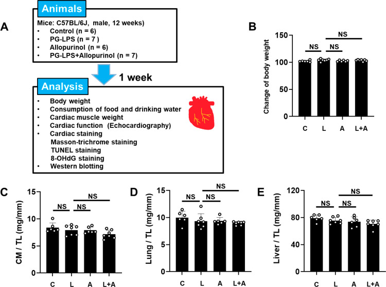
Oxidative stress is involved in the progression of periodontitis, independently of confounding factors such as smoking, and numerous studies suggest that periodontitis is associated with increased risk of cardiovascular disease. In this study, therefore, we examined the effects of the xanthine oxidase inhibitor allopurinol on cardiac dysfunction in mice treated with Porphyromonas gingivalis lipopolysaccharide (PG-LPS) at a dose (0.8 mg/kg/day) equivalent to the circulating level in patients with periodontal disease. Mice were divided into four groups: 1) control, 2) PG-LPS, 3) allopurinol, and 4) PG-LPS + allopurinol. After1 week, we evaluated cardiac function by echocardiography. The left ventricular ejection fraction was significantly decreased in PG-LPS-treated mice compared to the control (from 68 ± 1.3 to 60 ± 2.7%), while allopurinol ameliorated the dysfunction (67 ± 1.1%). The area of cardiac fibrosis was significantly increased (approximately 3.6-fold) and the number of apoptotic myocytes was significantly increased (approximately 7.7-fold) in the heart of the PG-LPS-treated group versus the control, and these changes were suppressed by allopurinol. The impairment of cardiac function in PG-LPS-treated mice was associated with increased production of reactive oxygen species by xanthine oxidase and NADPH oxidase 4, leading to calmodulin kinase II activation with increased ryanodine receptor 2 phosphorylation. These changes were also suppressed by allopurinol. Our results suggest that oxidative stress plays an important role in the PG-LPS-promoted development of cardiac diseases, and further indicate that allopurinol ameliorates Porphyromonas gingivalis LPS-induced cardiac dysfunction.


Conflict of interest statement

The authors have declared that no competing interests exist.

Figures

Fig 1
Fig 1. Schematic illustration of experimental procedure and comparison
of body weight, cardiac muscle weight, lung weight and liver weight in the four groups. (A-B) Control (C), PG-LPS (L), allopurinol (A) and PG-LPS+allopurinol (L +  A) groups all showed similar body weight at 1 week after the PG-LPS infusion. NS, not significantly diiferent from the Control (P >  0.05) by ANOVA/Tukey-Kramer. (C-E) Cardiac muscle (CM) weight per tibia length (TL) ratio (C), lung weight per TL ratio (D), and liver weight per TL ratio (E) were similar among the Control (C), PG-LPS (P), allopurinol (A) and PG-LPS +  allopurinol (L +  A) groups. all showed similar body weight at 1 week after the PG-LPS infusion. NS, not significantly different from the Control (P >  0.05) by non-parametric ANOVA/Steel-Dwass (C) or ANOVA/Tukey-Kramer (D and E).
Fig 2
Fig 2. Effects of allopurinol on PG-LPS-induced fibrosis in cardiac muscle.
(A) Representative images of Masson-trichrome-stained sections of cardiac muscle in the Control (upper left), PG-LPS (upper right), allopurinol (lower left), and PG-LPS +  allopurinol (lower right) groups. Scale bar: 100 μm (B) The area of fibrosis was significantly increased in the PG-LPS group (n =  6, **P <  0.01), but this increase was blocked in the PG-LPS +  allopurinol group (n =  7, **P <  0.01) by ANOVA/Tukey-Kramer. (C-D) Expression of collagen I (C) and collagen III (D) was significantly increased in the PG-LPS group, but these increases were blocked in the PG-LPS +  allopurinol group (n =  6 each). * P <  0.05, **P <  0.01 by non-parametric ANOVA/Steel-Dwass (C) or ANOVA/Tukey-Kramer (D). Data are presented as mean ±  SD and dots show individual data. Images of full-size immunoblots are presented in S1 and S2 Fig of S1 Data.
Fig 3
Fig 3. Effects of allopurinol on cardiac myocyte apoptosis induced by chronic PG-LPS infusion.
(A) Representative images of TUNEL-stained sections of cardiac muscle from the Control (upper left), PG-LPS (LPS) (upper right), allopurinol (lower left) and PG-LPS +  allopurinol (LPS +  Cap) (lower right) groups. Scale bars: 2 μm. (B) The number of TUNEL-positive cardiac myocytes area of fibrosis was significantly increased in the PG-LPS group (L) (P <  0.01 vs. Control), and this increase was significantly attenuated by allopurinol (L +  A). **P <  0.01 vs. Control (C) or **P <  0.01 vs. PG-LPS group (L) by ANOVA/Tukey-Kramer. (C) The Bax/Bcl-2 ratio was significantly increased in the PG-LPS group (n =  4), but this increase was blocked in the PG-LPS +  allopurinol group (n =  4). **P <  0.01 by ANOVA/Tukey-Kramer. Data are presented as mean ±  SD and dots show individual data. Images of full-size immunoblots are presented in S3 Fig of S1 Data.
Fig 4
Fig 4. Effects of allopurinol on chronic PG-LPS-induced oxidative stress in cardiac muscle.
(A) Representative immunohistochemical images of oxidative DNA damage (8-OHdG) in cardiac muscle from the Control (upper left), PG-LPS (LPS) (upper right), allopurinol (lower left) and PG-LPS +  allopurinol (L +  A) (lower right) groups. Scale bars: 2 μm (B) 8-OHdG-positive nuclei were significantly increased in the PG-LPS group (n =  5), but this increase was blocked in the PG-LPS +  allopurinol group (L +  A) (n =  4). **P <  0.01 by ANOVA/Tukey-Kramer. T (C) Expression of XO was significantly increased in the PG-LPS group (n =  5), and this increase was significantly blocked in the PG-LPS +  allopurinol group (L +  A) (n =  6). * P <  0.05 by ANOVA/Tukey-Kramer. Data are presented as mean ±  SD and dots show individual data. Images of full-size immunoblots are presented in S4 Fig of S1 Data.
Fig 5
Fig 5. Effects of allopurinol on PG-LPS-induced increases in NOX4
, p22phox, p91phox and 3-NT in cardiac muscle. (A) NOX4 expression was significantly increased in the PG-LPS group (L), and this increase was significantly attenuated in the PG-LPS +  allopurinol group (L +  A). **P <  0.01 vs. Control (C) or **P <  0.01 vs. L +  A by ANOVA/Tukey-Kramer. Images of full-size immunoblots are shown in S5 Fig of S1 Data. (B) p22phox expression was significantly increased in the PG-LPS group (L), and this increase was significantly attenuated in the PG-LPS +  allopurinol group (L +  A). * P <  0.05 vs. Control (C) or * P <  0.05 vs. L +  A by ANOVA/Tukey-Kramer. Images of full-size immunoblots are shown in S6 Fig of S1 Data. (C) p91phox expression was significantly increased in the PG-LPS group (L), and this increase was significantly attenuated in the PG-LPS +  allopurinol group (L +  A). **P <  0.01 vs. Control (C) or **P <  0.01 vs. L +  A by ANOVA/Tukey-Kramer. Images of full-size immunoblots are shown in S7 Fig of S1 Data. (D) Representative immunoblot showing expression levels of 3-NT in cardiac muscle from the Control (C), PG-LPS (L), allopurinol (A) and PG-LPS +  allopurinol (L +  A) groups. Images of full-size immunoblots are shown in S8 Fig of S1 Data. (E) 3-NT expression was significantly increased in the PG-LPS group (L), and this increase was significantly attenuated in the PG-LPS +  allopurinol group (L +  A). * P <  0.05 vs. Control (C) or * P <  0.05 vs. L +  A by ANOVA/Tukey-Kramer.
Fig 6
Fig 6. Effects of allopurinol on PG-LPS-induced phospho-CaMKII
, ox-CaMKII, phospho-RyR2 (Ser-2814), phospho-RyR2 (Ser-2808) and phospho-NFATc3 in cardiac muscle. (A) CaMKII phosphorylation (Thr-286) was significantly increased in the PG-LPS group (L), and this increase was significantly attenuated in the PG-LPS +  allopurinol group (L +  A). **P <  0.01 vs. Control (C) or * P <  0.05 vs. L +  A by ANOVA/Tukey-Kramer. Images of full-size immunoblots are shown in S9 Fig of S1 Data. (B) CaMKII oxidization (methionine-281/282) was significantly increased in the PG-LPS group (L), and this increase was significantly attenuated in the PG-LPS +  allopurinol group (L +  A). **P <  0.01 vs. Control (C) or **P <  0.01 vs. L +  A by ANOVA/Tukey-Kramer. Images of full-size immunoblots are shown in S10 Fig of S1 Data. (C) RyR2 phosphorylation (Ser-2814) was significantly increased in the PG-LPS group (L), and this increase was significantly attenuated in the PG-LPS +  allopurinol group (L +  A). **P <  0.01 vs. Control (C) or **P <  0.01 vs. L +  A by ANOVA/Tukey-Kramer. Images of full-size immunoblots are shown in S11 Fig of S1 Data. (D) RyR2 phosphorylation (Ser-2808) was significantly increased in the PG-LPS group (L), and this increase was significantly attenuated in the PG-LPS +  allopurinol group (L +  A). * P <  0.05 vs. Control (C) or **P <  0.01 vs. L +  A by ANOVA/Tukey-Kramer. Images of full-size immunoblots are shown in S12 Fig of S1 Data. (E) NFATc3 phosphorylation (Ser-265) was significantly increased in the PG-LPS group (L), and this increase was significantly attenuated in the PG-LPS +  allopurinol group (L +  A). **P <  0.01 vs. Control (C) or * P <  0.05 vs. L +  A by ANOVA/Tukey-Kramer. Images of full-size immunoblots are shown in S13 Fig of S1 Data.
Fig 7
Fig 7. Schematic illustration of the proposed role of XO and NOX4 in the heart of PG-LPS-treated mice.
PG-LPS induces expression of XO and NOX4, leading to ROS production, which mediates CaMKII activation and RyR2 phosphorylation (Ser-2814). We previously demonstrated that PG-LPS might induce myocardial ROS production and Ca2 + -mishandling via activation of the RAS [17] and cAMP/PKA signaling [64]. Our current study indicates that allopurinol might have a protective effect against PG-LPS-mediated cardiac dysfunction by blocking the increase of ROS generation by XO and NOX4 and Ca2 + leakage via altered RyR2 phosphorylation in mice. Solid black lines represent findings in this study and solid gray lines represent findings reported previously [17,64]. β-AR, β-adrenergic receptor; SR, sarcoplasmic reticulum; RyR2, ryanodine receptor 2; AT1, angiotensin II type 1 receptor; PKA, protein kinase A; ROS, reactive oxygen species; cAMP, cyclic AMP.

Similar articles

Cited by

References

    1. Genco R, Offenbacher S, Beck J. Periodontal disease and cardiovascular disease: epidemiology and possible mechanisms. J Am Dent Assoc. 2002;133 Suppl:14S–22S. doi: 10.14219/jada.archive.2002.0375 - DOI - PubMed
    1. Kataoka K, Ekuni D, Tomofuji T, Irie K, Kunitomo M, Uchida Y, et al.. Visualization of oxidative stress induced by experimental periodontitis in keap1-dependent oxidative stress detector-luciferase mice. Int J Mol Sci. 2016;17(11):1907. doi: 10.3390/ijms17111907 - DOI - PMC - PubMed
    1. Kumar J, Teoh SL, Das S, Mahakknaukrauh P. Oxidative stress in oral diseases: understanding its relation with other systemic diseases. Front Physiol. 2017;8:693. doi: 10.3389/fphys.2017.00693 - DOI - PMC - PubMed
    1. Cappola TP, Kass DA, Nelson GS, Berger RD, Rosas GO, Kobeissi ZA, et al.. Allopurinol improves myocardial efficiency in patients with idiopathic dilated cardiomyopathy. Circulation. 2001;104(20):2407–11. doi: 10.1161/hc4501.098928 - DOI - PubMed
    1. Berry CE, Hare JM. Xanthine oxidoreductase and cardiovascular disease: molecular mechanisms and pathophysiological implications. J Physiol. 2004;555(Pt 3):589–606. doi: 10.1113/jphysiol.2003.055913 - DOI - PMC - PubMed

MeSH terms