. 2025 Apr 3;11(1):135.
doi: 10.1038/s41420-025-02447-3.

The involvement of TRPV1 in the apoptosis of spermatogenic cells in the testis of mice with cryptorchidism

Affiliations

The involvement of TRPV1 in the apoptosis of spermatogenic cells in the testis of mice with cryptorchidism

Yanqiu Zhao et al. Cell Death Discov. .

Abstract

Cryptorchidism is associated with an increased risk of male infertility and testicular cancer. Persistent exposure to high temperature in cryptorchidism can lead to the apoptosis of spermatogenic cells. Transient receptor potential vanilloid 1 (TRPV1), a thermosensitive cation channel, has been found to have differential effects on various apoptosis processes. However, whether TRPV1 is involved in spermatogenic cell apoptosis induced by cryptorchidism remains unclear. Herein, we first observed the expression pattern of TRPV1 in the testes of mice with experimental cryptorchidism, and then investigated the role and mechanism of TRPV1 in spermatogenic cell apoptosis by using Trpv1-/- mice. The results showed that TRPV1 was highly expressed on the membrane of spermatocytes in mouse testis, and the expression increased significantly in the testis of mice with experimental cryptorchidism. After the operation, Trpv1-/- mice exhibited less reproductive damage and fewer spermatogenic cell apoptosis compared to the wild-type (WT) mice. Transcriptome sequencing revealed that the expression of apoptosis-related genes (Capn1, Capn2, Bax, Aifm1, Caspase 3, Map3k5, Itpr1 and Fas) was down-regulated in spermatocytes of cryptorchid Trpv1-/- mice. Our results suggest that TRPV1 promotes the apoptosis of spermatocytes in cryptorchid mice by regulating the expression of apoptosis-related genes.


Conflict of interest statement

Competing interests: The authors declare no competing interests. Ethics approval and consent to participate: All methods in this study were performed in accordance with the relevant guidelines and regulations. All animal care and experimental procedures were conducted according to the guidelines approved by the Institutional Animal Care and Use Committee of Air Force Military Medical University (IACUC-20220159).

Figures
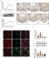
Fig. 1
Fig. 1. TRPV1 is mainly expressed on the cell membrane of mouse spermatocytes.
A, B TRPV1 mRNA and protein expression along testicular development. A mRNA expression was assessed using RT-qPCR (n = 3). Amplification of β-actin mRNA was used as an internal control. B Protein expression was determined using Western blot (n = 3). Protein expression levels were normalized to β-actin and densitometric analyses were performed with ImageJ software. C Localization of TRPV1 protein in testicular sections from adult mice (8-week-old) was assessed using IHC assay. IgG is shown in the right lower panel. SPG spermatogonium, SPC spermatocyte, rST round spermatid, eST elongated spermatid. D Co-stained of TRPV1 (red) with Sertoli cell marker SOX9 (green), spermatogonium marker PLZF (green), and spermatocyte marker SYCP3 (green) in testicular sections of WT mice (8-week-old) was detected by IF. Nuclei were stained with DAPI (blue). Dashed boxes showed localization of the enlarged images. E, F The expression profile of TRPV1 in spermatogenic cells. Different types of spermatogenic cells in WT testes (8-week-old) were isolated using the STA-PUT method. E, mRNA expression was assessed using RT-qPCR (n = 3). Amplification of β-actin mRNA was used as an internal control. F, Protein expression was assessed using Immunoblot analysis (n = 3). Protein expression levels were normalized to β-actin and densitometric analyses were performed with ImageJ software. Data were represented as mean ± SD from three independent experiments. *p < 0.05, **p < 0.01, ***p < 0.001, ****p < 0.0001 by Student’s t test.
Fig. 2
Fig. 2. Apoptosis of spermatogenic cells increases with time in wild-type (WT) mice after cryptorchidism.
A Schematic diagram of surgery-induced cryptorchidism in mice. Bilateral cryptorchidism was performed in 8-week-old mice. Comparison of the testicular size (B), testicular weight (C), testicular weight/body weight ratio (D) and the number of spermatozoa in cauda epididymis (E) on days 3, 6, 9, 12 and 15 after cryptorchidism (n = 5). F H&E staining of mouse testes and cauda epididymis on days 3, 6, 9, 12 and 15 after cryptorchidism. The arrows represent multinucleated giant cells. G Sections of testes on days 3, 6, 9, 12 and 15 after cryptorchidism were stained with the TUNEL probe (green), and nuclei were stained with DAPI (blue). Histogram showing the quantification of TUNEL positive tubules (H) and TUNEL positive cells per tubule (I) on days 3, 6, 9, 12 and 15 in cryptorchid testes (n = 3). Data were represented as mean ± SD from three independent experiments. *p < 0.05, **p < 0.01, ***p < 0.001 by Student’s t test.
Fig. 3
Fig. 3. TRPV1 expression increases in the testis on day 9 after cryptorchidism.
A, B TRPV1 mRNA and protein expression in the testis on day 9 after cryptorchidism (n = 3). A mRNA expression was assessed using RT-qPCR. Amplification of β-actin mRNA was used as an internal control. B Protein expression was determined using Western blot. Protein expression levels were normalized to β-actin and densitometric analyses were performed with ImageJ software. C IF staining was applied to detect TRPV1 (red) in the 9-day cryptorchid testis. Dashed boxes showed localization of the enlarged images. White arrows indicated cells with strong positive signals. Data were represented as mean ± SD from three independent experiments. **p < 0.01 by Student’s t test.
Fig. 4
Fig. 4. Knockout of Trpv1 does not affect sperm development and mice fertility.
A Protein expression of TRPV1 in the testes of WT and Trpv1−/− mice was determined using Western blot. β-actin served as a loading control. Comparison of average pups (B), testicular size (C), testicular weight (D), testicular weight/body weight ratio (E), and the sperm count in cauda epididymis (F) of 8-week-old Trpv1−/− and WT mice (n = 5). G H&E staining of testis and cauda epididymis of 8-week-old Trpv1−/− and WT mice. H H&E staining of spermatozoa from 8-week-old Trpv1−/− and WT mice. I Sections of testes from 8-week-old WT mice and Trpv1−/− mice were stained with a TUNEL probe (green), and nuclei were stained with DAPI (blue). Histogram showing the quantification of TUNEL positive tubules (J) and TUNEL positive cells per tubule (K) of 8-week-old WT mice and Trpv1−/− mice (n = 3). Data were represented as mean ± SD from three independent experiments. The data were analyzed with Student’s t test. ns not significant.
Fig. 5
Fig. 5. The apoptosis of spermatocytes decreases after 9 days of cryptorchidism in Trpv1−/− mice.
Comparison of the testicular size (A), testicular weight (B), testicular weight/body weight ratio (C), and the number of spermatozoa in cauda epididymis (D) on day 9 after cryptorchidism in 8-week-old WT mice and Trpv1−/− mice (n = 5). E H&E staining of testes and cauda epididymis in cryptorchid surgery groups and sham groups of 8-week-old WT mice and Trpv1−/− mice. F Sections of testes from 8-week-old WT mice and Trpv1−/− mice on day 9 after cryptorchidism were stained with a TUNEL probe (green), and nuclei were stained with DAPI (blue). Histogram showing the quantification of TUNEL positive tubules (G) and TUNEL positive cells per tubule (H) on day 9 of cryptorchid testis in 8-week-old WT mice and Trpv1−/− mice (n = 3). Data were represented as mean ± SD from three independent experiments. *p < 0.05, **p < 0.01, ***p < 0.001 by Student’s t test.
Fig. 6
Fig. 6. Trpv1−/− mice exhibit downregulated expression of apoptosis-related genes in spermatocytes after cryptorchidism.
A Schematic diagram depicting transcriptome analysis of spermatocytes. Briefly, total RNA from WT and Trpv1−/− spermatocytes of cryptorchid testes on day 9 were collected for the RNA-seq. B Heatmaps depicting significantly upregulated and downregulated genes in WT and Trpv1−/− spermatocytes. The genes with |log2 Fold change|≥1 and q < 0.05 were determined to generate the heatmap. C Volcano plot showing DEGs in WT and Trpv1−/− spermatocytes. The significant changed downregulated genes associated with apoptosis were labeled with gene name. D KEGG pathway analysis of 3176 downregulated genes in Trpv1−/− spermatocytes. The apoptosis pathway was labeled with asterisks. E, F Expression of CAPN1, CAPN2, BAX, AIFM1, Caspase 3, MAP3K5, ITPR1 and FAS in spermatocytes of WT and Trpv1−/− cryptorchid mice were detected by RT-qPCR and Western blot analysis (n = 3). Data were represented as mean ± SD from three independent experiments. *p < 0.05, **p < 0.01, ***p < 0.001 by Student’s t test. G Cryptorchidism induces the increase of TRPV1 expression in mouse spermatocytes, leading to the increase of [Ca2+]i, which participates in the endogenous mitochondrial apoptosis pathway through up-regulation of CAPN1, CAPN2, BAX, AIFM1, MAP3K5 and ITPR1 on the one hand, and in exogenous apoptosis pathway through up-regulation of FAS on the other hand, ultimately leading to spermatocyte apoptosis. This picture was drawn by figdraw.

Similar articles

References

    1. Aldahhan RA, Stanton PG. Heat stress response of somatic cells in the testis. Mol Cell Endocrinol. 2021;527:111216. - PubMed
    1. Gopalakrishnan NP, Chung IM. Alteration in the expression of antioxidant and detoxification genes in chironomus riparius exposed to zinc oxide nanoparticles. Comp Biochem Physiol B Biochem Mol Biol. 2015;190:1–7. - PubMed
    1. Hoang-Thi AP, Dang-Thi AT, Phan-Van S, Nguyen-Ba T, Truong-Thi PL, Le-Minh T, et al. The impact of high ambient temperature on human sperm parameters: a meta-analysis. Iran J Public Health. 2022;51:710–23. - PMC - PubMed
    1. Mieusset R, Bengoudifa B, Bujan L. Effect of posture and clothing on scrotal temperature in fertile men. J Androl. 2007;28:170–5. - PubMed
    1. Palnitkar G, Phillips CL, Hoyos CM, Marren AJ, Bowman MC, Yee BJ. Linking sleep disturbance to idiopathic male infertility. Sleep Med Rev. 2018;42:149–59. - PubMed

LinkOut - more resources