. 2025 Apr 3;16(1):3208.
doi: 10.1038/s41467-025-58493-z.

Sulphostin-inspired N-phosphonopiperidones as selective covalent DPP8 and DPP9 inhibitors

Affiliations

Sulphostin-inspired N-phosphonopiperidones as selective covalent DPP8 and DPP9 inhibitors

Leonard Sewald et al. Nat Commun. .

Abstract

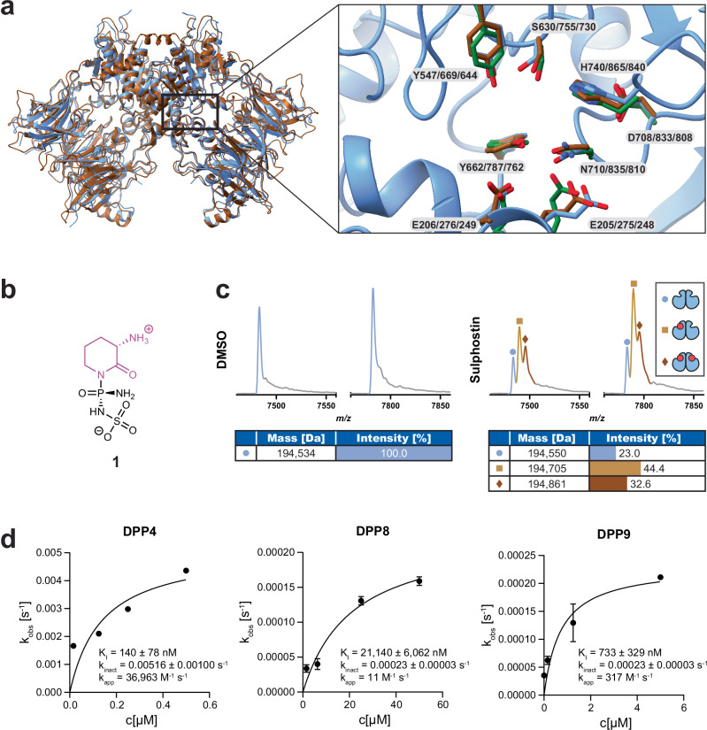
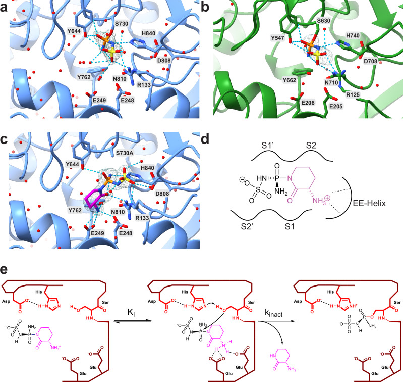
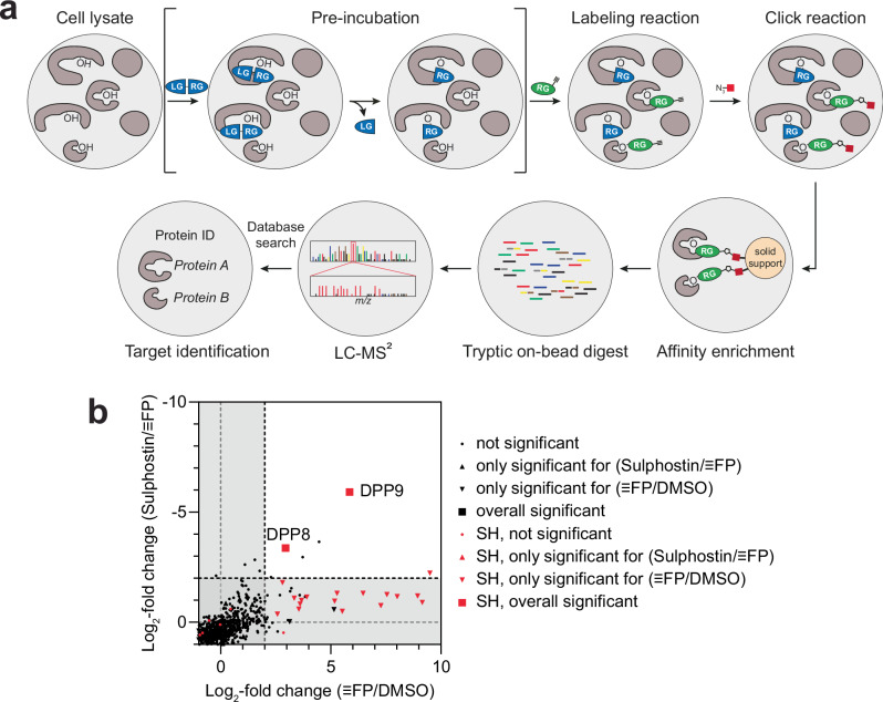
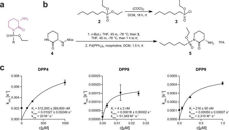
Covalent chemical probes and drugs combine unique pharmacologic properties with the availability of straightforward compound profiling technologies via chemoproteomic platforms. These advantages have fostered the development of suitable electrophilic "warheads" for systematic covalent chemical probe discovery. Despite undisputable advances in the last years, the targeted development of proteome-wide selective covalent probes remains a challenge for dipeptidyl peptidase (DPP) 8 and 9 (DPP8/9), intracellular serine hydrolases of the pharmacologically relevant dipeptidyl peptidase 4 activity/structure homologues (DASH) family. Here, we show the exploration of the natural product Sulphostin, a DPP4 inhibitor, as a starting point for DPP8/9 inhibitor development. The generation of Sulphostin-inspired N-phosphonopiperidones leads to derivatives with improved DPP8/9 inhibitory potency, an enhanced proteome-wide selectivity and confirmed DPP8/9 engagement in cells, thereby representing that structural fine-tuning of the warhead's leaving group may represent a straightforward strategy for achieving target selectivity in exoproteases such as DPPs.


Conflict of interest statement

Competing interests: A.L. is an employee of Proteros Biostructures GmbH. The remaining authors declare no competing interests.

Figures

Fig. 1
Fig. 1. Sulphostin binds to DPP proteins.
a The crystal structures of DPP8 and 9 in the apo state in an overlay, with DPP9 shown in blue and DPP8 in brown as a ribbon model. The active site was zoomed in, showing the active site residues of DPP4 (green), DPP8 (brown) and DPP9 (blue) as stick model. Backbone of DPP9 is shown in blue as ribbon model. Crystal structures from PDB: 1PFQ (DPP4), 6EOO (DPP8) and 6EOQ (DPP9). b Chemical structure of the natural product Sulphostin (1) containing a phosphosulfamate functional group (indicated in black) and the (S)−3-aminopiperidine-2-one group in purple. c Native MS spectrum of DPP9 in the dimeric state with or without Sulphostin bound. Peaks correspond to different charge states. The charge state distribution for the dimer without inhibitor bound is shown in blue, whereas the distribution for the DPP9 dimer with one Sulphostin molecule is shown in light brown and for two Sulphostin molecules in dark brown. d Kinetic analysis of DPP inhibition by Sulphostin. The pseudo-first order rate constant (kobs) was calculated from an exponential regression of progress curves and plotted against the inhibitor concentration. KI and kinact were obtained via fitting to a hyperbolic equation. All activity measurements were performed in triplicate (n = 3 technical replicates), mean values are shown and error bars indicate the standard error of the mean (SEM). Source data are provided as a Source Data file.
Fig. 2
Fig. 2. Sulphostin inhibits DPP proteins by a covalent binding mode.
a X-ray crystal structure of Sulphostin in complex with DPP9 refined to a final resolution of 1.89 Å. The phosphosulfamate group is covalently bound to the catalytically active serine S730 residue and is contoured at 1 σ with the refined 2Fo-Fc electron density map. Hydrogen bond interactions are depicted as dashed lines. b X-ray crystal structure of Sulphostin in complex with DPP4 refined to a final resolution of 2.38 Å. The phosphosulfamate group is covalently bound to S630 and is contoured at 1 σ with the refined 2Fo-Fc electron density map. Hydrogen bond interactions are depicted as dashed lines. c X-ray crystal structure of Sulphostin in complex with the S730A-DPP9 mutant (1.89 Å resolution) showing the non-covalent interactions prior to the covalent bond formation. The ligand is superimposed with the refined 2Fo-Fc electron density map contoured at 1 σ. Hydrogen bond interactions are depicted as dashed lines. d Sulphostin is coordinated by E248 and E249 from the EE helix with the free N-terminus of the (S)−3-aminopiperidine-2-one, which occupies two substrate binding sub-sites. e The proposed general binding mechanism of Sulphostin with DPP proteins. The stereogenic phosphorus of Sulphostin is attacked by the serine of the catalytic triad, whereby the (S)−3-aminopiperidin-2-one moiety functions as a leaving group.
Fig. 3
Fig. 3. Sulphostin shows promising selectivity within FP target proteins.
a Overview of a competitive ABPP workflow. A cell lysate is pre-incubated with an inhibitor, composed of a leaving group (LG) and a reactive group (RG), followed by a labeling reaction with a probe that covalently binds target proteins via its RG, which can be either an ABP such as FP or an alkyne-tagged ligand. After click attachment of a biotin moiety, labeled proteins are enriched via solid support, tryptic digested on-bead, and subsequently identified via LC-MS/MS analysis. b Cell lysates were treated with 50 µM Sulphostin or DMSO vehicle control prior to treatment with the ABP FP. ABPP of SHs without (FC plotted on x-axis) or after pre-treatment with 50 µM Sulphostin (competition experiment, FC plotted on y-axis) with 2 µM ≡FP after click chemistry (n = 4 biologically independent samples). Dashed lines indicate the FC ≥ 2 ( ≡FP/DMSO) or FC ≤ -2 (Sulphostin/≡FP) threshold. Black symbols indicate identified protein groups, red symbols highlight serine hydrolases (SHs). To identify statistically significant hits from the analysis (marked as a triangle or square), p-value ≤ 0.01 (two-sided Student’s t-test, permutation-based FDR with 250 randomizations and FDR = 0.05) was applied. Source data are provided as a Source Data file.
Fig. 4
Fig. 4. Sulphostin-inspired N-phosphono-(S)−3-aminopiperidine-2-ones inhibit DPPs.
a Chemical structure of Sulphostin-inspired N-phosphono-(S)−3-aminopiperidine-2-ones. The previously identified leaving group is shown in purple. b Representative synthesis route of N-phosphono-(S)−3-aminopiperidine-2-ones by activation of a phosphonate diethyl ester 2 with oxalyl chloride yielding a chloro-phosphonate intermediate 3, which then reacts with in situ lithiated Alloc-protected (S)−3-aminopiperidine-2-one 4 resulting in corresponding N-phosphono-(S)−3-aminopiperidine-2-one 5. c Kinetic analysis of DPP inhibition by N-phosphono-(S)−3-aminopiperidine-2-ones. The pseudo-first order rate constant (kobs) was calculated from an exponential regression of progress curves and plotted against the inhibitor concentration. KI and kinact were obtained via fitting to a hyperbolic equation. All activity measurements were performed in triplicate (n = 3 technical replicates), mean values are shown and error bars indicate the SEM. Source data are provided as a Source Data file.
Fig. 5
Fig. 5. N-phosphono-(S)−3-aminopiperidine-2-ones bind covalently to DPP9.
a X-ray structure of 16 in complex with DPP9 refined to a final resolution of 2.49 Å, shown from two different angles. The ligand is covalently bound to the catalytically active serine S730 residue and is contoured at 1 σ with the refined 2Fo-Fc electron density map. Hydrogen bond interactions are depicted as dashed lines. b ABPP without (FC plotted on x-axis) or after pre-treatment with 50 µM 8, 11 or 16 (competition experiment, FC plotted on y-axis) with 10 µM 9 after click chemistry (n = 4 biologically independent samples). Dashed lines indicate the FC ≥ 2 (9/DMSO) or FC ≤ -2 (Competitor/9) threshold. Black symbols indicate identified protein groups. To identify statistically significant hits from the analysis (marked as a triangle or square), p ≤ 0.01 (two-sided Student’s t-test, permutation-based FDR with 250 randomizations and FDR = 0.05) was applied. c Addition of N-phosphono-(S)−3-aminopiperidine-2-ones block the interaction between endogenous DPP9 and BRCA2. Quantification of PLAs showing MMC-induced DPP9-BRCA2 PLA events in HeLa wild-type cells, in the presence of 10 µM of the respective inhibitory compounds. Cells were treated with 300 nM MMC for 24 h and 10 µM of the indicated inhibitors for 1 h prior to fixation. Each dot represents the number of PLA events in a single cell. Data were analyzed by unpaired two-sided t-test comparisons (****p < 0.0001). To visualize the three biological replicates, each biological replicate is labeled differently: circle, triangle or square. n values indicate total number of cells in each analysis group from 3 independent biological replicates in total: n = 240 for BRCA2 Ab-Ctrl., n = 237 for DPP9 Ab-Ctrl., n = 222 for +Ctrl., n = 202 for 1G244, n = 231 for 8, n = 205 for 16. d Inhibition of DPP8/9 increases cellular sensitivity to genotoxic stress. HeLa wild-type (WT) cells were treated with 1 µM MMC and 10 µM of the respective inhibitors. Cell viability was measured after 72 h, and normalized to WT mock treated cells. DPP9KO cells and WT cells treated with 1G244 (10 µM) were analyzed as positive controls, whereas WT cells treated with Sitagliptin were used as a negative control. Dashed lines indicate the viability of the WT + MMC cells. The graph shows the mean and error bars indicate the SEM of all individual measurements (n = 18 (HeLa wild-type controls), 17 (HeLa DPP9KO control) or 9 (inhibitor-treated samples) independent biological replicates). Data were analyzed by a Tukey two-way ANOVA, using a mixed effect analysis (n.s. = not significant, *p = 0.0157, ****p < 0.0001). Source data are provided as a Source Data file.

Similar articles

Cited by

References

    1. Boike, L., Henning, N. J. & Nomura, D. K. Advances in covalent drug discovery. Nat. Rev. Drug Discov.21, 881–898 (2022). - PMC - PubMed
    1. Zhang, X. & Cravatt, B. F. Chemical proteomics–guided discovery of covalent ligands for cancer proteins. Annu. Rev. Cancer Biol.8, 155–175 (2024).
    1. Grams, R. J. & Hsu, K. L. Reactive chemistry for covalent probe and therapeutic development. Trends Pharm. Sci.43, 249–262 (2022). - PMC - PubMed
    1. Gehringer, M. & Laufer, S. A. Emerging and re-emerging warheads for targeted covalent inhibitors: applications in medicinal chemistry and chemical biology. J. Medicinal Chem.62, 5673–5724 (2019). - PubMed
    1. Faucher, F., Bennett, J. M., Bogyo, M. & Lovell, S. Strategies for tuning the selectivity of chemical probes that target serine hydrolases. Cell Chem. Biol.27, 937–952 (2020). - PMC - PubMed

MeSH terms

LinkOut - more resources