. 2025 Apr 3;16(1):3031.
doi: 10.1038/s41467-025-58036-6.

Inhibiting acute, axonal DLK palmitoylation is neuroprotective and avoids deleterious effects of cell-wide DLK inhibition

Affiliations

Inhibiting acute, axonal DLK palmitoylation is neuroprotective and avoids deleterious effects of cell-wide DLK inhibition

Xiaotian Zhang et al. Nat Commun. .

Abstract

Inhibiting dual leucine-zipper kinase (DLK) could potentially ameliorate diverse neuropathological conditions, but a direct inhibitor of DLK's kinase domain caused unintended side effects in human patients, indicative of neuronal cytoskeletal disruption. We sought a more precise intervention and show here that axon-to-soma pro-degenerative signaling requires acute, axonal palmitoylation of DLK. To identify potential modulators of this modification, we screened >28,000 compounds using a high-content imaging readout of DLK's palmitoylation-dependent subcellular localization. Several hits alter DLK localization in non-neuronal cells, reduce DLK retrograde signaling and protect cultured dorsal root ganglion neurons from neurodegeneration. Mechanistically, the two most neuroprotective compounds selectively prevent DLK's stimulus-dependent palmitoylation and subsequent recruitment to axonal vesicles, but do not affect palmitoylation of other axonal proteins assessed and avoid the cytoskeletal disruption associated with direct DLK inhibition. Our hit compounds also reduce pro-degenerative retrograde signaling in vivo, revealing a previously unrecognized neuroprotective strategy.


Conflict of interest statement

Competing interests: A Patent Application No. 16/631,969 (National Stage Application of PCT/US18/42620) related to the screening method used in this manuscript was jointly filed by Temple University and Shriners Hospitals for Children. Authors S.M.H., J.N. (co-inventors) and G.M.T. (inventor) are named in the patent application. The patent application is being overseen by Temple University in accordance with its appropriate policies. The remaining authors declare no competing interests.

Figures
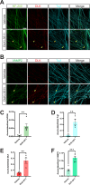
Fig. 1
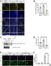
Fig. 1. DLK kinase domain inhibition disrupts axonal integrity in DRG neurons.
A Images of cultured DRG neurons that had been treated with DMSO vehicle or 500 nM DLK inhibitor GNE-3511 (DLKi-3511) for 4 hours and fixed and stained with the indicated antibodies. B As A, except that neurons were fixed to detect vesicle marker VAMP2 rather than NF-200. CF Quantified data from A and B confirm that DLK inhibition disrupts distribution of NF-200 (neurofilament heavy chain; 1C; 5 independent cultures per condition) and Tuj1 (neuron-specific tubulin; 1D; 4 independent cultures per condition) and leads to accumulations of DLK (1E, 4 independent cultures per condition) and VAMP2 (1F, 5 independent cultures per condition) in axons, suggestive of cytoskeletal dysregulation and/or impaired vesicle-based transport. Yellow arrowheads highlight examples of disruption/accumulation of the respective signals. Unpaired two-sided t tests reveal significant effects of DLKi-3511 vs. Vehicle treated conditions for NF-200 signal (p = 0.005), DLK signal (p  = 0.005), Tuj1 signal (p = 0.0025), and VAMP2 signal (p = 0.0005). Scale bar: 20 μm (all panels). Data are presented as mean values +/− SEM. Source data are provided in the Source Data file.
Fig. 2
Fig. 2. Trophic deprivation-induced recruitment of DLK to axonal vesicles and DLK-dependent retrograde signaling are both palmitoylation-dependent.
A Cultured DRG neurons were lentivirally infected to express myc-tagged wild type DLK (wtDLK-myc) and left untreated (+NGF) or were subjected to trophic factor deprivation (TD) in the presence of 2-bromopalmitate (2BP) or DMSO vehicle. Cultures were fixed 3 h post-TD and immunostained with the indicated antibodies. The bottom two rows of images (High Power Merge) show magnified views of the boxed region in the image directly above. Arrowheads in bottom row panels indicate DLK-myc vesicle-like puncta. Scale bars, 20 μm (all panels). B Quantified data from A confirm that the TD-induced increase in axonal DLK puncta (presumptive vesicles) is prevented by 2BP i.e. is palmitoylation-dependent. ***; p = 0.0008; ****; p < 0.0001, two-sided one-way ANOVA, Dunnett’s post hoc test. 5 independent cultures per condition. C Western blots of ABE (palmitoyl-) fractions of axonal lysates from DRG neuron spot cultures that had been treated as indicated prior to lysis. The righthand lane is a side-by-side exposure from a parallel control sample omitting the key ABE reagent NH2OH, run on the same gel, with intervening spacer lanes cropped. D Quantified data from C confirm that TD increases axonal DLK palmitoylation, which is prevented by 2BP. ****; p < 0.0001, two-sided one-way ANOVA, Dunnett’s post hoc test. 7 independent cultures per condition (+NGF/Vehicle, TD/Vehicle); 6 independent cultures (TD/2BP). E Images of cell body chambers of DRG microfluidic cultures, fixed and stained with the indicated antibodies 4 h after selective treatment of distal axonal compartments as indicated. Scale bars, 50 μm. F Quantified data from E confirm that TD-induced retrograde signaling requires acute palmitoylation in distal axons. ****; p < 0.0001, two-sided one-way ANOVA, Dunnett’s post hoc test. 3 independent cultures per condition. Data are presented as mean values +/− SEM. Source data are provided in the Source Data file.
Fig. 3
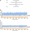
Fig. 3. An expanded High Content Imaging screen identifies compounds that inhibit the punctate localization of DLK-GFP.
A Experimental design of the high-throughput screen to identify compounds that inhibit DLK-GFP punctate localization, adapted from. B, C Evaluation of the effect of 26,677 compounds that passed cut-offs (of 28,400 compounds screened) from the Maybridge and Enamine Libraries™ on DLK-GFP puncta per transfected cell (Puncta/NLS) and average brightness of those puncta (Vesicle Average Intensity). Black dotted lines indicate 2 standard deviations (2 SD) above and below the mean of all determinations that passed cut-offs. Compounds that reduced both readouts by >2 SD (region highlighted in pale orange) were selected for further analysis. Dark orange dotted lines in B and C indicate the average reduction seen in each readout with 2-Bromopalmitate (2BP, tool compound positive control). A single biological replicate was run for each compound in this primary screen. Source data are provided in the Source Data file.
Fig. 4
Fig. 4. Multiple hit compounds reduce TD-induced c-Jun phosphorylation.
A Western blots of lysates from cultured DRG neurons that had been treated with vehicle (DMSO), GNE-3511 (3511) or the indicated hit compounds for 1 h at 5 days in vitro (DIV 5), followed by 2.5 h TD in the continued presence of the indicated compounds, or that had been left unstimulated (+ NGF). The secondary antibody used on the p-cJun blot also weakly recognizes residual anti-NGF IgG used during TD (indicated by asterisk). Subsets of compounds were assayed side-by-side, but in batches indicated by spaces between individual blots. B Quantified data from A, of p-cJun:tubulin, normalized to TD (vehicle) condition. Compounds whose effect on p-c-Jun:tubulin differed significantly from TD (vehicle) condition are highlighted with orange bars. Statistical significance versus TD (vehicle) control was as follows: +NGF (vehicle): p < 0.0001; 6: p = 0.0303; 8: p = 0.0014; 13: p = 0.0033; 20: p = 0.0309 25: p = 0.0005; 30: p = 0.0029, two-sided Kruskal Wallis tests with Dunn’s post hoc test for multiple comparisons. Data are plotted for the following number of independent cultures per condition: +NGF/Vehicle: 9; TD/Vehicle: 10; TD/1: 6; TD/1: 6; TD/2: 5; TD/3: 5; TD/4: 6; TD/5: 6; TD/6: 8; TD/7: 8; TD/8: 8; TD/9: 8; TD/10: 8; TD/11: 8; TD/12: 7; TD/13: 14; TD/14: 8; TD/15: 8; TD/16: 8; TD/17: 8; TD/19: 8; TD/20: 8; TD/21: 9; TD/22: 6; TD/23: 6; TD/24: 5; TD/25: 7; TD/26: 6; TD/27: 6; TD/28: 6; TD/29: 7; TD/30: 7; TD/31: 7; TD/32: 7; TD/33: 7. Data are presented as mean values +/- SEM. Source data are provided in the Source Data file.
Fig. 5
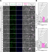
Fig. 5. A subset of hit compounds reduces TD-induced neurodegeneration.
A Phase contrast images (first column), Calcein-AM signal (2nd column) and Hoechst 33342 DNA signal (3rd column) of DRG neurons that were maintained in NGF ( + NGF) or subjected to TD for 45 h in the presence of vehicle (DMSO) or the indicated hit compounds that significantly reduced TD-induced c-Jun phosphorylation in Fig. 4. The fourth column shows an overlay of the phase contrast image with the Calcein/Hoechst double-positive signal (latter false-colored in magenta). The fifth column shows magnified views of the boxed region in the corresponding fourth column. Scale bars, 20 μm (all panels). B Number of Calcein/Hoechst double-positive cells per field (i.e. viable cell bodies), quantified from images from A. Magenta shaded bars and ns labels indicate hit compounds whose effect on cell body viability post-TD does not differ significantly from +NGF (vehicle) control condition. GNE-3511 and 2BP also protected cell bodies from the effects of TD. The slight increase in Calcein-AM signal in cultures subjected to TD is consistent with a prior report. Statistical significance versus +NGF (vehicle) control was as follows: 6: p = 0.0187; 20: p < 0.0001; 25: p = 0.0003; 30: p = 0.0375. TD (vehicle) condition also differed significantly from +NGF (vehicle) p < 0.0001. ns: not significant, two-sided one-way ANOVA, Dunnett’s post hoc test. Data are plotted for the following number of independent cultures per condition: +NGF/Vehicle: 4; TD/Vehicle: 4; TD/DLKi-3511: 3; TD/2BP: 3; TD/6: 4; TD/8: 3; TD/13: 3; TD/20: 3; TD/25: 4; TD/30: 3. C Axon integrity index, determined by counting the number of continuous elongated structures (i.e. unbroken axons) per unit area, quantified from phase contrast images (first column) in A. Magenta shaded bars and ns labels indicate hit compounds for which the axon integrity index post-TD did not differ significantly from the +NGF (vehicle) control condition. GNE-3511 and 2BP also protected axons from the effects of TD. Statistical significance versus +NGF (vehicle) control was as follows: 20: p = 0.0001; 25: p = 0.017; 30: p < 0.0001. TD (vehicle) condition also differed significantly from +NGF (vehicle) p = 0.0001. ns: not significant, two-sided one-way ANOVA, Dunnett’s post hoc test. Data are plotted for the following number of independent cultures per condition: +NGF/Vehicle: 4; TD/Vehicle: 4; TD/DLKi-3511: 3; TD/2BP: 3; TD/6: 4; TD/8: 3 TD/13: 3; TD/20: 3; TD/25: 4; TD/30: 3. Data are presented as mean values +/- SEM. Source data are provided in the Source Data file.
Fig. 6
Fig. 6. Neuroprotective hit compounds prevent TD-induced DLK recruitment to axonal vesicles and palmitoylation, and activation of DLK’s downstream target MKK4.
A Cultured DRG neurons were infected to express myc-tagged wild type DLK (wtDLK-myc) and left untreated or were subjected to TD in the presence of the indicated compounds or DMSO vehicle, prior to fixation and immunostaining with the indicated antibodies. The bottom two rows of images (High Power Merge) show magnified views of the boxed region in the image directly above. Arrowheads in bottom row panels indicate DLK-myc vesicle-like puncta. Scale bars, 20 μm (all panels). B Quantified data from A confirm that TD-induced increases in axonal DLK puncta (presumptive vesicles) are prevented by 8 and 13. Data for +NGF (vehicle), TD (vehicle) and TD (2BP) conditions are replotted from Fig. 2B. *; p = 0.0483; **; p = 0.0016, two-sided one-way ANOVA, Dunnett’s post hoc test. 5 independent cultures per condition. C Western blots of ABE (palmitoyl-) fractions of DRG axonal lysates that had been treated as indicated prior to lysis. The righthand lane is a side-by-side exposure from a parallel control sample omitting the key ABE reagent NH2OH, run on the same gel, with intervening spacer lanes cropped. D Quantified data from C confirm that 8 and 13 prevent the TD-induced increase in axonal DLK palmitoylation. Data for +NGF (vehicle), TD (vehicle) and TD (2BP) conditions are replotted from Fig. 2D. +NGF (vehicle) versus TD (vehicle): **; p = 0.0026; +NGF (vehicle) versus TD (2BP): **; p = 0.0054, two-sided one-way ANOVA, Dunnett’s post hoc test. Data are plotted for the following number of independent cultures per condition: +NGF/Vehicle: 7; TD/Vehicle: 7; TD/2BP: 6; TD/8: 6; TD/13: 6. E Western blots, detected with the indicated antibodies, of DRG lysates that had been treated as indicated prior to lysis. F Quantified data from E reveal that TD significantly increases pMKK4:total MKK4 ( + NGF (Vehicle) vs. TD (Vehicle); p  = 0.0219) while DLK inhibitor GNE-3511 (DLKi-3511) significantly reduces it (+NGF (Vehicle) vs. TD (DLKi-3511): p < 0.0001) but TD has no effect on pMKK4:total MKK4 in the presence of 8 or 13 (ns; non-significant). Two-sided one-way ANOVA, Dunnett’s post hoc test. Data are plotted for the following number of independent cultures per condition: +NGF/Vehicle: 8; TD/Vehicle: 8; TD/2BP: 8; TD/8: 7; TD/13: 7. Data are presented as mean values +/− SEM. Source data are provided in the Source Data file.
Fig. 7
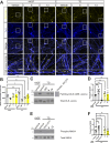
Fig. 7. Neuroprotective hit compounds do not phenocopy the effect of a DLK kinase domain inhibitor on axonal integrity.
A Images of cultured DRG neurons treated with DMSO vehicle, 500 nM DLKi-3511 or 10 μM of the indicated compounds for 4 hours and fixed and stained with the indicated antibodies. Yellow arrowheads highlight examples of disruption/accumulation of the respective signals. Scale bars, 20 μm (all panels). B As A, except that neurons were fixed to detect VAMP2 rather than NF-200. Quantified data from A and B confirm that DLKi-3511 causes disruptions of NF-200 (7C ***; p = 0.0006, 5 independent cultures per condition), Tuj1 (7D ****; p < 0.0001, 4 independent cultures per condition), DLK (7E ***; p = 0.0003, 4 independent cultures per condition) and VAMP2 (7F ***; p = 0.0001, 5 independent cultures per condition) in axons, but that 8 and 13 do not (n.s.; not significant). Two-sided one-way ANOVA with Dunnett’s post hoc test for (C-F). Data are presented as mean values +/- SEM. Source data are provided in the Source Data file.
Fig. 8
Fig. 8. Hit compounds reduce palmitoyl-DLK-dependent pro-degenerative retrograde signaling to a similar extent as a DLK kinase domain inhibitor in vivo.
A Images of retinas of mice that had been subjected to sham injury (1st column) or to optic nerve crush (ONC; 2nd-5th column), immediately prior to intravitreal injection with the indicated compounds or with DMSO vehicle. At 15 h post-ONC or sham injury, mice were perfused and fixed, and retinas were immunostained with the indicated antibodies. Scale bars, 20 μm (all panels). B Quantified data from A. DLKi-3511, 8 and 13 all inhibit ONC-induced c-Jun phosphorylation. ONC (vehicle) versus ONC (DLKi-3511); *; p = 0.0144;), ONC (vehicle) versus ONC (8): **; p = 0.0019; ONC (vehicle) versus ONC (13): *; p = 0.0181. Sham (vehicle) and ONC (vehicle) conditions also differ significantly (****; p < 0.0001), two-sided one-way ANOVA, Dunnett’s post hoc test. Data are plotted for the following number of mice per condition: Sham/Vehicle: 3; ONC/Vehicle: 6; ONC/DLKi-3511: 4; ONC/8: 7; ONC/13: 7. Data are presented as mean values +/- SEM. Source data are provided in the Source Data file.

Update of

Similar articles

References

    1. Köster, K. A., Dethlefs, M., Duque Escobar, J. & Oetjen, E. Regulation of the Activity of the Dual Leucine Zipper Kinase by Distinct Mechanisms. Cells1310.3390/cells13040333 (2024). - PMC - PubMed
    1. Jin, Y. & Zheng, B. Multitasking: Dual Leucine Zipper-Bearing Kinases in Neuronal Development and Stress Management. Annu Rev. Cell Dev. Biol.35, 501–521 (2019). - PMC - PubMed
    1. Tedeschi, A. & Bradke, F. The DLK signalling pathway-a double-edged sword in neural development and regeneration. EMBO Rep.14, 605–614 (2013). - PMC - PubMed
    1. Mata, M., Merritt, S. E., Fan, G., Yu, G. G. & Holzman, L. B. Characterization of dual leucine zipper-bearing kinase, a mixed lineage kinase present in synaptic terminals whose phosphorylation state is regulated by membrane depolarization via calcineurin. J. Biol. Chem.271, 16888–16896 (1996). - PubMed
    1. Ghosh, A. S. et al. DLK induces developmental neuronal degeneration via selective regulation of proapoptotic JNK activity. J. Cell Biol.194, 751–764 (2011). - PMC - PubMed

Substances

LinkOut - more resources