. 2025 Apr 3;15(1):11435.
doi: 10.1038/s41598-025-95449-1.

Peptides developed against receptor binding sites of the E glycoprotein neutralize tick-borne encephalitis virus

Affiliations

Peptides developed against receptor binding sites of the E glycoprotein neutralize tick-borne encephalitis virus

Patrícia Petroušková et al. Sci Rep. .

Abstract

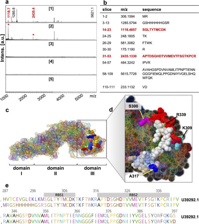
Infection caused by tick-borne encephalitis virus (TBEV) often manifests with meningitis or encephalitis. Domain III (DIII) of the envelope (E) glycoprotein on the TBEV surface facilitates virion attachment to the cell receptor and initiates cell entry. As a result, this research focused on the DIII to develop antiviral molecules that could prevent cell entry. We first identified two receptor-binding sites (RBS, 300SGLTYTMCDK309 and 317APTDSGHDTVVMEVTFSGTKPCR339) on DIII, that are involved in binding to human brain microvascular endothelial cells (hBMECs). Then we sought to isolate peptides from two combinatorial peptide-phage libraries that bind to the RBS and prevent protein E from attaching to hBMECs. Three cyclic peptides (CP2-CNSSKLHMC, CP20-CDGRPDRAC, CP23-CMKESIRGC) and a linear peptide (LP16-AFHPRQMETQMY) inhibited DIII binding to hBMECs. CP2 and CP20, both non-cytotoxic and hemocompatible, effectively neutralized a live virus. CP2 and CP20 deserve further attention for their potential application as anti-TBEV therapeutics that prevent viral cell entry.

Keywords: Blocking peptides; Combinatorial phage library; Domain III; PRNT; Phage display; Tick-borne encephalitis virus; Virus neutralization; hBMECs.


Conflict of interest statement

Declarations. Competing interests: The authors declare no competing interests.

Figures

Fig. 1
Fig. 1
Production of rDIII and its binding to hBMECs. (a) Amplicon of the gene fragment encoding DIII resolved on agarose gel. (b) Purified rDIII separated with LDS-PAGE. (c) The molecular mass of rDIII was confirmed by MALDI-TOF-MS. The observed mass of rDIII is [M + H]+. The observed molecular weight of rDIII (12 kDa) was consistent with the theoretical molecular mass predicted using Geneious Pro software. (d) Interaction between rDIII and proteins of hBMECs assessed by ELISA. The interaction was detected with the HisProbe-HRP conjugate. Underlined reagents were coated into the microtiter wells. Data present means of triplicates with ± S.D. A absorbance, hBMECs whole cell protein extract of hBMECs. As negative control wells were coated either coating buffer only (CB) or with cytoplasmic fraction of the hBMECs (Cyto) or with proteins from bovine turbinate endothelial cells (BT). As negative reaction, rDIII was excluded from the reaction (indicated by −). (e) Binding of rDIII on cultured hBMECs by immunocytochemistry. Bound rDIII was detected with FITC anti-6xHis tag antibody. Nuclei are stained with DAPI. The assay was performed in biological triplicates. Positive control—an unrelated protein known to interact with endothelial cells (MafA of Neisseria meningitidis); negative control—rDIII was excluded from the assay.
Fig. 2
Fig. 2
Receptor binding sites on DIII. (a) Isolation of RBSs of DIII using on-membrane limited tryptic digestion. Lane (1) MALDI-TOF-MS spectrum of in-solution limited tryptic digestion (60 min) of rDIII. Lane (2) hBMECs proteins were immobilized on PVDF membrane, allowed to interact with rDIII, complex was trypsinized, and bound peptides of rDIII to hBMECs were retrieved and identified on MALDI-TOF-MS. Lane 3–5. Negative controls were generated by omitting tryptic digestion of the complex (lane 3) or rDIII (lane 4) or hBMECs (lane 5) from the protocol. (b) List of theoretical peptides of DIII predicted by in silico tryptic digestion using mMass software V 1.0 (http://www.mmass.org/). Peptides identified from on-membrane limited proteolysis of rDII-hBMECs complex (in panel a *) matching with in-solution digestion of the ligand and theoretical masses are colored red in panels (a) and (b). Please note that the predicted masses of the peptides are [M + H]+. The observed masses of the peptides are also [M + H]+. No peptides were found to be leached in negative controls, which acknowledged the mapped RBSs on DIII. (c) Crystal structure of the E protein of TBEV (PDB accession number 1SVB), in which identified RBSs are highlighted. (d) Zoom in on the DIII. RBS1 is in red, RBS2 is in white. (e) Amino acid sequence alignment of rDIII TBEV used in the study (lower sequence in the alignment) with the sequence of the E protein of TBEV deposited under U39292.1 in the GenBank.
Fig. 3
Fig. 3
Production of RBSs and their binding to hBMECs. (a) Amplicons encoding tagged RBSs resolved on agarose gel. (b) Purified tagged RBSs resolved on LDS-PAGE. RBS-1 (panel c) and RBS-2 (panel d) detected by MALDI-TOF-MS. In both (c) and (d): I tagged RBS; II the N-terminal solubility tag after digestion with enterokinase; III purified RBS after removal of the tag. The observed molecular masses of tagged RBS-1 (7185.369 Da) and tagged RBS-2 (8630.471) were matched with theoretically predicted masses (7185 Da and 8630 Da respectively). The molecular masses of RBSs after removal of the tag correspond to the identified RBSs (~ 1118 Da and ~ 2435 Da). (e) Validation of the interaction between RBSs and protein of hBMECs assessed by ELISA. The interaction was detected with the HisProbe-HRP conjugate. Reagents in bold were coated into microtiter wells. hBMECs—protein extract of hBMECs; the tag—6.2 kDa solubility tag. As negative control wells were coated either coating buffer only (CB) or with cytoplasmic fraction of the hBMECs (Cyto) or with proteins from bovine turbinate endothelial cells (BT). Data present means of triplicates with ± S.D. (f) Validation of the binding of RBSs on cultured hBMECs by immunocytochemistry. Nuclei are stained with DAPI. Positive control—rDIII was incubated with the cells. Negative control—RBSs were excluded from the assay. The N-terminal solubility tag was incubated with the cells and no interaction was detected (this served as additional negative control). The assay was performed in triplicates. Scale bar—10 μm.
Fig. 4
Fig. 4
Peptides against RBSs isolated from combinatory phage libraries and production of best RBS-binders. (a) Phage clones separated from the last round of biopanning and sequenced. Amino acid sequences deduced from DNA sequences are aligned for clones from linear (upper panel) and cyclic peptide (lower panel) libraries. Sequences were grouped according to homology, and a representative sequence from each group is presented. The number of sequences present in each group are depicted in percentage. (b, c) Interaction of individual phage clones (displaying linear peptides—(b) and cyclic peptides—(c) with rDIII immobilized in ELISA wells. As a negative control, each phage clone was incubated in an empty well blocked with BSA. Values presented here are means of triplicates with ± S.D after subtraction of absorbance observed in negative control. (d) Amplicons encoding tagged peptides resolved on the agarose gel. (e) Purified tagged peptides on LDS-PAGE. (f,g) Molecular masses of the representative peptides (cyclic CP2—f and linear LP21—g) confirmed by MALDI-TOF-MS. In both (f) and (g) I N terminal tagged peptide; II the tag after enterokinase digestion; III purified peptide after removal of the tag. (h) The cyclic form of 7-mer peptides was confirmed. Any free thiols present in the tagged peptides were blocked with N-ethylmaleimide (NEM). Peptides were then either reduced (R) or maintained in oxidized form (O). Both R and O forms were incubated with thiol-reactive maleimide and then separated on non-reducing LDS-PAGE., If thiols in the peptide are occupied with the disulfide bond, they remain unblocked and get reduced with TCEP. Free thiols in reduced peptides are then labeled giving a green signal. In the oxidized form, no free thiols are available, thus no labeling occurs (no green signal).
Fig. 5
Fig. 5
Blocking of interaction between rDIII and hBMECs by cyclic and linear peptides. (a) Affinity of CPs and LPs peptides to rDIII was assessed by ELISA. Peptides (N-tag removed) were immobilized in CovaLink NH plates and allowed to interact with rDIII. The interaction was detected with HisProbe-HRP conjugate and TMB substrate. Data present the mean of triplicates with ± S.D after subtraction from negative control (rDIII was excluded from the assay). The statistical difference between the CPs and LPs was calculated using a two-tailed paired Student’s t-test (p < 0.01). No statistically significant difference was observed. Statistical analysis was performed using GraphPad Prism software v.8.4.3. CB—peptides were replaced by coating buffer in the assay (negative control) to see if rDIII is attaching to the well non-specifically. (b) Blocking of the interaction between proteins of hBMECs and rDIII. hBMECs proteins were coated in the ELISA well and then rDIII, preincubated with each LP or CP was added. Inhibition of the interaction was detected by HisProbe-HRP conjugate. Data present the mean of triplicates with ± S.D after subtraction from negative control (rDIII excluded from the assay). A statistically significant difference (p < 0.01, two-tailed paired Student’s t-test) was calculated compared to the positive control (+ interaction between rDIII and hBMECs proteins). **** < 0.0001, *** ≥ 0.0001; CP2: p < 0.0001; CP20: p = 0.0008; CP23: p = 0.0009; LP16: p = 0.0004; LP19: p = 0.0001; LP21: p = 0.0002. (c) Blocking of the adhesion of rDIII on cultured endothelial cells. 0.2 nM of rDIII was pre-blocked with a 10-fold molar excess of peptides before incubation with the cells. Any bound rDIII was detected with FITC anti-6xHis tag antibody. Positive control—rDIII was incubated with the cells. Negative control—rDIII was excluded from the assay. Nuclei are stained with DAPI. Bar scale in yellow—10 μm. The assay was performed in biological triplicates.
Fig. 6
Fig. 6
Toxicity of peptides and their ability to neutralize virus. (a) Cytotoxicity of peptides on endothelial cells assessed by XTT after 24 h of incubation at different concentrations (1 µM, 2 µM, 4 µM, and 6 µM) performed in replicates. The statistical difference between treated and untreated cells was calculated by two-tailed unpaired t-test with Welch’s correction (p < 0.01). None of the peptides showed significant cytotoxicity (cell viability < 70%). Statistical analysis was performed using GraphPad Prism software v.8.4.3. PBS—negative control (5 µL of PBS added to hBMECs). Triton x—positive control (cells treated with 0.1% Triton X-100). (b) Assessment of hemolytic activity of peptides. Suspension of sheep erythrocytes was incubated with peptides at two different concentrations (1 µM and 6 µM) at two different time points (after 1 h and 5 h). The release of oxyhemoglobin was measured at 414 nm. No hemolytic effect (no release of hemoglobin) of peptides was observed (hemolysis < 0.14%). PBS - negative control (5 µl of PBS added to sheep erythrocytes). Triton x—positive control (0.1% Triton X-100 was added). (c) Virus neutralization test. The virus was preincubated with 1 µg of CP2 or CP20 or hyperimmune serum (+ serum) or PBS (negat. cells) for 1 h and then allowed to infect BHK-21 cell monolayer for 3 days. Any plaques formed were counted under a microscope and compared with plaques observed in virus control wells (no virus was added to the monolayer). Assay performed in technical duplicate in BSL-3 biocontainment facility.
Fig. 7
Fig. 7
Peptides CP2 and CP20 efficiently inhibit TBEV replication in vitro. (a) Experimental design scheme showing the methodology of TBEV neutralization assays. CMC carboxymethylcellulose. Scheme drawing was prepared using Biorender (https://www.biorender.com). (b) Dose-dependent TBEV neutralization by C2 and C20 peptides in hBMEC. Presented are images of stained BHK21 cells infected with 3 dpi-hBMEC supernatants incubated with TBEV + C2, TBEV + C20, TBEV + TBEV-positive serum or TBEV + TBEV-negative serum in technical triplicates and three biological replicates (a representative plaque titration assay images are shown). NC TBEV-negative cells. The plots below the plaque images show respective mean plaque numbers and virus neutralization values, the calculation of which can be referred to in the Methods. The degree of the virus neutralization by the peptides and sera is expressed as a dose-dependent nonlinear fit curve. 50% neutralization threshold is indicated by a dashed line. TBEV re-titration is shown in a plot (n = 3 biological replicates) as an internal control of a virus dilution (20, 10 and 5 pfu/well) used if the assay. (c) The plot shows TBEV replication dynamic in hBMEC. The dots in the replication curve indicate mean TBEV titer (technical duplicates in two independent biological replications) in medium supernatants collected daily during 10 day-incubation period and tested in viral plaque assay using BHK21 cells. Error bars in the figure denote standard error of mean (SEM).

References

    1. Gritsun, T. S., Lashkevich, V. A. & Gould, E. A. Tick-borne encephalitis. Antiviral Res.57, 129–146 (2003). - PubMed
    1. Beaute, J., Spiteri, G., Warns-Petit, E. & Zeller, H. Tick-borne encephalitis in Europe, 2012 to 2016. Euro Surveillance 23 (2018). - PMC - PubMed
    1. Dobler, G. Zoonotic tick-borne flaviviruses. Vet. Microbiol.140, 221–228 (2010). - PubMed
    1. Wang, L. et al. Therapeutic peptides: Current applications and future directions. Signal. Transduct. Target. Ther.7, 48 (2022). - PMC - PubMed
    1. Al-Azzam, S. et al. Peptides to combat viral infectious diseases. Peptides134, 170402 (2020). - PMC - PubMed

MeSH terms

LinkOut - more resources